Corresponding author: [email protected]
The effect of
α
-terpineol on cell cycle,
apoptosis and Bcl-2 family protein
expression of breast cancer cell line MCF-7
Damiana Sapta Candrasari1*, Sofia Mubarika2, Mae Sri Hartati Wahyuningsih3
1 Faculty of Pharmcy, Universitas Sanata Dharma, 2Department of Histology and Cell Biology, 3Department of Pharmacology and Therapy, Faculty of Medicine, Universitas Gadjah Mada, Yogyakarta, Indonesia.
DOI: http://dx.doi.org/10.19106/JMedSci004702201502
ABSTRACT
The cytotoxic activity of α-terpineol on T47D and HeLa cancer cell lines have been
reported. This study was conducted to evaluate the effect of α-terpineol on cell cycle,
apoptosis and Bcl-2 as well as Bax expression on MCF-7 cell line. The cytotoxic activity of α-terpineol was determined using MTT cell assay. Cell cycle and apoptosis
were analysed using lowcytometry, whereas Bcl-2 and Bax expression were evaluated
using immunohistochemistry. The results showed that α-terpineol had cytotoxic effect
on the MCF-7 cell lines with an IC50 value of 33.0 ± 5.4 μg/mL. α-Terpineol induced
cell accumulation in Sub-G1 lead to apoptosis of the MCF-7 cell. Moreover, α-terpineol
inhibited Bcl-2 and induced Bax expressions. In conclusion, α-terpineol has potential
anticancer activity against MCF-7 cancer cell line trough through cells cycle inhibition and apoptosis stimulation.
ABSTRAK
Aktivitas sitotoksik α-terpineol pada sel T47D dan HeLa telah dilaporkan sebelumnya.
Penelitian ini dilakukan untuk mengkaji efek α-terpineol terhadap siklus sel, apoptosis
dan ekspresi Bcl-2 serta Bax pada sel MCF-7. Aktivitas sitotoksik α-terpineol ditetapkan
dengan metode MTT. Siklus sel dan apoptosis dianalisis dengan metode lowsitometri,
sedangka ekspresi Bcl-2 dan Bax ditetapkan dengan metode imunohistokimia. Hasil penelitian menunjukkan α-terpineol mempunyai aktivitas sitotoksik dengn nilai IC
50 33,0 ± 5,4 μg/mL. α-Terpineol menginduksi akumulasi sel MCF-7 pada fase Sub-G1 sehingga
menyebabkan apoptosis. Lebih lanjut terbukti α-terpineol menghambat ekspresi Bcl-2 dan
menginduksi ekspresi Bax. Dapat disimpulkan, α-terpineol mempunyai aktivitas antikanker
pada sel MCF-7 melalui penghambatan pada siklus sel dan pacuan apoptosis.
INTRODUCTION
Breast cancer is the most frequently diagnosed cancer and the leading cause of cancer deaths in females worldwide, accounting for 23% (1.38 million) of the total new cancer cases and 14% (458,400) of the total cancer deaths in 2008.1-3 In Indonesia in
2008, the incidence rate of breast cancer per 100,000 is 36.2, while the mortality rate per 100,000 is 18.6.4 In the 2007 data obtained
from Dharmais Hospital, National Cancer Center, Jakarta, it was reported that 437 breast cancer patients had been hospitalized among a total of 1,264 out patients.5
Chemotherapy is a treatment option for most types breast cancer. It is normally performed in conjunction with surgery and radiotherapy.6 However, chemotherapy owns
disadvantages ranging from adverse effects of drugs to patients’ death.7 In addition,
resistance to anticancer remains a major problem in chemotherapy.8 Face the problems
in this chemotherapy, efforts to ind a new anticancer that more sensitive and speciic is
urgently needed. Medicinal plant has been a source of new anticancer agents during last few decades. Many anticancers used in clinic have been developed from medicinal plants like vincristine, camptothecin, and docetaxel.9
α-Terpineol is, a monoterpenooid alcohol, one of natural agents that has potential
anticancer activity. α-Terpineol is the major
components of terpineol that has been isolated from a variety of plants such as Eucalyptus globulus (Eucalyptus) and Pinus merkusii
(Pinus).10,11 α-Terpineol is usually present in
a mixture of three isomers namely β-, γ- and
terpinen-4-ol.12 The potency of α-terpineol
as anticancer candidate has been reported by some authors. α-Terpineol has been proven to be able to induce cell cycle and apoptosis of colon cancer HCT-116 cells in vitro through
caspase activation, cytochrome C release and PARP cleavage.13 Whereas, Hassan et al.14
proved that α-terpineol is able to prevent the
MCF-7 and HeLa cells growth by supressing NF-kB signalling pathway.
In order to develop α-terpineol as a potential
anticancer, Budiman et al.11 synthesized
α-terpineol from α-pinene isolated from
Indonesian crude turpentine. Furthermore,
this α-terpineol synthesized has been proven
its cytotoxicity on T47D and HeLa cancer cell lines by induce the cell apoptosis and inhibit cell cycle.15,16 In this study we continued to
investigate the cytotoxicity of α-terpineol on
MCF-7 and its effect on cell cycle, apoptosis and Bcl-2 as well as Bax proteins expression.
MATERIALS AND METHODS
Materials
α-Terpineol was synthesized by Prof Arief Budiman from Department of Chemical Engineering, Faculty of Engineering, Universitas Gadjah Mada, Yogyakarta. MCF-7 cell line was kindly provided by Stem Cell and Cancer Institute Kalbe Farma, Jakarta.
Cell cultures
MCF-7 cells line were culture in tissue
culture lask 25 cm2 containing DMEM high
glucose, insulin, 10% FBS, 2% penisilin-streptomisin, and 0.5% fungizone. Cultures were maintained in 5% CO2 incubator at 37°C. After 24 hours, medium was replaced and
cells were grown until 70% - 80% conluency
for further experiments.
Cytotoxicity assay
Cytotoxicity of α-terpineol was evaluated
on MCF-7 cells using the MTT assay as
reported by Mosmann after modiication.17
mL of complete DMEM medium were added. The cell cultures were then incubated in 5% CO2 incubator at 37oC for 24 hours. After
incubation, the medium was removed and replaced with new complete DMEM medium
with various concentrations of α-terpineol. Cells culture and α-terpineol were incubated
again in 5% CO
2 incubator at 37
oC for 24
hours. After the incubation, the medium was removed and the cells were resuspended in DMEM medium, 10 µL of 5 mg/mL MTT [3-9,4,5-dimethylthiazole-2-yl2,5-diphenyltetrazolium bromide] and then further incubated for 4 hours. The reaction was stopped by adding 100 µL of 10% sodium dodecyl sulfate (SDS) in 0.01N HCl. Microplate was then shaken gently for 5 minutes, covered with aluminium foil and incubated at room temperature overnight. Absorbance of the microplate was measured in an ELISA plate reader at λmax 595 nm. The absorbance values were directly proportional to the number of live cells. The absorbance values in the
presence of α-terpineol were compared with that of control cultures without α-terpeniol to
obtain cells growth inhibition. For this MTT method, IC50 (inhibitory concentration of 50% cell growth) values were determined by probit analysis based on the relationship between log concentrations versus the percentage of cells growth inhibition.
Cell cycle analysis
Cells cycle analysis was performed by
lowcytometry. MCF-7 cell line were seeded in
6-well plates at 5x10⁵ cells/well and incubated
24 hours, 37°C, 5% CO2. After 24 hours, cells
were treated with α-terpineol in triplicate at 2
concentrations: ½ IC50 and IC50 for 24 hour. At the end of the incubation period, cells were collected and harvested. After centrifugation, cell pellets were washed twice with 500 µL of cold PBS and then added 1 mL of 70%
ice-cold ethanol and stored at -20°C for 30
minutes. The ixed cell centrifugated, the
pellet was washed once with PBS and then incubated with Propidium Iodide reagen for 10 minutes in 37°C and transferred to
lowcytotube. The cells were immediately analyzed by FACS Calibur lowcytometer to evaluate cell cycle proile. Flowcytometric
data were analysed using Cell Quest to evaluate the cells distribution at each phase of the cell cycle namely the sub G1 (apoptosis), S, G2/M, and the cells undergoing polyploidy. The cell cycle inhibition was observed by comparing the cells distribution at G0/G1 and G2/M phases of treated and untreated cells. Ethical approval of the study was obtained the Medical and Health Research Ethics Committee, Faculty of Medicine, Universitas Gadjah Mada, Yogyakarta.
Apoptosis
Cells cycle analysis was performed
by lowcytrometry. MCF-7 cell line were suspended at a inal concentration of 5 x
105 cells/well in complete DMEM medium
and distributed in 24 wells. The cells were then incubated for 24 hours, at 37°C in 5% CO2. After 24 hours of incubation, cells were
treated with α-terpineol in triplicate at 2
concentrations: ½ IC50 and IC50 for 24 hour. At the end of the incubation period, cells were collected, washed twice with 500 µL PBS, and then incubated with Annexin V Fluos-Propidium Iodide reagent for 10 minutes. The result of the staining was then detected using FACS Calibur.
Immunohistochemical analysis of Bcl-2 and Bax proteins expression
in 5% CO2 for 24 hours. Followed after
incubation, cells were treated with α-terpineol
in triplicate at 2 concentrations: ½ IC50 and IC
50 for 24 hour. At the end of incubation
period, cells were washed twice with PBS. The cells were incubated at 4oC for one hour
with dilution of primary antibodies against Bcl-2 (dilution 1:400) or Bax (dilution 1:400) and then were washed with PBS three times. The cells were subsequently incubated with
biotinylated secondary antibodies for ive
minutes, washed with PBS and incubated with HRP-conjugated streptavidin. The cells were washed with PBS and then visualized using 3,3’-diaminobenzidine (DAB) chromogen and counterstained with Harry’s haematoxylin. The cells were washed with PBS, dried, mounted in Canada balsam, and observed at
400 x magniication using an image analysis
system.
Statistical analysis
Data were expressed as the mean ± standard deviation (SD). Statistical comparisons were performed using Student’st-test. A p value less than 0.05 was considered to indicate
statistically signiicant.
RESULTS
Cytotoxicity of α-terpineol on MCF-7 cell line
The inhibition of MCF-7 cells growth
after incubation with α-terpineol in various
different concentrations for 24 hours is presented in TABLE 1. A dose-dependent manner in MCF-7 cells growth after incubation
with α-terpineol was observed. The maximum
growth inhibition (100 %) was observed after
incubation with α-terpineol in concentration
of 200 µg/mL. Probit analysis showed that the IC50 value of α-terpineol on MCF-7 cell line was 33.0 ± 5.4 µg/mL.
FIGURE 1. Growth inhibition of MCF-7 cells (% ± SD) after 24 hours
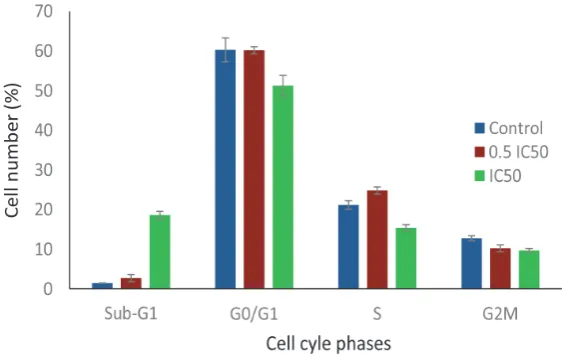
The effect of α-terpineol on MCF-7 cell cycle
The effect of α-terpineol on the MCF-7 cell
cycle changes was enalyzed by lowcytometry. FIGURE 2 shows the MCF-7 cell cycle proile after incubation with α-terpineol at 2 different
concentrations (16.5 and 33.0 µg/mL).
α-Terpineol at concentration of 33.0 µg/mL (IC50) induced MCF-7 cell accumulation until 18.59 % at Sub-G1 phase. This accumulation
was accompanied with the reduction of cycling cells in G0/G1 phase (51.24%), S phase (15.4%) and G2M phase (9.69%) as compared to control. At concentration of 16.5 µg/mL (½ IC50), α-terpineol induced MCF-7 cell accumulation until 24.78% at S phase, as compared to 21.15% in the control cells. The cell accumulation in S phase caused reduction of cycling cells in G2M phase.
FIGURE 2. Cell cycle analysis of MCF-7 cell line using lowcytometri afer 24 hours incubation with α-terpineol compared to control
The effect of α-terpineol on apoptosis
The effect of α-terpineol on the MCF-7 cell apoptosis was analysed by lowcytometry using Annexin V Fluos-Propidium Iodide staining. TABLE 1 shows MCF-7 cell distribution in four quadrant, whereas its percentage of early apoptotic cell after 24 hours
α-terpineol incubation compared to control
is presented in FIGURE 2. The percentage of apoptotic cell after 24 hours incubation with α-terpineol at concentration of 33.0 µg/ mL (5.92 ± 0.13%) was signiicantly higher than that at concentration 16.5 µg/mL (2.57 ± 0.12%) and that control (1.03 ± 0.06%) (p < 0.05).
TABLE 1. MCF-7 cell distribution in four quadrant (lower left/live, lower right/ early apoptotic cell, upper right/ necrotic cell, upper left/necrotic cell)
Treatment Concentration (µg/mL Cell percentage (% ± SD)
Live Early apoptotic Necrotic Necrotic
Control 0 96.42±0.43 1.03±0.06 0.59±0.08 1.95±0.33
Alpha terpineol 16.5 87.48±0.05 2.57±0.12 0.53±0.03 9.42±0.15
FIGURE 3. The effect of 24 hours α-terpineol incubation on apoptosis stimulation on MCF-7 cell line. *signiicantly different (p<0.05)
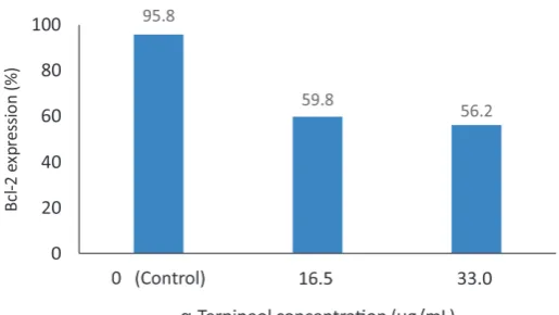
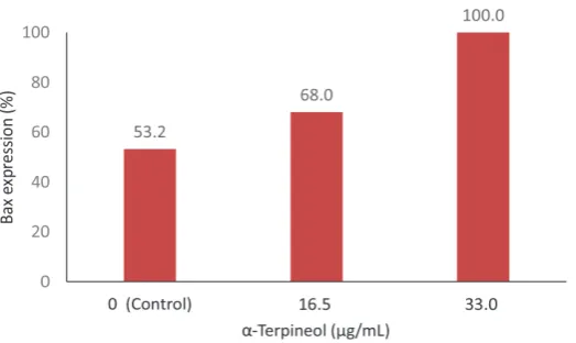
The effect of α-terpineol on Bcl-2 and Bax expressions
The effect of α-terpineol on Bcl-2 and Bax expressions of the MCF-7 cell were analysed by immunohistochemistry. The expression of Bcl-2 and Bax protein on MCF-7 cell line after 24 hours α-terpineol incubation are presented in FIGURE 4 and 5, respectively. The percentage of Bcl-2 expression after 24 hours
incubation with α-terpineol at concentration of 16.5 and 33.0 µg/mL (59.8 and 56.2%) were lower than that control (95.8%). Inversely, the percentage of Bax expression after 24 hours incubation with α-terpineol at concentration
of 33.0 µg/mL (100 %) was signiicantly
higher than that at concentration 16.5 µg/mL (68.0 %) and that control (53.2).
FIGURE 4. The effect of 24 hours α-terpineol incubation on Bcl-2
FIGURE 5. The effect of 24 hours α-terpineol incubation on Bax
expression of MCF-7 cell line
DISCUSSION
This study showed that α-terpineol
inhibited the MCF-7 cell growth in a dose-dependent manner with an IC50 value of 33.0 µg/mL. The cytotoxicity of α-terpineol on different cancer cell lines has been reported in the previous studies by some authors. Hasan
et al.14 reported that α-terpineol was most
active on NCI-H69 lung cancer cell with IC50 of 39.4 µg/mL among 14 human cancer cell lines tested. α-Terpineol was also reported active against T47D breast cancer cell line (IC50: 20.5 µg/mL)15 and HeLa cervical cancer
cell line (IC50: 12.5 µg/mL).16 Based on a
criteria proposed by America National Cancer Institute, α-terpineol can be classiied as a potential anticancer agent (IC50 about 30 µg/ mL).18
The effect α-terpineol on MCF-7 cell cycle in this study showed that α-terpineol
induced cell cycle arrest in the cell line tested in a dose-dependent manner. This result is consistent with the previous reports which
showed that α-terpineol is active in inducing
cell cycle arrest. Itani et al.13 reported that Salvia libanotica essential oil containing three bioactive compounds i.e. linalyl acetate,
terpeniol and camphor caused signiicant
growth suppression of colorectal cancer cell lines HCT116 (p53+/+) in pre G1 phase and in
p53-/- cells caused cell accumulation in pre G1
and G2/M phases. Moreover, Hassan et al.14
also reported that α-terpineol inhibited the
proliferation of lymphoma U937-GTB cancer cells in G0/G1 phase lead to reduction in the number of cells in the later stages of cell cycle (S, G2 and M) of the cells.
This study also showed that α-terpineol
induced apoptosis of the cell line tested in a dose-dependent manner. The apoptosis
induced by α-terpineol may be through
Bcl-2 protein family as demonstrated by the decrease of Bcl-2 ad the increase of Bax expressions in this study. The effect of
α-terpineol on cancer cell apoptosis has been
demonstrated previously. Itani et al.13 reported
that α-terpineol, linaly acetate and camphor
synergised to induce cell cycle arrest and apoptosis, mainly via mitochondrial damage (cytochrome crelease), caspase activation, and PARP cleavage, in human colorectal cancer cells. Furthermore, Hassan et al.14
reported that α-terpineol exhibited a potential anticancer which acts by suppressing NF-κB
which signals various cancer cells line.
are involved in the control of inlammatory responses, developmental processes, cellular growth and apoptosis.18 α-Terpineol inhibits
NF-κB translocation and activity and down-regulates the expression of several
NF-κB-related genes such as IL-1β and IL1R1
resulting in the inhibition of cancer cells growth.14
CONCLUSION
In conclusion, α-terpineol has potential anticancer activity against MCF-7 cancer cell line trough through cells cycle inhibition and apoptosis stimulation. The cells cycle inhibition is indicated by the induction of cell accumulation in Sub-G1, whereas cells apoptosis induction may be through Bcl-2 protein family as demonstrated by the decrease of Bcl-2 ad the increase of Bax expressions.
ACKNOWLEDGMENTS
We would like to thank Prof. Ir. Arief Budiman, MS, D.Eng. who kindly provided
α-terpineol. We also would like to Stem Cell
and Cancer Institute, Kalbe Farma, Jakarta who provided MCF-7 cell line. This study was funded by Ministry of Health, Republic of Indonesia through Iptekdok Risbin Research Grand 2011.
REFERENCES
1. Ferlay J, Shin HR, Bray F, Forman D, Mathers C, Parkin DM. Estimates of worldwide burden of cancer in 2008: GLOBOCAN 2008. Int J Cancer 2010 ; 127(12):2893-917. http:// dx.doi.org/10.1002/ijc.25516
2. Bray F, Ren JS, Masuyer E, Ferlay J. Global estimates of cancer prevalence for 27 sites in the adult population in 2008. Int J
Cancer 2012; 132(5):1133-45. http://dx.doi. org/10.1002/ijc.27711
3. Jemal A, Bray F, Center MM, Ferlay J, Ward E, Forman D. Global cancer statistics. Ca Cancer J Clin 2011;61:69–90. http://dx.doi. org/10.3322/caac.20107
4. Kimman M, Norman R, Jan S, Kingston D, Woodward M. The burden of cancer in member countries of the Association of Southeast Asian Nations (ASEAN). Asian
Paciic J Cancer Prev13; 411-20. http://
dx.doi.org/10.7314/APJCP.2012.13.2.411 5. Bidang Rekam Medis RSKD. 10 besar kanker
tersering di RS Kanker Dharmais rawat jalan (kasus baru) tahun 2007 [serial online].2009 [cited 2015 January 22]. Available from URL: http://www.dharmais.co.id/index.php/ statistic-center.html
6. Michaud LB, Espirito JL, Esteva FJ. Breastcancer. In: Dipiro JT, Talbert RT, Yee GC, Matzke GR, Wells BG, Posey LM, Eds. Pharmacotherapy: a pathophysiologic approach, 7th ed., NewYork:The McGraw- Hill Companies, Inc., 2008: 2121-56.
7. Medina PJ, Fausel C. Oncologic disorders, cancer treatment and chemotherapy. In: DipiroJT, Talbert RT, Yee GC, Matzke GR, Wells BG, Posey LM, Eds. Pharmacotherapy: a pathophysiologic approach, 7th ed., New York: The McGraw-Hill Companies, Inc., 2008: 2099.
8. Partridge AH, Burstein HJ, Winer EP. Side effects of chemotherapy and combined chemohormonal therapy in women with early-stage breast cancer. J Natl Cancer Inst Monogr 2001;30:135-42.
http://dx.doi.org/10.1093/oxfordjournals. jncimonographs.a003451
10. Moreira MR, Cruz GMP, Lopes MS, Albuquerque AAC, Leal-Cardoso JH. Effects of terpineol on the compound action potential of the rat sciatic nerve. Braz J Med Biol Re 2001; 34: 1337-40. http://dx.doi.org/10.1590/ S0100-879X2001001000015
11. Budiman A, Arifta TI, Dina, Sutijan.
Continuous production of α-terpineol from α-pinene isolated from Indonesian crude
turpentine. Modern Appl Sci 2015; 9(4): 225- 32. http://dx.doi.org/10.5539/mas.v9n4p225 MSDS. 2009. Terpineol. [serial online] 2009 [cited, 2015 January 25th] Available from: http:// www.sciencelab.com/msds. php?msdsId=9925180
12. Itani WS, El-Banna SH, Hassan SB, Larsson RL, Bazarbachi A, Gali-Muhtasib HU. Anticolon cancer components from lebanese sage (Salvia libanotica) essential oil. Cancer Biol Ther 2008; 7(11):1765-73. http://dx.doi. org/10.4161/cbt.7.11.6740
13. Hassan SB, Galib-Muhtasib H, Goransson H, Larsson R. Alpha terpineol: a potential anticancer agent which acts through
suppressing NF-κB signalling. Anticancer
Res 2010; 30:1911-20.
14. Indrasetiawan P, Astuti I, Mustofa.
Activity of α-terpineol as a potential
anticancercandidate:cytotoxicity, pro apopto-tican dan tiproliferative evaluation in TD47 cell lines. J Med Sci 2012; 44(1):10-7. 15. Rasuane N, Astuti I, Mustofa. Cytotoxicity of
α-terpineol in HeLa cell line and its effects
to apoptosis and cell cycle. J Med Sci 2014; 46(1):1-9.
16. Mossman T. Rapid colorimetric assay for cellular growth and survival: application to proliferation and cytotoxicity assays. J Immunol Methods 1983; 65(1-2):55-63. http:// dx.doi.org/10.1016/0022-1759(83)90303-4 17. Suffness M, Pezzuto JM. Assays related to
cancer drug discovery. In: Hostettmann K editor. Methods in plant biochemistry: assays for bioactivity, vol. 6. London: Academic Press, London, 1990: 71–133.