PUBLISHED BY THE AMERICAN ARACHNOLOGICAL SOCIETY
Whip spiders of the genus
Sarax
in the Papuan region, with description of two new species
(Amblypygi: Charinidae)
Cahyo Rahmadi: Natural History Laboratory, Faculty of Science, Ibaraki University, Mito, 310-8512 Japan and Museum Zoologicum Bogoriense, Research Center for Biology, Indonesian Institute of Sciences (LIPI) Jl. Raya Jakarta-Bogor Km. 46, Cibinong, 16911 Indonesia. E-mail: [email protected]
Jun-ichi Kojima: Natural History Laboratory, Faculty of Science, Ibaraki University, Mito, 310-8512 Japan
Abstract. Three species of the genus Sarax are recognized in the Papuan region. Among them, two species, Sarax newbritainensis, new species, from New Britain, andS. monodenticulatus, new species, from Waigeo Island are described.
Sarax newbritainensisresembles S. willeyi in having the same number of denticles on the pedipalpal tarsus, but they distinctly differ from each other in body size, form of carapace, length of legs and number and arrangement of the trichobothria of tibia of leg IV.Sarax monodenticulatusis distinguished from the other two Papuan species by possessing a single denticle on the pedipalpal tarsus. The taxonomic status and the natural history of the species are discussed.
Keywords: Taxonomy, Waigeo Island, Batanta Island, New Britain, Papua New Guinea, Indonesia
Whip spiders, or Amblypygi, are flattened arachnids characterized by their raptorial pedipalps and the first legs modified into extremely elongate antenniform appendages (Weygoldt 2000). They are mostly active during the night and hide under stones and fallen trees during the day. A total of 143 species belonging to 17 genera in five families are currently known (Harvey 2002, 2003; Armas & Gadar 2004; Weygoldt 2002; 2006; Rahmadi & Harvey 2008). Of these, ten species and two subspecies belong to the charinid genusSaraxSimon 1892, which is known from the Oriental and Papuan region (India to the Solomons) (Harvey 2003; see also Weygoldt 2005:12).
Kraepelin (1895) recorded Sarax sarawakensis (Thorell 1888) from New Guinea and Solomon Islands, and Pocock (1898) recorded the species from New Britain based on specimens procured by Dr. A. Willey, the director of the Colombo Museum at that time. Gravely (1915) describedS. willeyibased on the two specimens ‘‘preserved in the Indian Museum’’ and ‘‘collected by Dr. Willey in New Britain.’’
The other charinid genus occurring in the Papuan region is CharinusSimon 1892. The genus occurs pantropically with 35 species and in the Papuan region only one species,Charinus papuanusWeygoldt 2006, is known, from the type locality near Port Moresby, Papua New Guinea (Weygoldt 2006).
During a study on whip spiders collected during the ‘‘Ekspedisi Widya Nusantara (e-WIN Expedition)’’ to Raja Ampat Island, West Papua (organized by the Indonesian Institute of Sciences in 2007 and 2008), and based on the examination of some additional specimens from Papua New Guinea and New Britain and the holotype of Charon
sarawakensis(5S. sarawakensis), we recognized three distinct species of Sarax in the Papuan region. Two of them are described here as new to science, and the taxonomic status of S. willeyiis discussed.
METHODS
Specimens from the e-WIN expedition examined in this study were preserved in 90–95% ethanol and others in 70– 80%. General terminology and pedipalpal spination follow Weygoldt (2000), and pedipalpal terminology follows Harvey and West (1998). We made the measurements (in mm) and drawings using, respectively, an ocular micrometer and a drawing tube mounted on a stereoscopic dissecting microscope (Olympus SZX12). To prepare the digital images of the carapace, we compiled multiple focal planes taken with a digital microscope (KEYENCE VH-5500) using the software Helicon Focus 4.70 (Helicon Soft Ltd. 2009: online at http:// www.heliconsoft.com/heliconfocus.html). In order to examine the structure of genitalia we lifted the genital operculum. The acronyms of the museums/institutions are as follows: MCSG, Museo Civico di Storia ‘‘Giacomo Doria,’’ Genova, Italy; MCZ, Museum of Comparative Zoology, Cambridge, Mas-sachusetts, USA; MZB, Museum Zoologicum Bogoriense, Cibinong, Indonesia; MNHN, Museum National d’Histoire Naturelle, Paris, France.
The holotype female ofSarax sarawakensis(Thorell 1888), lodged in MCSG and labeled ‘‘Sarax saravakensis Thor. Sarawak, Viag Doria and Beccari’’ was examined to compare with the threeSaraxspecies treated below.
KEY TO THE CHARINID WHIP SPIDERS OF THE PAPUAN REGION
1. Abdominal sternite III without ventral sac covers . . . Charinus papuanusWeygoldt Abdominal sternite III with ventral sac covers . . . Sarax…2 2. Body reddish-brown. Tibia of leg IV with 17 trichobothria (Fig. 4d); pedipalpal tarsus with one denticle (Fig. 4b) . . . .
. . . .Sarax monodenticulatusnew species Body pale brown or dark greenish-brown. Tibia of leg IV with 17 or 19 trichobothria; pedipalpal tarsus with two denticles (Figs. 2c, 3b) . . . 3
2010. The Journal of Arachnology 38:475–484
3. Body pale brown. Legs elongate; tibia of leg IV with 19 trichobothria (Fig. 3e); trichobothrium bt close to distal margin; metatarsus of leg I 1.5 times as long as the first tarsal segment . . . .Sarax newbritainensisnew species Body dark greenish-brown. Legs not elongate; tibia of leg IV with 17 trichobothria (Fig. 2d); trichobothriumbtlocating at about mid-length of fourth basitibial segment; metatarsus of leg I as long as the first tarsal segment . . . Sarax willeyiGravely
TAXONOMY
Family Charinidae Quintero 1986 GenusSaraxSimon 1892
Sarax Simon 1892:43, 48; Kraepelin 1895:45; Kraepelin 1899:250; Pocock 1900:131; Gravely 1915:441; Mello-Leita˜o 1931:55; Werner 1935:471; Weygoldt 2000:25; Harvey 2003:7.
Phrynichosarax Gravely 1915:437; Mello-Leita˜o 1931:52 (as Phrynicosarax [sic]); Werner 1935:470; Weygoldt 2000:25 (synonymized withSarax); Weygoldt 2002:146.
Type species.—Sarax: Sarax brachydactylusSimon 1892, by
original designation.
Phrynichosarax: Phrynichosarax cochinensis Gravely 1915, by original designation.
Diagnosis.—Small to medium-sized whip spiders; adult body length 4–10 mm. Pedipalpal patella with three large primary spines, the distal spine largest, subsequent spines becoming shorter proximally; ventral sac covers present on abdominal sternite III. Lateral eyes close to lateral margin of carapace. Basitibia of leg IV consisting of three or four segments.
Remarks.—The family Charinidae currently consists of the following three genera: Sarax,CatageusThorell 1889 with a single species from Myanmar, and Charinusconsisting of 35 species with a circum-tropical distribution (Harvey 2003; Weygoldt & Van Damme 2004; Weygoldt 2006).Saraxis, in general, similar to Charinus, especially in the pedipalpal patella spination, but clearly distinguished by the presence/ absence of the ventral sac cover; inSaraxthe ventral sac cover is present, while it is absent in Charinus. Catageus can be distinguished from the other two genera by the second spine of the pedipalpal patella being the largest (in other two genera, the first is the largest) and the proximal spine of the antero-dorsal pedipalpal tibia being larger than the distal one (Weygoldt 2000).
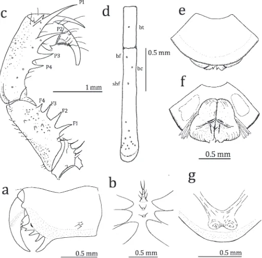
Sarax willeyiGravely 1915 (Figs. 1a, 2a–g)
Sarax willeyi Gravely 1915:441, fig. 7; Mello-Leita˜o 1931:55; Werner 1935:471; Kraus 1970, figs. 10–11; Harvey 2003:9.
Material examined.—INDONESIA: West Papua Province: 3 females (MZB.Ambl.119–120 and 121 [ovigerous]), under stone in forest near Gua Eleg (00u53.519S, 130u40.069E, 156 m asl.), Wailebet, Raja Ampat Regency, Batanta Island, 1 May 2008, C. Rahmadi; 4 males (MZB.Ambl.122, 125–127), 1 female (MZB.Ambl.123, ovigerous with 7 eggs), under stone in small limestone forest near Gua Umso (00u49.899S, 130u53.829E), Yenanas, Raja Ampat Regency, 27 April 2008, C. Rahmadi; 1 male (MZB.AMbl.128), Solol Village, Salawati Island, 7 May 2008, C. Rahmadi. PAPUA NEW GUINEA:Madang Prov-ince: 2 males, 3 females (MZB.AMbl. 129–133), 2 males, 2 females (MCZ DNA104752), Madang Bitabag Reserve, (05u08919.30S, 145u46928.20E, 109 m asl.), 28 March 2006, R.M. Clouse, Ulai & Nataniel.
Diagnosis.—Sarax willeyi differs from other congeneric species from the region by its small size (adult body length about 4.0–6.2 mm) and dark greenish-brown body. The legs are proportionally shorter than other species. The pedipalpal tibia has two spines on the antero-dorsal margin; the proximal spine more than half as long as the distal one. Pedipalpal tarsi with two denticles; proximal denticle about half as long as distal one. Tibia of leg IV with 17 trichobothria; trichoboth-riumbcmuch closer tobfthan tosbf, andbtat about mid-length of the fourth basitibial segment (Fig. 2d). The metatarsal segment of leg I as long as the subsequent two segments together. Description.—Male: Color in alcohol. Carapace dark
greenish-brown with yellow marks centrally. Pedipalps and legs green except as follows: major spines of pedipalps light brown; tibia and tarsus of leg I with yellow annulations; distal and proximal margins of basitibiae of Legs II–IV brown; distibiae and tarsi of Legs II–IV light green. Abdomen green with brown spots on each tergite.
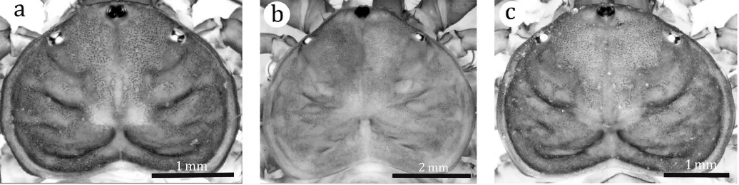
Carapace (Fig. 1a): Width about 1.5 times length; surface finely granulate, without setiferous tubercles; median sulcus deep in posterior one-fourth of the carapace, three sulci running laterally on each lateral half of carapace; flange wide and bend upward; anterior margin rounded, with six fine frontal setae. Median eye tubercle black, without apical setae, slightly emarginated antero-medially to form heart shape; median eyes facing antero-laterally. Lateral eye close to lateral margin of carapace.
Chelicera (Fig. 2a): Dorsum smooth, with one fine frontal seta and three fine lateral setae. Basal segment with four teeth: lowermost tooth largest, uppermost tooth bicuspid, with upper cusp larger than lower cusp; inner surface with seven setae arranged in vertical row; outer surface with small blunt tooth opposite bicuspid tooth and four setae near proximal margin. Movable hand with three teeth about equal in size.
Sternum (Fig. 2b): First sternite (5tritosternum) elongate, with paired apical, median and strong basal setae; second
Figure 2.—Sarax willeyi, from Batanta Island. a. External view of left chelicera; b. Sternal area of carapace, ventral view; c. Antero-dorsal view of left pedipalp; d. Arrangement of trichobothria on the fourth basitibial segment and distibia of leg IV; e, f. Male genitalia (e. ventral view; f. dorsal view); g. Dorsal view of female gonopods.
and third sternites rounded and flattened, with paired apical setae.
Pedipalp (Fig. 2c): Short and stout. Trochanter with four setiferous tubercles arranged in a row along antero-dorsal margin, one spine medially and four setiferous tubercles on antero-ventral margin; ventro-anterior apophysis equipped with ten setiferous tubercles present on distal margin of trochanter. Femur with four major spines, some setiferous tubercles and small tubercles in the antero-dorsal part, length of spine F1.F2.F3.F4; area without setiferous tubercles or small tubercles forming narrow band running lengthwise from proximal to distal margin; four major spines, several minor spines and small tubercles on antero-ventral margin; one spine present dorsally of FI and as long as 3/4 length of FI, length of spine FI.FII.FIII, three minor spines between FI and FII. Patella with four major spines, several minor spines, several setiferous tubercles and small tubercles on antero-dorsal margin; length of spine P1.P2.P3.P4, two minor spines (one in several specimens) between P1 and distal margin of patella; three major spines, several setiferous tubercles and small tubercles on antero-ventral margin, length of spines PI.PII.PIII. Tibia with two major spines on antero-dorsal margin, length of proximal spine more than half that of distal one; one major spine on antero-ventral margin close to distal margin of tibia; outer surface of the tibia with several setiferous tubercles, finely granulate. Tarsus completely divided (claw clearly demarcated by articulation), with two denticles on antero-dorsal margin; proximal denticle about 3/4 as long as distal denticle; cleaning organ ventrally with about 30 modified hairs; apotele present.
Legs (Fig. 2d): Femora of Legs I–IV with small tubercles bearing setae. Tibia and tarsus of leg I consisting of 23 and 41 segments, respectively; tibiae of Legs II and III two-segmented; basitibia of leg IV four-segmented, fourth segment with one trichobothrium (value in parentheses: ratio of the distance from the trichobothrium to the proximal margin of the segment against the length of the segment),bt(0.50); distitibiae of Legs II–IV each with 16 trichobothria (Fig. 2d),bf(0.07),sbf(0.31), bc(0.17),btat about mid-length of the segment,bcclose tobf than tosbf. Tarsi of Legs II–IV four-segmented; first segment about as long as length of subsequent three segments combined; second segment with light-yellow transverse line; fourth segment without oblique slit; pulvilli present.
Genitalia (Figs. 2e, f): Covered ventrally by genital opercu-lum; paired apically-pointed small median projections present at posterior margin; two brown marks present near the base of projections (Fig. 2e). In dorsal view, paired anteriorly-rounded brown bands present; paired weakly-sclerotized brown markings present medially (Fig. 2f).
Female:Similar to the male. Gonopods with paired finger-like apically pointed projections (Fig. 2g).
Measurements.—Male (n57), [female (n59)]; values for segments of the appendages are their lengths. Body length (excluding chelicera) 4.00–6.20 [4.04–6.95]. Carapace: median length 1.75–2.40 [1.48–2.70]; width 1.50–3.60 [2.08–3.75]; median eyes to anterior margin of carapace 0.04–0.05 [0.04– 0.05]; distance between lateral eyes 0.92–1.72 [0.80–1.75]; lateral eye to anterior margin of carapace 0.25–0.40 [0.20– 0.40]; lateral eye to lateral margin of carapace 0.08–0.25 [0.04–0.25]. Pedipalps: trochanter 0.40–0.80 [0.32–0.75];
femur 1.00–2.28 [0.70–1.90]; patella 1.00–2.60 [1.00–2.40]; tibia 0.40–1.25 [0.28–1.00]; tarsus 0.40–1.00 [0.60–1.25]. Leg I: femur 2.50–4.60 [4.60]; patella 0.35–0.50 [0.32–0.50]. Leg II: femur 1.75–3.20 [1.48–3.25], patella 0.48–0.70 [0.40–0.75]; basitibia 2.08 [0.80–2.25]; distitibia 1.60 [0.84–1.50]; metatar-sus+ tarsus 1.60 [0.88–1.50]. Leg III: femur 2.25–3.60 [1.68– 3.75]; patella 0.48–0.70 [0.40–0.75]; basitibia 1.15–2.80 [1.20– 3.00]; distitibia 1.28–1.80 [1.00–2.00]; metatarsus+ tarsus 1.05–1.60 [1.00–2.00]. Leg IV: femur 1.85–3.20 [1.52–3.50]; patella 0.35–0.70 [0.40–0.70]; basitibia 1.60–3.00 [1.20–3.15]; distitibia 1.00–1.60 [0.80–1.65]; metatarsus +tarsus 0.95–1.60 [0.80–1.75].
Remarks.—Sarax willeyi was described by Gravely (1915) based on two specimens collected by Dr. A. Willey in New Britain, which were stated to be in the Indian Museum in Calcutta. Pocock (1898) examined the specimens collected by Dr. A. Willey in New Britain, compared them with Sarax specimens from Luzon Island and Andaman Islands, and having agreed with Kraepelin (1895), he concluded that only the single species, S. sarawakensis, was recognized in Sarax. Although Gravely (1915) did not refer to Pocock (1898), the specimens collected by Dr Willey in New Britain that they examined may have been the same.
Gravely (1915) distinguishedS. willeyifromS. sarawakensis by the proximal spine of the pedipalpal tarsus being more than half as long as the distal one (less than half inS. sarawakensis). The specimens from Batanta Island, Salawati Island, and Madang in Papua New Guinea that we examined have the proximal spine of the pedipalpal tibia longer than half the length of the distal spine. The holotype of Charon sarawa-kensis (5 Sarax sarawakensis), on the other hand, has the pedipalpal tibia with the proximal spine shorter than half the length of distal one. In addition, the holotype of C. sarawakensis has the pedipalpal tarsus armed with two very small denticles, while all of our specimens identified as S. willeyi have the pedipalpal tarsus with two rather long denticles. All the distribution records of S. sarawakensis in the Papuan regions including Solomon Islands and Bismarck Archipelago, such as reported by Kraepelin (1899), need reconfirmation. At this moment, it is reasonably considered that S. sarawakensis is restricted, in its distribution, to the Oriental region.
Natural history.—We found this whip spider most often under stones, and when specimens were collected during the day, they were found in a resting position attached to the underside of a stone on forest floor. We also collected some individuals under fallen trees in Salawati Island. According to the collection data, the specimens from Madang, Papua New Guinea were collected under rotten logs.
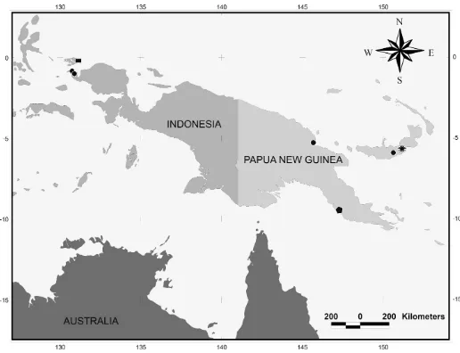
Distribution.—This species occurs in New Britain (Gravely 1915), and is here newly recorded from the West Papua Province of Indonesia, and in the Madang Province of Papua New Guinea (Fig. 5).
Sarax newbritainensisnew species
(Figs. 1b, 3a–h)
(MNHN.Am.6). Paratype: 1 female, same locality data as the holotype (MZB.Ambl.134).
Etymology.—The specific name refers to the island where the type locality is located.
Diagnosis.—Sarax newbritainensis differs from all other congeneric species in the region by the large adult body length (about 8.25–9.5 mm) and the pale brown body. The carapace is proportionally less wide, without distinct lateral sulci, and the eyes are reduced in size. The pedipalpal tarsus has two rather long denticles, separated from each other by about twice the basal diameter of the denticle; the proximal denticle is about half as long as the distal one. The legs are elongate; metatarsi of leg II–IV are longer than the length of the subsequent three tarsal segments combined. Tibia of leg IV
with 19 trichobothria, arranged with bc situating near the middle of bfandsbfandbtclose to the distal margin of the fourth basitibial segment.
Description.—Male:Color in alcohol: Carapace light brown
with darker marks; pedipalps brown but pedipalpal tarsus light brown. Legs I–IV yellowish brown, without annulations except for tibia and tarsus of leg I having white annulations; patella dark brown.
Carapace (Fig. 1b): Width about 1.3 times the length; surface finely granulate, sparsely with small tubercles, without setiferous tubercles; sulcus deep and distinct on posterior one-fourth of carapace. Flange present from level of lateral eyes to posterior margin, wide and bent upward along lateral margin, narrow on posterior margin. Anterior margin of carapace
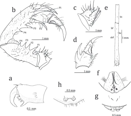
Figure 3.—Sarax newbritainensis, new species, from New Britain, Papua New Guinea. a. External view of left chelicerae; b. Antero-dorsal view of left pedipalp; c. Antero-ventral of left pedipalpal trochanter; d. Left pedipalpal tibia; e. Arrangement of trichobothria on fourth basitibial segment and distibia of left leg IV; f, g. Male genitalia (f. dorsal view; g. ventral view); h. Dorsal view of female gonopods.
rounded, with six frontal setae and four small fine setae close to each antero-lateral corner; slightly concave in part anterior to lateral eyes. Median eye tubercle black, without apical seta, slightly reduced in size, slightly emarginated antero-medially to form heart shape; eyes facing antero-laterally. Lateral eyes close to lateral margin of carapace, distance between them about the diameter of the lateral eye, normal pigmentation and tapetum. Frontal process triangular, visible from above.
Chelicera (Fig. 3a): Dorsum smooth, with three setiferous tubercles and two fine frontal setae. Basal segment with four teeth: the lowermost tooth largest, the uppermost tooth bicuspid, with upper cusp larger than lower cusp; inner surface with 14 setae arranged in vertical row near proximal margin; outer surface with small blunt tooth opposite the bicuspid tooth, and with five setae arranged in vertical row. Movable hand with five teeth; second tooth largest.
Sternum: First sternite (5 tritosternum) elongate, with paired apical and strong median setae, five setae between apical and median setae and 12 small setae at base. Second and third sternites rounded, with paired apical setae; second with six basal setae; third with five basal setae.
Pedipalp (Figs. 3b–d): Short and stout, with several setiferous tubercles. Trochanter with a row of seven setiferous tubercles on antero-dorsal margin, six setiferous tubercles dorsally and one median spine on antero-ventral margin, medially with one spine and eight setiferous tubercles; ventral anterior apophysis equipped with several setiferous tubercles in basal part present on distal margin of trochanter (Fig. 3c). Femur with four major spines, several setiferous tubercles and small tubercles on antero-dorsal part, length of spine F1.F2.F3.F4; four major spines, several setiferous tubercles and small tubercles on antero-ventral margin, one spine present dorsally of FI and as long as 3/4 length of FI, length of spine FI.FII.FIII.FIV, one minor spine between FI and FII. Patella with four major spines, several setiferous tubercles and small tubercles on antero-dorsal margin, length of spine P1.P2.P3.P4; one setiferous tubercle and one spine between P1 and distal margin of patella, the spine as long as half length of P1; three major and one minor spines, several setiferous tubercles and small tubercles on antero-ventral margin, length of spines PI.PII.PIII. Tibia with two major spines on antero-dorsal margin, length of proximal spine more than half the length of distal one (Fig. 3d); antero-ventral margin with one major spine; outer surface finely granulate, with several setiferous tubercles. Tarsus completely divided (claw clearly demarcated by articulation), with two denticles on antero-dorsal margin: proximal denticle slightly longer than half the length of distal one, distance between them about three times basal diameter of proximal denticle; cleaning organ ventrally with 28–30 modified hairs; several blunt setae on inner surface of tarsus; apotele present.
Legs (Fig. 3e): Femora of Legs IIV with small setiferous tubercles. Tibia and tarsus of leg I with 23 and 41 segments, respectively; tibiae of leg II and III two-segmented; basitibia of leg IV four-segmented: fourth segment with one trichobothrium (value in parentheses as forS. willeyi),bt(0.57); distitibiae of Legs II–IV with 18 trichobothria,bf(0,13),sbf(0.32),bc(0.23), btclose to distal margin,bcat the middle ofbfandsbf, (Fig. 3e). Tarsi of Legs II–IV four–segmented; first segment slightly
longer than length of subsequent three segments combined; second segment with light yellow transverse line; fourth segment without oblique slit; pulvilli present.
Genitalia (Figs. 3f, g): Covered ventrally by genital oper-culum, of which posterior margin is equipped with paired setae; posteriorly with paired ventral and dorsal lobes, the dorsal lobe smaller than ventral one (Fig. 3f). In dorsal view, submedian brown bands running from anterior margin to the middle; inner margin of median lobe with brown region weakly sclerotized; brown spot present on each median lobe. In ventral view, base of ventral lobe with a narrow brown band (Fig. 3g).
Female: Similar to the male but differing as follows: carapace slightly darker; chelicera with three frontal fine setae; meta-sternum slightly more elongate, with two apical setae; tibia and tarsus of leg I respectively with 26 and 42 segments. Gonopods soft, cone-shaped, with several setae on margin of genital operculum (Fig. 3h).
Measurements.—Male (holotype, MNHN.Am.6) [female (paratype, MZB.Ambl.134)]; values for segments of the appendages are their lengths: Body length (excluding chelicera) 9.50 [8.25]. Carapace: median length 3.75 [3.50], width 5.00 [5.00]; median eye to anterior margin 0.05 [0.05], distance between lateral eyes 2.60 [2.25], lateral eye to anterior margin 0.60 [0.65], lateral eye lateral margin 0.20 [0.25]. Pedipalps: trochanter 1.00 [1.15], femur 3.20 [2.50], patella 3.25 [3.00], tibia 1.50 [1.25], tarsus 1.60 [1.50]. Leg I: femur 10.00 [10.15], patella 0.75 [0.75]. Leg II: femur 6.00 [5.75], patella 1.00 [1.00], basitibia 5.00 [4.90], distitibia 3.00 [2.90], metatarsus+tarsus 2.55 [2.60]. Leg III: femur 7.25 [6.50], patella 1.10 [1.25], basitibia 6.35 [6.25], distitibia 3.50 [3.25], metatarsus+tarsus 2.95 [3.00]. Leg IV: femur 6.50 [5.75], patella 1.00 [1.00], basitibia 7.25 [6.75], distitibia 2.95 [2.75], metatarsus+tarsus 3.00 [3.00].
Remarks.—Sarax newbritainensisis distinguished from the otherSaraxspecies known from New Britain,S. willeyi, by the generally larger body, the distinctly paler coloration, the carapace proportionally less wide (about 1.3 times as wide as long, while about 1.5 times inS. willeyi) and without distinct lateral sulci, the eyes reduced in size, the strongly elongated legs, and the number and arrangement of the trichobothria as given in the key.
Natural history.—We collected the specimens of S. new-britainensisfrom the caves called Resurgence of Lali Bairaman in New Britain. The species has characteristics typical of cave dwellers such as small eyes, elongate legs, and pale body color. Distribution.—This species is known only from the type locality in New Britain (Papua New Guinea) (Fig. 5).
Sarax monodenticulatusnew species
(Figs. 1c, 4a–h)
Etymology.—The specific name refers to the presence of one denticle on the pedipalpal tarsus.
Diagnosis.—Sarax monodenticulatus differs from other congeneric species in the region by being small- to medium-sized (adult body length 2.5–6.5 mm) and with a reddish-brown carapace. The pedipalpal tarsus has a single denticle. Metatarsus of leg I as long as the subsequent two segments together; short- and long-segment alternating combination present in second to eleventh tarsal segments.
Description.—Male: Color in alcohol: Carapace dark
red-dish-brown with darker marks; pedipalp dark brown but
pedipalpal tarsus light brown. Legs I–IV brown, without annulations, but tibia and tarsus of leg I yellowish brown, with white annulations. Basitibiae of Legs II–IV brown; their distibiae and tarsi greenish-brown. Abdomen dark brown; each tergite with yellow marginal line and light-brown spots.
Carapace (Fig. 1c): Width about 1.5 times its length; surface finely granulate; frontal area with dense, small tubercles, without setiferous tubercles; median sulcus present in poste-rior one-fourth of the carapace. Flange present in area posterior to lateral eyes, wide but narrow in posterior margin, bent upward. Anterior margin of carapace rounded, with six
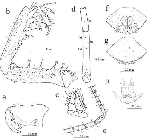
Figure 4.—Sarax monodenticulatus, new species, from Waigeo Island, Indonesia. a. External view of left chelicerae; b. Antero-dorsal view of left pedipalp; c. Antero-ventral view of left pedipalpal trochanter; d. Arrangement of trichobothria on fourth basitibial segment and distibia of left leg IV; e. Apical four tibial and basal five tarsal segments of antenniform leg I; f, g. Male genitalia (f. dorsal view; g. ventral view); h. Dorsal view of female gonopods.
frontal setae and some fine setae close to antero-lateral corner. Median eye tubercle black, without apical setae, slightly emarginated antero-medially to form heart shape; eyes facing antero-laterally. Lateral eye large, with normal pigmentation and tapetum, separated from lateral margin of carapace by about its diameter. Frontal process triangular, visible from above.
Chelicera (Fig. 4a): Dorsum smooth, with two fine frontal setae and several setae on dorsal and lateral parts of outer margin. Basal segment with four teeth: lowermost tooth largest, uppermost tooth bicuspid, with upper cusp larger than lower one; inner surface with eight setae arranged in a vertical row close to proximal margin; outer surface with a small blunt tooth and five setae near proximal margin. Movable hand with two teeth close to the proximal margin.
Sternum: First sternite (5 tritosternum) elongate, with paired apical and median setae, paired small setae between apical and median setae, and four small setae basally. Second and third sternites rounded, each with paired apical setae.
Pedipalp (Figs. 4b, c): Strong and slender, with several setiferous tubercles and small tubercles. Antero-dorsal margin of trochanter with four setiferous tubercles dorsally, two spines and seven setiferous tubercles ventrally; antero-ventral margin with ventral anterior apophysis basally equipped with several setiferous tubercles; ventral anterior apophysis with one spine medially and one spine dorsally (Fig. 4c). Femur with four major spines, several setiferous tubercles and small scales on the antero-dorsal margin, length of spine F1.F2.F3.F4; five major spines, two minor spines, four setiferous tubercles and small scales present on antero-ventral margin, one spine present dorsally of FI and about as long as 3/4 length of FI, length of spine FI.FII.FIII.FIV, single minor spine present between FII and FIII and between FIII and FIV. Patella with four major spines, several setiferous tubercles and small scales on antero-dorsal margin, length of spine P1.P2.P3.P4, one minor spine present between distal margin and P1 and as long as half of the length of P1, two spines and ons setiferous tubercle present between P4 and the proximal margin; three major spines and one minor spine, several setiferous tubercles and small scales on antero-ventral margin, length of spine PI.PII.PIII, one minor spine between PIII and the proximal margin. Tibia with two major spines on antero-dorsal margin: proximal spine slightly longer than half of distal one; one major spine on antero-ventral margin close to distal margin; outer surface finely granular, with setiferous tubercles arranged in three rows. Tarsus completely divided (claw clearly demarcated by articulation), with one denticle on antero-dorsal margin; blunt setae present on inner surface; cleaning organ ventrally with 27–28 modified hairs; apotele present.
Legs (Figs. 4d, e): Femora of Legs I–IV with small scales and setae forming a longitudinal row. Tibia and tarsus of leg I with 23 and 41 segments, respectively (Fig. 4e); tibiae of legs II–III two-segmented; basitibia of leg IV four-segmented (in some specimens left basitibia three-segmented): fourth (third in specimen with three basitibial segments) segment with one trichobothrium (value in parentheses as for S. willeyi) bt (0.61); distitibiae of legs II–IV each with 16 trichobothria (Fig. 4d),bf(0.10),sbf(0.30),bc(0.17),bcabout the middle of bfandsbf,btclose to distal margin. Tarsi of legs II–IV
four-segmented; first segment about as long as length of the subsequent three segments combined; second segment with light yellow transverse line; fourth segment without oblique slit; pulvilli present.
Genitalia (Figs. 4f, g): Covered ventrally by genital oper-culum equipped with several setae; distally with paired small submedian lobes. In dorsal view, with paired large median lobes, of which distal margins are brown; four longitudinal sclerotized bands present posterior to median lobes (Fig. 4f). In ventral view, brown band present near base of distal lobe (Fig. 4g)
Female: Similar to the male. Gonopods soft and cone-shaped (Fig. 4h)
Measurements.—Male (n 55) [female (n 54)]; values for segments of the appendages are their lengths: Body length (excluding chelicera) 4.88–6.48 [2.48–5.60]. Carapace: median length 1.88–2.60 [2.00–2.48], width 2.60–3.72 [3.00–3.60]; median eyes to anterior margin 0.04–0.08 [0.04–0.08], distance between lateral eyes 1.20–1.80 [1.40–1.72], lateral eye to anterior margin 0.28–0.44 [0.36–0.40], lateral eye to lateral margin 0.12–0.16 [0.08–0.16]. Pedipalps: trochanter 0.40–1.00 [0.60–0.80], femur 1.20–3.08 [1.40–2.20], patella 1.40–3.52 [1.60–2.64], tibia 0.60–1.20 [0.68–1.00], tarsus 0.08–1.12 [0.60–1.20]. Leg I: femur 3.08–5.40 [3.40–4.80], patella 0.52– 0.60 [0.40–0.48]. Leg II: femur 2.00–3.40 [2.40–3.20], patella 0.48–0.64 [0.48–0.72], basitibia 1.36–2.40 [1.60–2.40], distitibia 1.12–1.60 [1.20–1.60], metatarsus+tarsus 1.00–1.60 [1.20–1.36]. Leg III: femur 2.40–4.00 [2.80–3.60], patella 0.48–0.72 [0.56– 0.60], basitibia 1.88–3.32 [1.88–3.00], distitibia 1.20–1.80 [1.36–1.68], metatarsus+tarsus 1.20–1.60 [1.20–1.44]. Leg IV: femur 1.20–3.48 [2.40–3.28], patella 0.60–2.80 [0.44–0.60], basitibia 0.48–3.20 [2.16–3.04], distitibia 1.40–2.80 [1.40– 1.60], metatarsus+tarsus 1.04–1.80 [1.20–1.52].
Remarks.—This species is the only Papuan Sarax with a single denticle on the pedipalpal tarsus. The other Sarax species that have the single denticle on the pedipalpal tarsus areS. javensis(Gravely 1915) distributed in West Java andS. cochinensis(Gravely 1915) known from the Western Ghats in Cochin, India. Gravely (1915) distinguished the two species by the length of the denticle; the denticle ofS. cochinesisis long and distinct, while in S. javensis it is minute (see Gravely 1915:figs. 2, 3).Sarax monodenticulatusis distinguished from these two species by having four-segmented basitibia (single-or two-segmented inS. cochinensisand three-segmented inS. javensis[see Gravely 1915:437]).
Natural history.—We collected specimens of this species mostly singly under stones in limestone forests. During the exploration of cave fauna in Waigeo Island, it was never found within the caves.
Distribution.—Sarax monodenticulatusis known only from Waigeo Island (Indonesia) (Fig. 5).
DISCUSSION
sarawakensis from New Britain and, following the view of Kraepelin (1895), remarked on its occurrence in Borneo, the Philippines, and New Guinea. Gravely (1915), on the other hand, described a species from New Britain as distinct fromS. sarawakensisunder the nameSalax willeyi, possibly based on the specimens that Pocock (1898) identified asS. sarawakensis (see remarks in the section of S. willeyi). Consequently, the occurrence of S. sarawakensis in the Papuan region needs reconfirmation. Among the specimens from the Papuan region we have so far examined, we have not recognized any specimens ofS. sarawakensis.
Sarax newbritainensisis very similar toS. willeyi, but can be distinguished fromS. willeyiby the number and arrangement of the trichobothria. Compared with S. willeyi and S. monodenticulatus, both of which live outside caves, S. newbritainensis is larger in adult body size, has a pale body color, smaller median and lateral eyes, and strongly elongate legs, all characteristics highly adapted to cave environments. Such troglomorphic features have not so far been reported
amongst Sarax, while several species of a similar genus, Charinus, are known to have such features (Baptista & Giupponi 2002; Weygoldt et al. 2002; Weygoldt & Van Damme 2004).
ACKNOWLEDGMENTS
Mark Judson (MNHN, Paris, France) and Giuliano Doria (MCSG, Genova, Italy) arranged the first author’s access to the specimens in their respective institutes. Franck Brehier (Toulouse, France) provided the first author with an opportunity to examine the specimens from New Britain. Collections ofS. willeyiby R. M. Clouse, Ulai and Nataniel in Papua New Guinea were funded by Putnam Expedition Grants (MCZ) for R. M. Clouse (Harvard University) who arranged a loan of the specimens. Daisy Wowor and Hari Nugroho (Museum Zoologicum Bogoriense, Bogor, Indone-sia) organized the zoological team in the E-WIN Expedition to Waigeo Island and Batanta Island. Matjaz Kuntner critically
Figure 5.—Map showing the distribution of charinid species in the Papuan region. Symbols used: circle,Sarax willeyifrom Batanta Island, Salawati Island (Indonesia), Madang and New Britain (Papua New Guinea); flower,Sarax newbritainensis, new species from New Britain; rectangle, Sarax monodenticulatus, new species from Waigeo Island (Indonesia); polygon, Charinus papuanus from Port Moresby (Papua New Guinea).
read an earlier draft of the manuscript and provided us with comments and suggestions to improve the manuscript.
LITERATURE CITED
Armas, L.F. de & Y. Gadar. 2004. Nueva especie de Phrynus
Lamarck, 1801 (Amblypygi: Phrynidae) de Chiapas, Me´xico. Revista Ibe´rica de Aracnologı´a 10:133–136.
Baptista, R.L.C. & A.P. de Lea˜o Giupponi. 2002. A new troglo-morphic Charinus from Brazil (Arachnida: Amblypygi: Charini-dae). Revista Ibe´rica de Aracnologia 6:105–110.
Gravely, F.H. 1915. A revision of the Oriental subfamilies of Tarantulidae order Pedipalpi. Records of the Indian Museum 11:433–455.
Harvey, M.S. 2002. The neglected cousins: what do we know about the smaller arachnid orders. Journal of Arachnology 30:357–372. Harvey, M.S. 2003. Catalogue of the Smaller Arachnid Orders of the
World: Amblypygi, Uropygi, Schizomida, Palpigradi, Ricinulei and Solifugae. CSIRO Publishing, Collingwood, Victoria, Aus-tralia.
Harvey, M.S. & P.L.J. West. 1998. New species of Charon
(Amblypygi, Charontidae) from northern Australia and Christmas Island. Journal of Arachnology 26:273–284.
Kraepelin, K. 1895. Revision der Tarantuliden Fabr. Verhandlungen des Naturwissenschaftlichen Vereins in Hamburg 13(3):3–53. Kraepelin, K. 1899. Scorpiones und Pedipalpi. Pp. i–xviii, 1–265.In
Das Tierreich. 8. Arachnoidea. (F. Dahl, ed.). R. Friedlander und Sohn Verlag, Berlin.
Kraus, O. 1970. Genitalmorphologie und systematique der Ambly-pygi (Arachnida). Bulletin du Muse´um National d’Histoire Naturelle, Paris (2)41(Supplement 1):176–180.
Mello-Leita˜o, C. 1931. Pedipalpos do Brasil e algumas notas sobre a ordem. Archivos do Museu Nacional 33:7–72.
Pocock, R.I. 1898. Scorpions, Pedipalpi and spiders collected by Dr. Willey in New Britain, the Solomon Islands, Loyalty Islands, etc. Pp. 95–120.In Zoological Results Based on Material from New Britain, New Guinea, Loyalty Islands and Elsewhere, Collected During the years 1895, 1896 and 1897, Part 1 (A. Willey, ed.). Cambridge University Press, Cambridge, UK.
Pocock, R.I. 1900. The Fauna of British India, Including Ceylon and Burma. Arachnida. (W.T. Blanford, ed.). Taylor and Francis, London.
Rahmadi, C. & M.S. Harvey. 2008. The first epigean species of
Stygophrynus (Charontidae, Amblypygi) from Java and adjacent Islands with notes on S. dammermani Roewer, 1928. Raffles Bulletin of Zoology 56:281–288.
Simon, E. 1892. Arachnides. InEtude sur les Arthropodes caverni-coles de ıˆle Luzon, Voyage de M. E. Simon aux ıˆles Philippines. Mars et avril 1890. (A. Raffray, I. Bolivar & E. Simon, eds.). Annales de la Socie´te´ Entomologique de France 61:35–52. Thorell, T. 1888. Pedipalpi e Scorpioni dell’Arcipelago Malese
conservati nel Museo Civico di Storia Naturale di Genova. Annali del Museo Civico di Storia Naturale di Genova 26:327–428. Werner, F. 1935. Klasse: Arachnoidea, Spinnentiere. Pedipalpen.
Pp. 317–490. In Klassen und Ordnungen des Tierreichs, Volume 5(IV).(8)(3). (H.G. Bronn, ed.). Akademische Verlagsge-sellschaft, Leipzig.
Weygoldt, P. 2000. Whip Spiders: Their Biology, Morphology and Systematics. Apollo Books, Stenstrup, Denmark.
Weygoldt, P. 2002. Sperm transfer and spermatophore morphology of the whip spidersSarax buxtoni,S. brachydactylus(Charinidae),
Charon cf. grayi, and Stygophrynus brevispina nov. spec. (Char-ontidea). Zoologischer Anzeiger 241:131–148.
Weygoldt, P. 2005. Biogeography, systematic position, and repro-duction ofCharinus ioanniticus(Kritscher 1995) with the descrip-tion of a new species from Pakistan (Chelicerata, Amblypygi, Charinidae). Senckenbergiana Biologica 85:43–56.
Weygoldt, P. 2006. New Caledonian whip spiders: Notes onCharinus australianus, Charinus neocaledonicus and other south-western Pacific species of the Charinus species group (Chelicerata, Amblypygi, Charinidae). Verhandlungen des naturwissenschaftli-chen Vereins Hamburg 42:5–37.
Weygoldt, P., H. Pohl & S. Polak. 2002. Arabian whip spiders: four new species of the generaCharinus andPhrynichus (Chelicerata: Amblypygi) from Oman and Socotra. Fauna of Arabia 19:289–309.
Weygoldt, P. & K. Van Damme. 2004. A new troglomorphic whip spider of the genus Charinus (Amblypygi: Charinidae) from Socotra Island. Fauna of Arabia 20:327–334.