P-ISSN : 1978-225X; E-ISSN : 2502-5600 DOI: https://doi.org/10.21157/j.ked.hewan.v12i1.9288
TOTAL BACTERIA AND IDENTIFICATION OF PROTEOLYTIC
RUMINAL AND RETICULUM BACTERIA FROM LOCAL
KACANG GOAT
Safika1*,Darmawi2, Fitria Sari Ramadhani3, Nurhaspika3, and Moliwati3
1
Department of Veterinary Science and Public Health, Faculty of Veterinary Medicine, Bogor Agricultural University, Bogor, Indonesia 2
Microbiology Laboratory, Faculty of Veterinary Medicine, Syiah Kuala University, Banda Aceh, Indonesia 3
Study Program of Veterinary Education, Faculty of Veterinary Medicine, Syiah Kuala University, Banda Aceh, Indonesia *Corresponding author: [email protected]
ABSTRACT
This study aimed to calculate the total bacteria and to identify the proteolytic bacteria in rumen and reticulum of local Kacang goat. The samples used were the rumen and reticulum fluids of five goats at abattoirs in Banda Aceh. Isolation of proteolytic bacteria was carried out using skim milk agar with pour plate method and incubated at 39° C for 48 hours. The bacterial colonies morphology was observed and the total bacterial count was recorded. DNA of the widest proteolytic index colony was isolated, amplified, and sequenced. The results showed that the dominant colonies morphology was white with position inside the agar. The average of total proteolytic bacteria in goat rumen fluid and reticulum fluid were 6.416x10⁶ CFU/mL and 2,382x107 CFU/mL, respectively. Isolates with the widest proteolytic index was Ru3 (2.5 mm) in the rumen which homology and phylogenetic tree analysis of 16S rRNA showed 93% sequence similarity with Bacillussubtilis, while in reticulum was Re1 (2.0 mm) which has 92% sequence similarity to Tatumella. It is concluded that the number of proteolytic bacteria in reticulum is greater than the number of proteolytic bacteria in the rumen of local kacang goat. Homology analysis in this study proved that the Ru3 and Re1 isolate were probably either a new species or unconfirmed species.
____________________________________________________________________________________________________________________ Key words: goat, proteolytic index, reticulum, rumen, bacteria
ABSTRAK
Penelitian ini bertujuan menentukan jumlah dan mengidentifikasi bakteri proteolitik di rumen dan retikulum kambing kacang lokal. Sampel yang digunakan berupa cairan rumen dan retikulum dari lima ekor kambing kacang di tempat pemotongan kambing Banda Aceh. Isolasi bakteri proteolitik menggunakan media skim milk agar dengan metode tuang (pour plate) dan diinkubasi pada suhu 39° C selama 48 jam. Selanjutnya koloni bakteri yang tumbuh dihitung jumlah bakteri dan diamati morfologinya. Koloni dengan indeks proteolitik terluas diisolasi DNA nya dan dilakukan amplifikasi serta sekuensing. Hasil penelitian menunjukkan bahwa morfologi koloni dominan bewarna putih dan posisi di dalam agar. Rata-rata jumlah bakteri proteolitik pada cairan rumen kambing kacang 6,416x106 CFU/ml dan cairan retikulum adalah 2,382x107 CFU/ml. Isolat dengan indeks proteolitik terluas yaitu Ru3 (2,5 mm) pada rumen menunjukkan homologi 93% dengan 16S rRNA Bacillus subtilis sedanghkan Re1 (2,0 mm) pada retikulum menunjukkan homologi 92% dengan Tatumella punctata. Dari hasil penelitian dapat disimpulkan bahwa jumlah bakteri proteolitik retikulum kambing kacang lebih tinggi dibandingkan jumlah bakteri proteolitik pada rumen. Hasil identifikasi 16S rRNA menunjukkan bahwa kemungkinan isolat Ru3 dan Re1 merupakan species baru atau belum dikonfirmasi.
____________________________________________________________________________________________________________________
Kata kunci: kambing, indeks proteolitik, rumen, retikulum, bakteri
INTRODUCTION
Kacang goat is one of the original Indonesian goat whose population quite high and widespread in Indonesia. These goats have a small body with shoulder height of about 50 cm to 60 cm. Some of the benefits of Kacang goat are: efficient in the conversion of grass into meat, resistant to disease, rapid breeding, relatively easy maintenance, not requiring extensive land, the meat is in great interest after beef (Fitra et al., 2009; Sudewo et al., 2012) and able to adapt to tropical environment such as in Aceh. However, this potential is not yet optimal due to the slow growth of the goats and posing a challenge in the effort to improve their productivity, particularly their growth.
Some factors influence the growth of goats, such as the quality and quantity of feed, as well as the process of feed degradation by microbes in the gastrointestinal tract. In farmer community of Aceh, goat feed was not generally given additional concentrate; the forage contain only cellulose, protein, and other nutrients. The crude protein content (CP) of forage varies considerably, ranging from 30 g/kg of dry weight in old grass, to more than 300 g/kg in well-grown young grass (Haryanto,
2012; Yang et al., 2014). The feed was degraded in the rumen, reticulum, and goat omasum, where the digestion in the reticulorumen is very intensive. Protease enzymes, produced by proteolytic microbes, hydrolyze feed proteins in the rumen (Kim et al., 2011; McSweeney and Mackle, 2012; Muslim et al., 2014).
Protease is a group of enzymes which hydrolyzes polypeptide bonds to oligopeptides and amino acids, and then hydrolyzed further to produce ammonia (NH3). Ammonia is utilized by microbes as the primary source of nitrogen for protein synthesis (Kumar et al., 2012). According to Genzenbu and Tesfay (2015), bacteria capable of utilizing nitrogen in the rumen are classified as proteolytic bacteria. Bacteria are found in largest population inside the rumen, and 38% of the total bacteria in the rumen are proteolytic bacteria. The availability and ability of proteolytic bacteria to degrade proteins will affect the amount of amino acids that enter the bloodstream.
Proteus vulgaris, Streptobacillus, Staphylococcus, Streptococcus, Clostridium butylicum, Clostridium spoogerns, Lactobacillus pentosus, Propionibacterium
pentosaceum, Corynebacteria, Bacteroides
amiylophilus, Bacteroides ruminicola, Butyrivibrio fibrisolvens, Butyrivibrio alactacidigens, Selenomonas
ruminantium var. ruminantium, Selenomonas
ruminantium var. lactilytica, Fibrobacter
succinogenes, Butyrivibrio fibrisolvens, Ruminobacte
amylophilus, and Streptococcus bovis (Kamra, 2005;
McSweeney and Mackle, 2012; Hungate, 2013; Krause
et al., 2014). Until present, no information on the total
and types of proteolytic bacteria in Kacang goat in Aceh has been reported, requiring studies to be conducted on the number and types of proteolytic bacteria in rumen and reticulum of Kacang goat.
MATERIALS AND METHODS
Isolation of Proteolytic Bacteria and Total of Proteolytic Bacteria
Five goats were slaughtered at slaughter house and had its rumen and reticulum fluid sampled. Subsequently the liquid sample was diluted with a 10-1 to 10-5 dilution. One ml solution of each dilution was inserted into sterile petri dish, and then 20 mL of skim milk agar medium was added by pour plate method in duplicate before incubated for 48 hours at 39° C.
Total proteolytic bacteria were calculated from the bacterial colonies that grow from the 10-2 to 10-5 dilution by multiplying the number of colonies with the dilution factor. The colonies that grow was Gram stained and its clear zone was measured. The pure colonies with the largest proteolytic index were cultured into liquid medium and incubated for 48 hours. The incubation result was centrifuged at 7000 g and its supernatant was disposed. The DNA of the cell pellet was then isolated.
Total DNA Isolation
Total DNA isolation used PrestoTM Mini gDNA Bacteria kit (Geneaid). Cell pellet was added with 180 µL extraction buffer and 20 µL Proteinase K and was then incubated at 60° C. About 200 µL GB buffer is added to the sample. The sample was mixed and incubated at 70° C. Afterward, 200 µL of absolute ethanol was added and the sample was inserted into the tube column and centrifuged at 14000-16000 x g for 1 minute. A total of 400 µL of buffer W1, was added into the tube column and centrifuged 14000-16000 x g for 30 seconds. The supernatant was discarded and the column was added with 600 µL buffer wash, centrifuged at 14000-16000 x g for 30 seconds and its supernatant discarded. The column was then added with 30-50 µL elution buffer and incubated at room temperature for 3-5 minutes, and then centrifuged at 14000-16000 x g for 1 minute.
Amplification of 16S rRNA Gene
The gene was amplified with PCR. One of the primers (BacF) used was a complement to conserved
areas in the bacterial domain, and the other primary domains (UniB) is based on universal conserved area of the 16S rRNA E. coli gene (Baker et al., 2003). A total of 25 μL PCR reaction mixture was used for DNA amplification. The PCR cycle was 1 minute for denaturation at 94° C, 1 minute for annealing at 50° C and 2 minute for primary elongation at 72° C. The final stages of primary elongation were carried out for 8 minute at 72° C (Safika et al., 2013). Electrophoresis was then performed to the PCR result on 1% agarose gel.
Determination of DNA Sequence
DNA sequencing is performed through commercial services at Macrogen Inc., Korea. The process of determination is using the Dye Terminator method (3'-dye labelled dideoxynucleotide triphosphate) which includes several stages, namely preparation of templates, sequencing reactions, PCR product purification, and electrophoresis with scanning fluorescence.
Phylogenetic Analysis
The sequencing results are compared using program Basic Local Alignment Search Tool (BLAST) program at NCBI http://www.ncbi.nlm.nih.gov. Homology analysis of the 16S rRNA gene sequence used the data in GenBank. The phylogenetic tree was constructed using a neighbor-joining model matrix from the MEGA 7.0 program (Kumar et al., 2016), with the maximum composite likelihood substitution method (Tamura et al., 2004) with a bootstrap analysis 1000 times.
RESULTS AND DISCUSSION
The Morphology of Proteolytic Bacteria
The growth of proteolytic bacteria colonies from rumen and reticulum fluid of Kacang goat were seen spreading inside the skim milk agar (SMA) medium. Colonies that grow in the media indicated that the bacteria are facultative anaerobic bacteria. The growing colonies have a variety of morphologies. The result of isolation of proteolytic bacteria can be seen in Figure 1. Proteolytic colonies were observed its morphologies, such as, color, shape, position, fringe, and colony diameter. Based on the observations made, bacterial colonies that grow were generally white with rounded shape, and smooth edges, with varying proteolytic index (IP) as can be seen in Table 1.
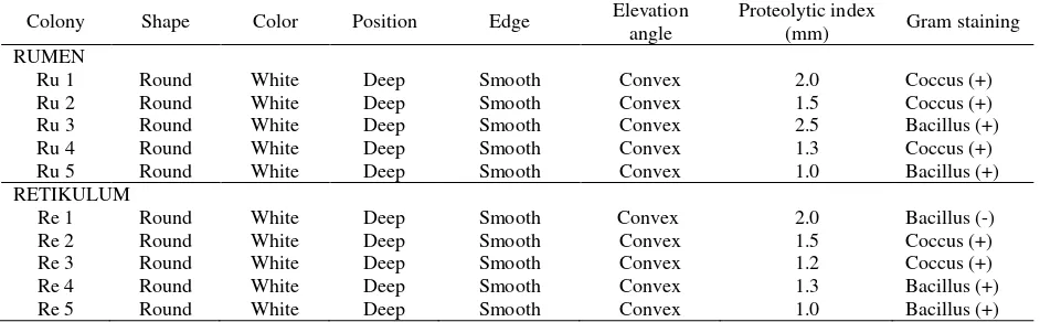
was used to show the hydrolytic activity of proteases (Susanti, 2003; Yuanita and Wikandari, 2014). Increased enzyme activity is evident with the increase in the diameter of the clear zone present around bacterial colonies on skim milk media. Based on the morphological observations of Gram staining (Figure 2), the bacteria found are generally gram-positive coccus or basil bacteria and Gram-negative basil bacteria in both the rumen and the reticulum fluid (Table 1).
Total of Proteolytic Bacteria
Colonies of proteolytic bacteria on all petri dish from each dilution was counted. Dilution reduced the quantity of bacteria that grows on the media. The greater the dilution result in the lesser the number of colonies growing on the media. Dilution can also help facilitate the calculation of the colony. The total of proteolytic bacteria from the rumen fluid and kacang goat reticulum grown on skim milk agar after incubation for 48 hours can be seen in Table 2.
In this study, the number of proteolytic bacteria in the local kacang goat reticulum was greater (2.382x10⁷ CFU/mL) than in the rumen (6.416x10⁶ CFU/mL). The total of bacterial results of this study is smaller than Eschenlauer et al. (2002), which stated that the total of proteolytic bacteria in the rumen is 109 cells/g, but the total of proteolytic bacteria in goat or cow reticulum
has not been reported. The existence of a number of varying bacteria is influenced by the type and content of feed consumed by the livestock. In general, nutrient content in forages consists of cellulose (35-50%), hemicellulose (20-35%), lignin (50-30%) and protein (18-30%) (Kamra, 2005; Haryanto 2012; Yang et al., 2014). High-fiber feeding, such as forage in large quantities without supplementation with protein-rich feed, causes fewer proteolytic bacteria or microbes in the rumen of Kacang goat. When more animals are fed rough fiber feeds, the populations of fiber-degrading microbe will be dominant (Sari et al., 2017). In addition, according to Jones et al. (1994) and McSweeney et al. (1999), forage feeds such as legume contain many protein-tannin complexes (polyphenols). This tannin reduces the availability of nitrogen in the rumen and inhibits the growth of dominant bacteria in the rumen, especially proteolytic bacteria, due to lack of nitrogen. This is presumed to be one of the causes of the low number of proteolytic bacteria in Kacang goat in Aceh, where the feed was given in the form of forage without the addition of concentrate.
Based on this study, bacteria proteolytic index in rumen is higher, but the number of bacteria is lower than in the reticulum. This is because the rumen is where protein degradation, such as protein hydrolysis by proteinase enzyme into peptide and amino acid, takes Figure 1. Colonies of bacteria growing on the Skim Milk Agar medium. A= Colonies of rumen fluid at dilution 105; B= Colonies of the reticulum fluid at dilution 104
Table 1. Morphology of bacterial colonies on skim milk agar medium
Colony Shape Color Position Edge Elevation
angle
Proteolytic index
(mm) Gram staining RUMEN
Ru 1 Round White Deep Smooth Convex 2.0 Coccus (+)
Ru 2 Round White Deep Smooth Convex 1.5 Coccus (+)
Ru 3 Round White Deep Smooth Convex 2.5 Bacillus (+)
Ru 4 Round White Deep Smooth Convex 1.3 Coccus (+)
Ru 5 Round White Deep Smooth Convex 1.0 Bacillus (+)
RETIKULUM
Re 1 Round White Deep Smooth Convex 2.0 Bacillus (-)
Re 2 Round White Deep Smooth Convex 1.5 Coccus (+)
Re 3 Round White Deep Smooth Convex 1.2 Coccus (+)
Re 4 Round White Deep Smooth Convex 1.3 Bacillus (+)
place. The peptide and amino acid can be utilized directly by microflora or further degraded by peptidase and
deaminase enzyme into short chain fatty acid and ammonia. Rumen and reticulum are connected by a channel called reticuloruminal fold, so the food ingesta can flow from the rumen to the reticulum or vice versa. Feed that has not been digested in the rumen will be digested in the reticulum. Proteins consumed by ruminants are not entirely degraded in the rumen. It takes longer for the protein to be digested in the rumen, so undigested proteins are transported to the reticulum for further processing (Nuswantara, 2003; Chiba, 2014; Safika et al., 2017). The presence of proteins in the reticulum causes an increase in number of proteolytic bacteria to hydrolyze proteins by producing protease enzymes.
DNA Bacteria and Amplicon Gen 16S rRNA Isolation of bacterial DNA is an important step in molecular analysis in the field of microbiology. The result of DNA isolation electrophoresis can be seen in Figure 3a. The isolated DNA is then amplified using PCR and the result of can be seen in Figure 3b. In
Figure 3b, the intact and non-fragmented DNA bands indicated the existence of specific bacteria DNA and
have a good purity. The amplified band is parallel to the 1500 bp marker band.
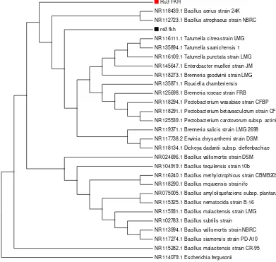
Homology of Bacteria
Homology analysis from BLAST revealed that the DNA sequence of proteolytic bacteria in the rumen of Kacang goat had 93% sequence similarity with Bacillus
aerus. While the Re1 isolate of Kacang goat reticulum
had 92% similarity with Tatumella punctata. Phylogenetic analysis using MEGA 7.0 Ru3 isolates was close to the Bacillus group and the Re1 isolate was close to Tatumella (Figure 4).
Identification of bacteria using the 16S rRNA gene sequence is a widely practiced technique, although with limitations for closely related taxa members (Kim et al., 2011). If the sequence has a similarity of greater than 98% with a known bacterial 16S rRNA gene, this species is considered a member of the species, but if less than 97% then it is considered different species (Vandamme et al., 1996; Janda and Abbott, 2007). The homology of 92% sequence for Ru3 and 93% for Re1 Figure 2. Gram staining results on colonies of proteolytic bacteria enlarging 10 x 100. A-B= Rumen goat bacteria; C-D= Goat reticulum bacteria. A= Gram-positive focus; B= Gram-positive bacillus; C= Short Gram-negative bacillus; D= Gram positive bacillus
Table 2. The average number of proteolytic bacteria in the rumen and peanut goat's reticulum
No Kacang goat Bacteria total count
Rumen Reticulum
1 Goat 1 3.55 x 10⁶ 3.07 x 10⁷
2 Goat 2 6.35 x 10⁶ 1.24 x 10⁷
3 Goat 3 9.40 x 10⁶ 2.82 x 10⁷
4 Goat 4 6.53 x 10⁶ 1.95 x 10⁷
5 Goat 5 6.25 x 10⁶ 2.83 x 10⁷
in this study indicated that both isolates are either new or unconfirmed. Further research is required to determine whether the isolates are new species such as DNA-DNA hybridization analysis, G + C content and
free fatty acid analysis.
Microbial protease enzymes account for about 60% of the total production of enzymes in the industry. Protease is one of the most important groups of industrial enzymes with extensive applications including animal feed, pharmaceuticals, detergent
manufacture, cheese making, bread making, waste management and silver recovery. The industrial sector often uses Bacillus subtilis for the production of various enzymes. All proteases, especially the alkaline
proteases produced by the Bacillus species, are very important in the industry because they have a stable temperature and a high pH. Bacillus is a major producer of extracellular proteases. The isolation and characterization of new strains of Bacillus in the enzyme production process are promising. This Figure 3. Result of agarose gel electrophoresis. A= Total DNA of proteolytic bacteria from rumen fluid (1) and reticulum (2) kacang goat, B= Results amplification of 16S rRNA gene with amplicon length ± 1500 bp. 1= Rumen fluid of kacang goat, 2= kacang goat reticulum; 3= DNA marker 1kb
Ru3 FKH
NR 118439.1 Bacillus aerius strain 24K
NR 112723.1 Bacillus atrophaeus strain NBRC
re3 fkh
NR 116111.1 Tatumella citrea strain LMG
NR 135894.1 Tatumella saanichensis 1
NR 116109.1 Tatumella punctata strain LMG
NR 145647.1 Enterobacter muelleri strain JM
NR 118273.1 Brenneria goodwinii strain LMG
NR 135871.1 Rouxiella chamberiensis
NR 125698.1 Brenneria roseae strain FRB
NR 118294.1 Pectobacterium wasabiae strain CFBP
NR 118291.1 Pectobacterium betavasculorum strain CFBP
NR 125539.1 Pectobacterium carotovorum subsp. actinidiae
NR 119371.1 Brenneria salicis strain LMG 2698
NR 117738.2 Erwinia chrysanthemi strain DSM
NR 118134.1 Dickeya dadantii subsp. dieffenbachiae
NR 024696.1 Bacillus vallismortis strain DSM
NR 104919.1 Bacillus tequilensis strain 10b
NR 116240.1 Bacillus methylotrophicus strain CBMB205
NR 118290.1 Bacillus mojavensis strain ifo
NR 075005.1 Bacillus amyloliquefaciens subsp. plantarum
NR 115325.1 Bacillus nematocida strain B-16
NR 115931.1 Bacillus malacitensis strain LMG
NR 102783.1 Bacillus subtilis strain
NR 113994.1 Bacillus vallismortis strain NBRC
NR 117274.1 Bacillus siamensis strain PD-A10
NR 115282.1 Bacillus malacitensis strain CR-95
NR 114079.1 Escherichia fergusonii
bacteria are rod-shaped, resistant to extreme environmental conditions, aerobic or facultative anaerobes and include normal flora although some species are pathogenic (Suganthi et al., 2013; Pant et al., 2015).
Tatumella spp. is also produce proteases which can
grow at pH 5.0-7.0 (Rodarte et al., 2011). Tatumella is a Gram-negative, short-shaped, facultative anaerobes, negative oxidase, and catalase-positive bacteria. This bacteria included in family Erwiniaceaae, order Enterobacteria, class Gammaproteobacteria and phyla Proteobacteria. Types of bacteria Tatumella spp. can grow at pH 5.0-7.0 and is also known to produce protease (Rodarte et al., 2011).
.
CONCLUSION
The amount of proteolytic bacteria with protease activity in reticulum fluid of Kacang goat is higher the Tatumella bacteria (92%). There is a possibility that both isolate is a new or unconfirmed species.
REFERENCES
Baehaki, A., Rinti, and A. Budiman. 2011. Isolasi dan karakterisasi protease dari bakteri tanah Rawa Indralaya, Sumatera Selatan. Jurnal Teknologi dan Industri pangan. XXII(1):10-16. Baker, G.C., J.J. Smith, and D.A. Cowan. 2003. Review and
re-analysis of domain specific 16S primers. J. Microbiol. Meth. 55: 541-555.
Chiba, L.I. 2014. Animal Nutrition Hand Book. 3rd Revision. Department of Animal Sciences 303C Upchurch Hall Auburn University, Alabama, USA.
Eschenlauer, S.C.P., N. McKain, N.D. Walker, N.R. Mc Ewan, C.J. Newbold, and R.J. Wallace. 2002. Ammonia production by ruminal microorganisms and enumeration, isolation, and characterization of bacteria capable of growth on peptides and amino acids from sheep rumen. Appl. Environ. Microbiol. 68(10):4925-4931.
Fitra, A.P., A. Batubara, M. Doloksaribu, and E. Sihite. 2009. Petunjuk Teknis Potensi Plasma Nutfah Kambing Lokal Indonesia. Pusat Penelitian dan Pengembangan Peternakan. Deli Serdang.
Genzenbu, D. and G. Tesfay. 2015. The role of bacteria in nitrogen metabolism in the rumen with emphasis of cattle. Res. J. Agric. Management. 4(7):282-290.
Haryanto, B. 2012. Perkembangan penelitian nutrisi ruminansia. Wartazoa. 22(4):169-177.
Hungate, R. E. 2013. The Rumen and Its Microbes. Elsevier-Academic Press, New York.
Janda, J.M. and S.L. Abbott. 2007. 16S rRNA gene sequencing for bacterial identification in the diagnostic laboratory: Pluses, perils, and pitfalls. J. Clin. Microbiol. 45(9):2761-2764. Jones, G.A., T.A. McAllister, K.J. Cheng, and A.D. Muir. 1994.
Effect of sainfoin (Onobrychis viciifolia Scop) on growth and proteolysis by four strains of rumen bacteria: Resistance of Prevotella (Bacteroides) ruminicola B14. Appl. Environ. Microbiol. 60:1374-1378.
Kamra D.N. 2005. Rumen microbial ecosystem. Current Science.
89(1):124-135.
Kim, M., M. Morrison, and Z. Yu. 2011. Status of the phylogenetic diversity census of ruminal microbiomes. FEMS Microbiol. Ecol. 76:49-63.
Krause, D.O., T.G. Nagaraja, A.D. Wright, and T.R. Callaway. 2014. Board invited review: Rumen microbiology: Leading the way in microbial ecology. J. Anim. Sci. 91(2):331-342.
Kumar, M.D.J., P. Venkatachalam, N.N. Govindarajan, M.D. Balakumaran, and P.T. Kalaichelvan. 2012. Production and purification of alkaline protease from Bacillus Sp. MPTK 712 isolated from dairy sludge. Global Vet. 8(5):433-439.
Kumar, S., G. Stecher, and K. Tamura. 2016. MEGA7: Molecular Evolutionary Genetics Analysis version 7.0 for bigger datasets. Molecular Biology and Evolution. 33:1870-1874.
Mcsweeney, S.C., B. Palmer, R. Bunch, and O.D. Krause. 1999. Isolation and characterization of proteolytic ruminal bacteria from sheep and goats fed the tannin-containing shrub legume Calliandra calothyrsus. Appl. Environ. Microbiol. 65(7):3075-3083. McSweeney, C. and R. Mackle. 2012. Microorganisms and
Ruminant Digestion: State of Knowledge, Trends and Future Prospects. Background Study Paper. (61).
Muslim, G., J.E. Sihombing, S. Fauziah, A. Abrar, and A. Fariani. 2014. Aktivitas proporsi berbagai cairan rumen Deep mengatasi tannin dengan tehnik in vitro. Jurnal Peternakan Sriwijaya.
3(1): 25-36.
Nuswantara, L.K. 2003. Ilmu Makanan Ternak Ruminansia (Sapi Perah). Kannisius. Semarang.
Pant, G., A. Prakash, J.V.P. Pavani, S. Bera, G.V.N.S. Deviram, A. Kumar, M. Panchpuri, and R.G. Prasuna. 2015. J. Taibah Univ. Sci. 9:50-55.
Rudwitasari, Y.N. and P.R Wikandari. 2014. Aktivitas bakteri proteolitik yang diisolasi dari sumber air panas singgahan, Tuban. J. Chem. 3(3):183-188.
Rodarte, M.P., D.R. Dias, D.M. Vilela, and R.F. Schwan. 2011. Proteolytic activities of bacteria, yeasts and filamentous fungi isolated from coffee fruit (Coffea Arabica L.). Acta Scientiarum. Agronomy Brazil Maringá. 33(3):457-464.
Safika, F. Madayanti, P. Aditiawati, and Akhmaloka. 2013. Succession culture independent bacterial during manure composting process. J. Pure Appl. Microbiol. 7(13):269-276. Safika, S.W. Matondang, Darmawi, M. Abrar, Erina, and M.
Jalaluddin. 2017. Total colony of cellulolitic bacteria in the rumen of aceh cattle. J. Med. Vet. 11(1):51-58.
Sari, W.N., Safika, Darmawi, and Y. Fahrimal. 2017. Isolation and identification of a cellulolytic Enterobacter from rumen of Aceh cattle. Vet. World. 10(12):1515-1520.
Suganthi, C., A. Mageswari, S. Karthikeyan, M. Anbalagan, A. Sivakumar, and K.M. Gothandam. 2013. Screening and optimization of protease production from a halotolerant Bacillus licheniformis isolated from saltern sediments. J. Genet. Eng. Biotechnol. 11:47-52.
Susanti, E. 2003. Isolasi dan karakterisasi protease dari Bacillus subtilis. Biodiversitas. 4(1):12-17.
Sudewo, A.T.A., S.A. Santosa, and S. Agus. 2012. Produktivitas kambing peranakan etawah berdasarkan litter size, tipe kelahiran dan mortalitas di village breeding centre Kabupaten Banyumas. Prosiding Seminar Nasional Pengembangan Sumber Daya Pedesaan dan Kearifan Lokal Berkelanjutan II. 1-7. Tamura, K., M. Nei, and S. Kumar. 2004. Prospects for inferring very
large phylogenies by using the neighbor-joining method. Proceedings of the National Academy of Sciences. USA. 101:1030-11035.
Vandamme, P., B. Pot, M. Gillis, and P. Vos De. 1996. Polyphasic taxonomy, a consensus approach tobacterial systematics. Microbiol. Rev. 60:407-438.
Yang, W., F. Meng, J. Peng, P. Han, F. Fang, L. Ma, and B. Cao. 2014. Isolation and identification of a cellulolytic bacterium from the Tibetan pig's intestine and investigation of its cellulase production. Electron. J. Biotechn. 17: 262-267.