Dexamethasone Activities toward Population of B cells, Gr-1, and TNF-
α
cytokine in Mice
(
Musmusculus
) Balb/c Biliary Atresia Model
Riza Rahmawati1), Muhaimin Rifa’I 2)1,2 Animal Physiology Laboratorium, Department of Biology, Mathematic and Natural Science Faculty
Brawijaya UniversityMalang
1riezha.swann@gmail.comand 2rifa123@ub.ac.id
ABSTRACT
Biliary atresia iscondition caused by Rotavirus (RRV) infection. The aims of this study were to know the immune responses of mice model of biliary atresia treated with corticosteroid.Mice were splitinto 3 treatment groups: control (K), RRV injection (R), and RRV injection in the present of dexamethasone(R+D). In R treatment, the baby mice born in <24 hours were injected with 20 µl of phosphate buffered saline containing 1.5 x 10⁶ fluorescence-forming units Rhesus Rotavirus (RRV). First termination was performed in the day 7 to 14, while second termination was done in the day 14 to 21. The dosage of dexamethasone which is applied in this experiment is 0.5mg/kg body weight.Immunocompetent cells were isolated from spleen, and cell surface molecules were then analyzed by flowcytometry. The data was tested by SPSS 16.0 for Windows program. The results showed that dexamethasone given as corticosteroid for biliarry atresia theurapy couldsuppress TNF-αproduction as well as Gr-1 proliferation. In the other hand dexamethasonecan promote B220+ cell proliferation inrotavirus infected mice.
Keywords : Baby mice, biliary atresia, dexamethason, flowcytometry, rotavirus
INTRODUCTION
Biliaryatresia is the most of general cause in
pathologic jaundiceand acholic stools disease in
children around the world. BA (Biliary Atresia)
entails a progressive, inflammatory injury of one
or
any
bile
ducts,
leading
to
fibrosis
andobliteration of both the extrahepatic and
intrahepatic bile ducts. The function of bile
systems aredisposing metabolic waste from liver
and transport of bile salts which required to digest
fat in the small intestine. In biliary atresia, a
blockage of bile flow take placefrom the liver to
the gallbladder. It may lead to liver damage and
cirrhosis of the liver, which if left untreated could
be fatal [17].
Causes of biliary atresia is derived from
various things such as: viruses, especially rotavirus
infection, genetic disorders, toxic materials which
interfere the growth of the biliary tract as well as
the bile duct damage during delivery of perinatal.
There are three types of biliary atresia: Type I,
atresia of the common bile duct; type II, atresia of
the hepatic ductswhereas type III, obstruction or
blockage of the bile duct to upstream the porta
hepatis and above the porta hepatis.Most patients
included in biliary atresia type III, which reached
90% [14].
This disease occurs in 1/14.000 up to
1/10.000 live births. The ratio of biliary atresia in
girls and boys is 4: 1. From 904 cases of biliary
atresia were enrolled in over 100 institutions,
biliary atresia widely experienced by Caucasians
(62%), blacks (20%), Hispanic (11 %), Asia
(4.2%) and American Indians (1.5%) [15].
At the time of diagnosis, a Kasai
portoenterostomy isperformed in an attempt to
re-establish
bile
flow.
Despite
thissurgical
intervention, the intrahepatic bile duct injury
continues,leading to cirrhosis and the need for liver
transplantation duringchildhood in the majority of
patients.This surgery must carried out before the
baby is 2 months old[9]. Originated from an
attempt to treat infants with biliary atresia (BA)
after
hepatic
portoenterostomi
(HPE)
[6],
Corticosteroids (steroids) became therapies which
commonly used in the post-portoenterostomi and
believed to improve clinical outcomes in BA.
Basic theory of the use of steroids are
anti-inflammatory effect that has aimed to reduce viral
infection of the biliary tract by increasing the bile
flow, as well as reducing inflammation and edema
periduktal [5].
therapy in the treatment of biliary atresia in infants
that could be known the effectiveness their use
compared with other treatments.
METHODS
Research Design
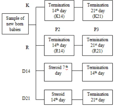
Treatments in this study were divided into
three treatments, with K treatment as Control, R
treatment as RRV infection, and D14 n D21
treatment as RRV infection+ Dexamethasone
injection.
Grouping of Mice
Table 1.Grouping of mice in each treatment
Termination
14
thday
Termination
21
stday
Control
treatment
6 mice babies
6 mice babies
R
treatment
9 mice babies
9 mice babies
D14
treatment
6 mice babies
-
D21
treatment
-
mice babies
Total
21 mice
babies
21 mice
babies
RRV(Rhesus rotavirus) Injection
Baby mice BALB / c born in <24 hours is
taken to be experimental animals. Control mice (C)
were not given any treatment, while the treatment
group mice R and D (D14 and D21) were injected
with 20
µ
l 106 PFU RRV strain MMU 18006
subcutaneously.
DexamethasoneInjection
Dexamethasone dose which injected in baby
mice is 0.5 mg / kg body weight of mice babies.
D14 group given steroids from day 7 and
terminated after observed until day 14. D21 group
given steroids from day 14 and terminated after
observed until day 21.
Lymphocytes Cell Isolation
Spleen were crushed with wire, by adding
1ml PBS to obtain homogenates. Thehomogenates
then moved into propylene tubes, added PBS till
10 ml. Then centrifugated in 2500 rpm, 4ºC for 5
minutes. Supernatant removed and obtained pellet
was homogenized with 1 ml PBS, taken 40
µ
l,
moved into microtube and add with500
µ
l PBS,
then centrifugated again in 2500 rpm, 4ºC for 5
minutes.Pellets added with cytofix / cytosperm
100
µ
l then incubated for 20 min, 4 º C. Then add 1
ml washperm then centrifuged 2500 rpm, 4 º C, 5
minutes. Pellets ready to immunostaining.
Flowcytometry Analysis
Isolated lymphocytes cells of spleen
thenadded with antibody staining, FITC
anti-mouse CD4 clone GK 1.5, PE rat anti-anti-mouse
CD8a clone 53-6.7, PE anti-mouse TNF-
α
clone
MP6-XT22 and FITC Rat anti-mouse Ly-6G and
Ly-6C
clone
RB6-8C5,
PE
anti-mouse
CD45R/B220 clone RA3-6B2, PE/Cy7 anti-mouse
IgM clone RMM-1. Conjugated results incubated
for 15 minutes inice box. The sample then added
with 350
µ
l PBS placed inside flowcytometer
cuvette.
Then
choose
acquire
and
the
RESULT AND DISSCUSSION
Population of CD4
+TNF-
α
+cells on Spleen
Organ
The results of CD4
+TNF-
α
+in the RRV
induction treatment and control not only on the
first termination but also on the second termination
(Figure 1) showed difference in the absolute
number of cells.In first termination, there were
amounted to 61.560 cells in the control,while the
RRV induction treatment was 65.100 cells.Then in
D14 treatmentwere lowerthan RRV induction.In
second termination, there were amounted to 65.190
cells in the control,while the RRV induction
treatment was 215x 103cells.
Figure1.Absolute cell number of CD4+TNF-α+ at first and second termination (C= Control, R= Infection, R+D= Infection+ Dexamethasone)
RRV (R) treatment able to increase TNF-
α
expression by CD4+ cells.Immunity responses for
virus infectioncommonly mediated by T helper
cells type 1 (Th1) as inflammation process where
IFN
γ
and TNF-
α
play a main role in this case [2].
About 1-2 weeks after RRV infection, CD4+ cells
produce IFN-
γ
which found in baby mice hepar
with rising of macrophage quantity that produce
TNF-
α
[7]. The increasing of quantity and size of
Kupffer cells is caused by fibrosis forming in
heparrelated to the increasing of IL-18 serum on
Biliary atresia. IL-18 is macrophage derived
cytokine which work together with IL-12 cytokine
to increase proliferation and differentiation of Th1
cells which will cause T cells release
pro-inflammatory cytokine[18].
Population of Gr-1cells on Spleen Organ
The result of D14 treatment and D21
treatment showing the decreased of
CD4+TNF-α
+population when compared with control.This
could caused by corticosteroid activity in biliary
atresia theurapy with induce bile to enhance
bile-salt flow with induce Na+K+ATPase enzyme to
increase electrolyte transport in canaliculi as well
as inhibit inflammation response [16]. Besides
that,
Corticosteroid
able
to
work
as
immunosupressan which have ability to inhibit
lymphocyte and macrophage migration to bile
duct, enhance gen coding transcription for
anti-inflammation protein, and regulate IL-4, IL-10,
and IL-13 production by Th2 cells. By this
mechanism, Th1 expression will be pressing so T
helper will work on Th2 cells, where could
suppress TNF-
α
cytokine secretion eventually[3].
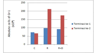
Figure 2.Absolute cell number ofGr-1at first and second termination (C= Control, R= Infection, R+D= Infection+ Dexamethasone)
The results of Gr-1 cells in the RRV
induction treatment and control not only on the
first termination but also on the second termination
(Figure 2) showed difference in the absolute
number of cells. In first termination, there were
amounted to 718x103 cells in the control,while the
RRV induction treatment was 980x103 cells.Then
the RRV + DexamethasoneInduction were lower
than RRV induction. In second termination, there
were amounted to 662x103 cells in the
control,while the RRV induction treatment was
2,1x 10⁶cells.
Total absolute cell number of Gr-1 in
control are higher than R treatment at first
treatment as well as second treatment. This can
caused by neutrophils and macrophage activation
in R treatment which is innate immunity strong
responses
against
Rotavirus
infection.
in the area of infection which is one of non-spesific
immune responses[11, 12]. Migration of
neutrophils during an infection and inflammation
influenced the presence of chemotactic signals in
the form of KC/CXCL1, MIP-2/CXCL2/3 and
CXCR2 receptors. Secretion of substances that
resemble toxins by rotavirus can stimulate CXC
chemokine, so neutrophils moving towards the
area of inflammatory [1].
The absolute cells number of D14 and D21
treatment decreased when compared with the
treatment of R. This may caused by
anti-inflammatory
and
immunosuppressive
of
Dexamethasone which potentially inhibits the
migration of lymphocytes and macrophages into
the bile duct to suppress the cytokine IL-3 which
has the potential to trigger the proliferation of a
variety of hematopoietic cells into myeloid
progenitor cells, which trigger proliferation of
various cells including myeloid cells that one of
them is granulocytes. IL-3 plays a role in various
cellular activities, such as cell growth, cell
differentiation and apoptosis. Generally, IL-3
secreted by activated helper T cells as immune
response to stimulate the T cells producing from
bone marrow [19], so that it may reduce the
absolute number of Gr-1.
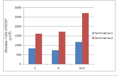
Population of B220
+cells on Spleen organ
The results of B220
+cells in the RRV
induction treatment and control on the first
termination (Figure 3) showed difference in the
absolute number of cells. In first termination, there
were
amounted
to
8,3x10
6cells
in
the
control,while the RRV induction treatment was
7,4x10
6cells.Then
the
RRV
+
DexamethasoneInduction were higher than RRV
induction. In second termination, there were
amounted to 1,6x10
6cells in the control,while the
RRV induction treatment was 1,7x 10
6cells.In
R+D treatment, the absolute cells were higher than
RRV induction treatment.
Figure 3.Absolute cell number ofB220+ at first and second termination (C= Control, R= Infection, R+D= Infection+ Dexamethasone)