prevalence is increasing by age.1
Several risk factors have been associated with the occurrence of CVI, such as older age, female, family history, obesity, standing occupation, and pregnancy. CVI prevalence in women is 1% -40% in some areas, while in men 1% - 17%. Physical activity and ergonomic position contribute to the occurrence of CVI. Orthostatic type of work increase the prevalence and severity of CVI. Women with jobs that require standing for a long time about 4 hours will have a 2.7 times higher risk of going CVI.2
This case report will discuss the clinical manifestations, classifi cation and management of CVI on 60 years old women with several risk factors including standing occupation.
position at work, more often felt on her left leg. Swelling and stiffness often appeared in the afternoon. Patient then got pain on her foot. There was no worsening of pain during walking. Her blood glucose was checked and the result was normal. History of previous swollen at the feet was denied. History of leg trauma, blockage in the leg veins were also denied. She was referred to Sardjito Hospital for vascular examination and revealed a CVI. The risk factors of CVI that can be identifi ed are: aged of 60 years, a teacher occupation with a long time standing position (about 4-5 hours) every day, obesity (her BMI was more than 30 kg/ m2), and lack of physical activities.
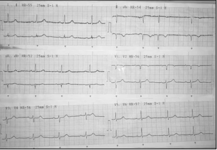
[image:1.595.125.472.490.731.2]On physical examination, the general status was good and full of consciousness. Blood
pressure 120/80 mm Hg, regular heart rate at 80 beats/min, respiratory rate 20 breaths/min, and her temperature was of 36.5 ° C. Head examination was normal. There were no elevated jugular venous pressure or lymphadenopathy. Chest examination was normal, lung sound was vesicular, symmetric and no lag of motion. Examination of the heart revealed no abnormalities. Examination of the abdomen was within normal limits, there were no tenderness and liver or spleen enlargement. On extremity examination, there were strong arterial pulsation, warm skin, and found a scar and ulceration on the medial aspect of the left foot. There were no edema in both legs. Peripheral arterial oxygen saturation by pulse oximetry on inferior extremity was around 98% - 99%. An electrocardiographic examination showed sinus rhythm with the heart rate was 55 bpm (Figure 1).
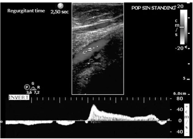
Duplex ultrasonography examination found a venous regurgitation with regurgitation time was 2480 msec in the right femoral vein, 830 msec in left femoral vein, and 2500 msec in the left popliteal vein. Then patient was confi rmed that she got a severe venous valve insuffi ciency on bilateral femoral vein and in the left popliteal vein. Arterial and venous fl ow were normal. Patients then treated with the installation of stocking compression on her right and left inferior extremity and given a micronized purifi ed fl avonoid fraction (Ardium®) 500 mg twice daily.
Discussion
Physiologically, vein serves primarily as a conduit to travel blood back from the capillaries to the heart. In addition, it also serves as a blood reservoir, vascular capacity regulator, part of peripheral pump, as well as involved in the regulation of body temperature. The vein is divided into superfi cial and deep vein. This division is based on the location of that vasculature. The superfi cial veins has large diameter, thick wall, muscular characteristic, and located beneath the skin. Deep vein generally runs along the artery and the diameter is about three times larger than the adjacent artery. Perforate veins connects superfi cial veins with the deep veins. One of the most important structures of the veins is a one way bicuspid valve that prevents blood refl ux.1
[image:2.595.144.460.81.308.2]Our patient came with symptomatic manifestation as oedema and pain in the leg especially in the afternoon. The clinical manifestations of venous disorders range from asymptomatic to symptomatic, such as dilatation of the superficial veins until skin wounds. Some of the most frequent manifestation are teleangiectasis, varicose vein and CVI. Intramuscular or intermuscular deep veins are important in determining the occurrence of CVI. There are three pairs of intermuscular veins named in accordance with the arteries that accompany the posterior tibial vein behind the medial malleolus, Figure 2. Pulse wave Doppler (DUS) examination showed a refl ux at the
veins by muscular contraction. The effectiveness of the pump is then determined by the conditions of competent venous valves. Competent venous valves have two functions, to prevent transmission due to high pressure in the superfi cial veins and capillaries during muscle contraction and prevents muscle contraction at the end of backfl ow into the superfi cial veins. When it goes well, mean venous pressure in the superfi cial veins is around 20-30 mmHg. If the average venous pressure rises to 60-90 mmHg, it will cause venous hypertension and may alter the anatomy, physiology, and histopathology associated with CVI.3
Epidemiology and prevalence of varicose veins in women is two times higher than in men. Increasing age is a risk factor for CVI and varicose veins. CVI prevalence rises to 21.2% in men aged > 50 years and 12% in women> 50 years. The presence of family history of varicose veins will increase the risk of varicose veins. Forty-two percent of women in Japan with varicose veins have family history, as compared to 14% if the downregulation of desmulin gene will affect the smooth muscle cells in the the saphenous vein wall. A job that requires standing position in a long period of time is an independent factor for CVI occurrence. It was found that the prevalence CVI in standing profession is 36% compared to 27% in others. 4 Body mass index (BMI) > 30 kg/ m2 increase the risk of CVI. In this patients, she had several risk factors that can induce the occurance of CVI, such as women, age> 50 years, working with long-duration of standing position up to 4-5 hours per day, obesity with BMI> 30 kg/ m2, while family history was denied.
Professions requiring long period of time in standing position were associated with increased of CVI compared to professions with various
Some of the factors that infl uence the occurrence of CVI are venous stasis, venous hypertension, fi brin cuff, water hammer effect, and trapping leukocytes. Venous stasis occurs due to accumulation of the stagnant blood in the veins that become tortuous, wider and lead to tissue anoxia and cell death, causing skin changes and ulcerations. Venous hypertension causes muscle pump dysfunction and venous ulceration. The hydrostatic pressure between the deep veins and superfi cial veins at rest and standing position is the same. During the contraction of the calf muscles, deep vein pressure will be higher than the superfi cial veins. Valve closure needed to prevent transmission of pressure to the superfi cial vein. But in the incompetent valve and pump dysfunction, pressure was transmitted to the superfi cial veins, causing CVI symptoms and ulcerations. Pericapillary blockage by fi brin (fi brin cuff) was related to restriction of oxygen diffusion that passes through the vessel. It will cause edema and dermatoslerotic changes in the skin. Water hammer effect theory stated that refl ux was transmitted mainly through the perforator into the superfi cial veins which time a break of 20 -25% in patients with normal venous pressure but venous hypertension will occur by the induction of valsava maneuver.6
manifestation which usually appears in the medial malleolus, where there is a maximum venous pressure caused by the presence of large vein perforation. Lipodermatosclerosis is skin discoloration on the lower extremity including capillary proliferation, fat necrosis, and fi brosis of the skin in the subcutaneous tissue. The skin becomes red and brown due to hemosiderin deposition of the red blood cells. CVI clinical course was consistent with progressivity of chronic venous stasis and hypertension. Swelling is caused by refl ux and obstruction. The oedema type is pitting. Flushed skin discoloration or brownish is because CVI is localized along the medial part of the lower leg. Unhealed ulcer localized to the medial part of the lower extremity can be a symptom of CVI.7 On this patient, the initial complaints were swelling in the legs and soreness of the foot, especially in the afternoon. Later she complained about her injured left leg below the medial portion that was consistent to the CVI ulcer characteristic.
Patients with CVI and ulcerations may also have an arterial disease at the same time. Thus in patients with ulceration, measurements of ankle-brachial index (ABI) is also required. Distinguishing CVI ulceration from arterial disease can be seen in table 1.3 According to this table it can be concluded that the characteristics of the wound in our patient was venous-associated ulceration, with ABI score 0.9 still in normal ranges.
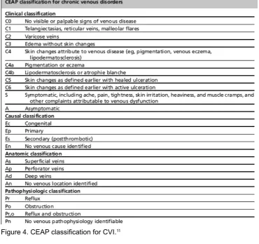
[image:4.595.97.508.85.282.2] [image:4.595.71.530.329.466.2]There are several criteria used for the classification and severity of CVI. The CEAP (Clinical, etiology, anatomic, Pathophysiology) is a classifi cation module that is used to report, diagnose and treat CVI. As a note, CVI is a progressive disorder so serial CEAP classifi cation was necessary. For report writing, a basic and advanced format CEAP can be used, where the difference is in the advanced CEAP classifi cation, venous anatomy segment was included.9 Clinical manifestations in our patient were skin changes with healed ulceration, no history of post Table 1. Differences in characteristics related injuries veins, arteries.8
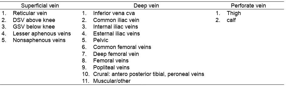
Table 2. Classifi cation of venous anatomy segment10
Superfi cial vein Deep vein Perforate vein
Reticular vein 1.
DSV above knee 2.
GSV below knee 3.
Lesser aphenous veins 4.
Nonsaphenous veins 5.
Inferior vena cva 1.
Common iliac vein 2.
Internal iliac veins 3.
Esternal iliac veins 4.
Pelvic 5.
Common femoral veins 6.
Deep femoral vein 7.
Femoral veins 8.
Popliteal veins 9.
Crural: antero posterior tibial, peroneal veins 10.
Muscular/other 11.
Thigh 1.
Figure 4. CEAP classifi cation for CVI.11
Figure 5. Severity score of venous disorders.12
thrombotic, located in the femoral and popliteal veins, with the refl ux pathophysiology. Based on CEAP classifi cation, our patients is C5; Ep; Ad; Pr 13, 14, level II.
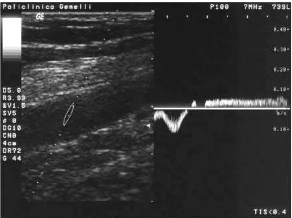
examination was done while lying and standing by using color Doppler and pulsed wave Doppler. The existence of a refl ux is the predominant fi nding in primary CVI. To identify CVI, refl ux may be provoked by performing compression on the calf and Valsalva maneuver.14 In our patients doppler examination, the regurgitation time measured on the right femoral vein was 2480 msec, on the left femoral vein was 830 msec and on the left popliteal vein was 2500 msec.
Management of CVI is intended to reduce symptoms and repair the cause of the abnormality. Ranging from the use of drugs, use of compression such as compression stockings, and surgery. Some drugs used to relieve symptoms in patients with CVI were divided into 4 groups, those are venoactives, hemorrheologic agents, antiplatelet agents, and other agents. Venoactive drugs suchn as phlebotonic agent works to increase the venous tone and capillary permeability. Hemorrheological such as Pentoxifylline, can be used to treat CVI by inhibiting cytokines and reducing white blood cell adhesion to the endothelium and releasing free radicals. Other agents such as zinc supplements, prostaglandin E1 was found to be ineffective to overcome ulcer due to venous disorders.9 The use of compression stockings is intended to put pressure 30-40 mmHg or 40-50 mmHg on the ankle and gradually reduce pressure on the proximal part of the foot to restore venous flow pattern both on the superficial and deep veins. Putting the foot in higher position when
sitting or lying down will reduce the pressure on the veins and reduce edema.15 In patients with symptomatic CVI who fail conservative therapy, invasive advance management might be required, as in CVI classifi cation CEAP class 4 to 6 with recurrent or unhealed ulceration and progressive infection and lymphedema. Superfi cial venous reflux can be healed with a cool touch laser ablation, RFA therapy, venous sclerotherapy, and venous phlebectomi. Deep venous reflux valve can be healed by reconstruction surgery or valvuloplasty. It also can be done by valve replacement and valve transposition procedure to patient whose valvuloplasty cannot be done. Subfascial endoscopic perforator surgery (SEPS) can be done in case of any incompetence of the perforator vein. In case of CVI with venous obstruction, endovascular procedure such as stenting on iliac vein can be done.6
In our patients, conservative therapy with the use of medications and compression stockings were conducted. Evaluation report showed a good results thus invasive intervention did not required. Education regarding control of risk factors such as reducing the time associated with long-duration of standing during working and reducing weight had been done to improve the outcome.
Conclusion
[image:6.595.151.443.84.302.2]We reported 60 years old women with severe chronic venous insuffi ciency (CVI) present with Figure 6. Doppler reflux pulse wave examination result with
Refferences
Sumadikarya, I.K. 2005. Insufisiensi Vena 1.
Kronik Patofi siologi dan Jenis Gangguan yang Sering Terjadi. Meditek; 13(33): 17-24. Beebe-Dimmer, J.L., Pfeifer, J.R., Engle, J.S., 2.
Schottenfeld, D. 2005. The Epidemiology of Chronic Venous Insuffi ciency and Varicose Veins. Ann Epidemiol; 15(3): 175 -184. Alguire, P.C., Mathes, B.M., Mills, J.L., Collin, 3.
K.A. 2015. Diagnostic Evaluation of Chronic Venous Insuffi ciency. Available from: URL: http://www.uptodate.com/contents/diagnostic-evaluation-of-chronic-venous-insuffi ciency. Boer, E.M., Krijnen, R.M.A. 1997. Venous 4.
Insuffi ciency in Male workers with standing Profession. Dermatology; 194: 121-126. Tuchsen, F., Krause, N., Hannerz, H., Burr H., 5.
Kristensen, T.S. 2000. Standing at Work and Varicose Veins. Scand J Work Environ Health; 26(5):414-420.
Gujja, K., Wiley, J., Krishnan, P. 2014. Chronic 6.
Venous Insuffi ciency. Intervent Cardiol Clin;3: 593 -605.
Weiss, R., James, W.D., et al. 2015.
7. Venous
Insuffi ciency Clinical Presentation. Available from: http://emedicine.medscape.com/ article/1085412-clinical
Gujja, K., Wiley, J., Krishnan P. Chronic 11.
Venous Insuffi ciency. Intervent Cardiol Clin. 2014;3: 593 -605. Table 1, CEAP classifi cation for chronic venous disorders;p.597
Gujja, K., Wiley, J., Krishnan, P. Chronic 12.
Venous Insuffi ciency. Intervent Cardiol Clin. 2014;3: 593 -605. Table 2, Revised venous clinical severity score; p.598
Cina, A., Pedicelli, A., Distasi, C., Porcelli, A., 13.
Fiorentina, A., Cina, G., Rulli, F., Bonomo, L. 2005. Color-Doppler Sonography in Chronic Venous Insuffi ciency: What the Radiologist should Know. Curr probl diagn Radiol; 34:51-62. Figure 5, Incompetence of the deep system. In a patient with postthrombotic syndrome, the PW Doppler shows prolonged inversion of the flow in the superficial femoral vein during Valsalva maneuver; p.54
Cina, A., Pedicelli, A., Distasi, C., Porcelli, A., 14.
Fiorentina, A., Cina, G., Rulli, F., Bonomo, L. 2005. Color-Doppler Sonography in Chronic Venous Insuffi cinecy: What the Radiologist should Know. Curr probl diagn Radiol ;34:51-62
Weiss, R., James, W.D.
15. Venous Insuffi ciency