Effect of
â
-carotene on cell proliferation and differentiation of
adipose-derived stem cells into endothelial progenitor cells
Wahyu Widowati1, Caroline Tan Sardjono1, Laura Wijaya2, Dian Ratih Laksmitawati3, Jeanne Adiwinata
Pawitan4*, Ferry Sandra5
1Medical Research Center, Faculty of Medicine, Maranatha Christian University, Bandung, (INDONESIA) 2Stem Cell and Cancer Institute, Jakarta, (INDONESIA)
3Faculty of Pharmacy, Pancasila University, Jakarta, (INDONESIA)
4Department of Histology, Faculty of Medicine, Universitas Indonesia, Jakarta, (INDONESIA)
5Department of Biochemistry and Molecular Biology, Faculty of Dentistry, Trisakti University, Jakarta, (INDONESIA) E-mail: [email protected]; [email protected]
F
ULL
P
APER
A
BSTRACT
Aim: To evaluate the ability of â-carotene to protect oxidation effect of
H2O2, and to enhance proliferation and differentiation into AD-MSCs into EPCs. Experimental: AD-MSCs were induced by EGM Bullet Kit medium with or without â-carotene. The proliferation of EPCs was determined by viable cell number counts. Differentiation into EPCs was characterized by the following surface markers: CD 34, CD133 and vascular endothelial growth factor receptor 2 (VEGFR-2). The protective effect of â-carotene
was measured base on the level of intracellular reactive oxygen species (ROS) by fluorescence with 2’,7’- dichlorofluorescein diacetate (DCF-DA)
using flow cytometry. Results: â-Carotene enhanced cell proliferation and differentiation of AD-MSCs into EPCs, and also decreased the accumulation of H202-induced intracellular ROS in EPCs. Conclusion:â
-Carotene plays a role in cell proliferation and differentiation of AD-MSCs to EPCs that might be due to decreased intracellular ROS level.
2014 Trade Science Inc. - INDIA
K
EYWORDS
Endothelial progenitor cell; Oxidative stress; Reactive oxygen species (ROS);
â-carotene;
Antioxidant; Endothelium.
INTRODUCTION
Mesenchymal stem cells (MSCs) appear to be both multipotent and immune privileged, which make them particularly attractive for stem cell therapy[1,2]. Mesen-chymal stem cells have improved heart function in both
cal studies of patients with heart failure[1,3]. The mecha-nism of improvement involved their differentiation into smooth muscle and endothelial cells, which caused neovascularization and improved cardiac function[4]. Cell population, termed processed lipoaspirate (PLA) cells, can be isolated from human lipoaspirate, and can
dif-BTAIJ, 9(10), 2014 [407-412]
BioTechnology
An Indian Journal
F
ULLP
APERchondrogenic lineages. Processed lipoaspirate cells ex-pressed multiple CD marker antigens similar on MSCs[5].
Endothelial dysfunction plays a major role in the development and clinical complications of heart failure. Endothelial progenitor cells (EPCs) have been shown to provide an endogenous repair mechanism to coun-teract detrimental risk factor-induced effects and re-place dysfunctional endothelium. Enhancing the num-ber and function of EPCs with targeted interventions may elicit functional improvement in individuals with heart failure due to cardiovascular disease (CVD)[6].
Free radical formation is associated with the nor-mal natural metabolism of aerobic cells[7]. The resulting reactive oxygen species (ROS) serve as secondary in-tracellular messengers and affect the overall redox sta-tus of a cell. The intracellular redox environment has a critical role in controlling apoptosis, proliferation, self-renewal, senescence, and differentiation. Dysregulation of any of these processes in EPCs will alter endothelial cell (EC) function, predisposing to the development of vascular pathology[8]. To contribute to tissue repair, EPCs and stem cells have to be equipped with anti-oxidative defense system to survive[9].
In cohort studies, high intake of â-carotene is as-sociated with a lower incidence of and mortality from CVD[10]. Moreover, epidemiological studies have shown that a high intake of â-carotene is associated
with a decreased risk for coronary artery disease[11]. Dietary intake of â-carotene or high level â-carotene
in serum and adipose tissue shows inverse associa-tions with CVD[12]. Beta carotene is a lipid-soluble antioxidant that rapidly scavenges reactive oxygen in-termediates[11], and ROS[13], which protects LDL from oxidation[14]. Antioxidants also counterbalance the pro-duction of ROS that may cause oxidative damage to cells and modify cell growth regulatory pathways [15-17]. Further, previous studies suggested the role of â -carotene in the regulation of endothelium differentia-tion[18].
Therefore, the aim of this study is to evaluate the ability of â-carotene to enhance cell proliferation of AD-MSCs and their differentiation into EPCs. In addition, to evaluate â-carotene protection effect on H2O2 oxi-dation in AD-MSCs.
EXPERIMENTAL
This is a descriptive experimental study that was done in the Stem Cell and Cancer Institute, Jakarta, from July 2010 through December 2010. All protocols were reviewed and approved by the Stem Cell and Cancer Institute Institutional Review Board prior to the study.
MSC isolation from lipoaspirates
Lipoaspirates were obtained with informed con-sent from individuals undergoing tumescent liposuction surgery. Lipoaspirates were stored at 2–8°C for no
longer than 24 hours. Methods used to isolate the MSCs from lipoaspirate were adopted from Sardjono et al. (2009)[19].
The raw lipoaspirates (120ml) were diluted with equal volume of Phosphate Buffered Saline (PBS) and divided in 50ml-tubes. The diluted lipoaspirates were centrifuged at 430×g for 10 minutes continuously at
20oC. After centrifugation, the target cell
–containing lipid
phase was removed from the top and transferred into new tubes and diluted with an equal volume of PBS. This washing step was repeated twice followed by fur-ther an equal volume dilution of cell-containing lipid frac-tion with pre-warmed (37oC) 0.075% collagenase type I (Sigma C-9722) in PBS. Enzyme digestion was done by incubation at 37 °C for 30 minutes on an orbital
shaker. After digestion, enzyme activity was neutralized by adding equal volume of 10% fetal bovine serum (FBS [Invitrogen 26140]) containing DMEM (Gibco 11965-092). Digested product was then subjected to centrifu-gation at 600×g for 10 minutes. Pellet was re-suspended
in DMEM with 10% PBS, and filtered through a 100-µm strainer mesh that was attached to a vacuum-pump to remove cellular debris. Collected cells after filtration were then ready for culture[19].
AD-MSC culture
Isolation of MSCs from other contaminating cells was done by allowing the cells to adhere on plastic-surfaced culture dish (Nunc). Cells were seeded with a density of 40,000 cells/cm2 in MesenCult® basal me-dium (Stem Cell Technology 5401), which was supple-mented by MSC stimulatory supplement (Stem Cell Technology 5402 [final concentration 10%]), 100 unit/ ml penicillin/0.1 mg/ml streptomycin (Sigma P4333), then kept in 37oC, 5% CO
2. After 4 days, unwanted cells (non-MSC cells and debris) were removed by two washes of medium and expanded to reach 80% confluence. In another 6-7 days, adherent cells were detached using 0.25% trypsin EDTA solution, then DMEM + 20% FBS was used to inactivate the trypsin. Detached cells with fibroblast-like morphology were cultured in a 25 cm2 flask (Nunc) for 1 week or until confluence was achieved, and the cells were used for further experiments.
AD-MSC differentiation into EPC
To differentiate the MSCs into EPCs, cells were maintained in EGM-2MV Bullet Kit medium (Cambrex CC-155; CC-4176; CC-3162), which contained 10% (v/v) heat-inactivated FBS, and 1% (v/v) penicillin–
streptomycin. To determine the effect of â-caroteneon differentiation, the culture medium was supplemented with 20 µg/mL â-carotene (Sigma Aldrich CAS no. 7235-40-7) compared to culture medium without â -carotene supplement. Culture was done in a humidified atmosphere of 95% air and 5% CO2 for four, and seven days, and then harvested for further analysis. Mesen-chymal stem cells were plated on a 24-well plate (Nunc) precoated with fibronectin (1 µg/cm2 [Roche 10-838-039-001]) at a density of 104 cells/well for prolifera-tion/viability analysis or 105 cells/well on a 6-well fibronectin coated plate (for EPC marker and ROS level analysis).
Cell proliferation/viability analysis
The effect of â-carotene 20 µg/mL on cell viability in EGM-2MV Bullet Kit medium was determined by cell proliferation/viability analysis. Briefly, cultured cells were dissociated using trypsin, incubated for 3 minutes in 37oC, harvested and washed using DMEM + 20% FBS followed by centrifugation at 300g, for 10
min-utes. Cell pellet was resuspended, and cell count was done by trypan blue exclusion method. The experiment was done in duplo. Percentage of the total viable cell number was computed and noted. The mean of per-centages and standard deviation of viable cells in four and seven day culture with and without â-carotene were calculated and compared.
Flowcytometry Assay of EPC markers
To confirm the effect of 20 µg/mL â-carotene in MSC differentiation, flowcytometry assays to detect EPC markers were conducted. Cultured cells were dis-sociated using TrypLE™Select (Gibco ME 080181),
incubated for 5 minutes in 37oC, harvested and washed using DMEM + 20% FBS followed by centrifugation at 300xg, for 10 minutes. The cells were washed for 3 times using PBS + 2% FBS and centrifuge at 300xg, for 10 minutes. Cell pellet was collected and incubated in PBS + 2% FBS + FcR Blocking Reagent, at room temperature, dark condition, for 15 minutes. After-wards, mIgG1 anti-CD34-PE/CD45-FITC (BD) 20µl,
mIgG1 anti-CD133-PE (Miltenyi Biotech) 10µl, and
mIgG1 anti-KDR/VEGFR-2-PE (R&D system) 15µl
was added separately to cells, followed by appropriate incubation (CD 34 and CD 133 were incubated for 15 minutes, and KDR was incubated for 40 minutes, in 4 0C and in darkness). The same procedure was done for the respective isotype antibodies (20µl mIgG1-PE/
mIgG1-FITC [BD] as CD34/CD45 control, 3.3 µl
mIgG1-PE [BD] as CD133 control, and 9 µl
mIgG1-PE [BD] as KDR control). The cells were analyzed by flowcytometry using FACSCalibur (BD Biosciences), the CD counts were computed and noted. Flowcytometry assay was done in duplo. The mean and standard deviations of CD counts in four and seven day cultures with and without â-carotene were calcu-lated and compared.
Effect of â-carotene on ROS level
Quantification of intracellular ROS level was done
in duplo by fluorescence assay using 2’,7’
DCF-F
ULLP
APERDA for 30 minutes at 37 °C. After the incubation, the
excess DCF-DA was washed out with PBS + KCl. Effect of â-caroteneon ROS level was examined by incubating the cells with â-carotene (20 µg/mL) for 30
minutes, followed with H2O2 (final concentration 100
µM) for one hour, compared to control (without â
-caro-tene pre-treatment). The intracellular ROS levels were measured using FACSCalibur flowcytometer(BD Bio-sciences). The measured ROS level values were ex-pressed as a percentage compared to control ROS level values.
RESULTS
Effect â-carotene on cell proliferation
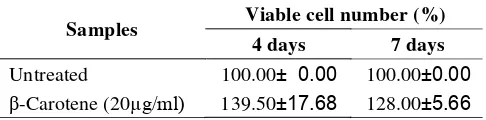
The effect of â-carotene treatment on cell prolif-eration can be seen in TABLE 1, which showed that â
-carotenesupplementation could increase cell prolifera-tion both in four and seven day culture. However, higher cell number was observed after four days compared to seven days of incubation.
Effect of â-caroteneon AD-MSCs differentiation into EPCs
TABLE 2 showed the effect of â-carotene on AD-MSCs differentiation into EPCs. Cells from culture with
â-carotenesupplementation showed higher EPC marker
expression compared to those without supplementa-tion. Marked KDR expression was observed after seven days of incubation with supplementation of â
-carotene.
Effect of â-caroteneon EPC ROS level
The effect of â-carotene on ROS intracellular level in EPCs can be seen in Figure 1, which showed that â
-carotene decreased ROS intracellular level; thus de-creased oxidative damage in -EPCs both in after four and seven days of incubation.
TABLE 1 : The effect of â-carotene on cell proliferation
Viable cell number (%) Samples
4 days 7 days
Untreated 100.00± 0.00 100.00±0.00
â-Carotene (20µg/ml) 139.50±17.68 128.00±5.66
TABLE 2 : The effect of â-caroteneon various EPC markers
CD34/45 (%) CD133 (%) VEGFR-2 (%)
Samples
4 days 7 days 4 days 7 days 4 days 7 days
Untreated 0.13±0.00 0.13±0.00 0.00±.00 0.00±0.00 0.00±0.00 0.00±0.00
â-carotene 0.17±0.03 0.16±0.04 0.01±0.00 3.40±0.78 0.00±0.00 2.78±0.12
Figure 1 : Effect â-carotene supplementation on ROS level of EPCs at different incubation periods
UT= untreated
DISCUSSIONS
EGM Bullet Kit medium is a special commercial medium for endothelial cell lineage, and therefore might not support the survival and proliferation of adipose-derived stem cells, but benefited the endothelial cell lin-eage; a study showed that endothelial cell lineage might
be present in earlier passages of adipose-derived stem cells[22]. Compared to culture without â-carotene, a marked increase in cell number was observed in cul-ture with â-carotene supplementation after four days. The increase in proliferation might be due to the capac-ity of â-carotene to scavenge ROS, which production
is inherent in cell growth due to oxygen consumption. Interaction of ROS with lipid produces new free radi-cals[7] that are involved in the production of prostaglan-dins, which modulate cell growth. Free radicals them-selves appear to have a down regulatory effect on cell proliferation[23]. Therefore, the increase in proliferation due to â-carotene on day-4 in our study was in line
with other findings, in term that â-carotene scavenge
the free radicals, and thus counteract down regulation of cell proliferation[7,23].
vascular endothelial growth factor receptor-2 [VEGFR-2]/KDR). VEGFR-2 is a marker to indicate endothe-lial characteristics, whereas CD34 and CD133 are markers to indicate cell plasticity (stem cell character-istics). Endothelial progenitor cells have originally been defined by their cell surface expression of hematopoi-etic marker proteins (CD133 and CD34), and endot-helial marker (VEGFR-2), and their capacity to differ-entiate into endothelial cells in situ, and to induce neovascularization[4,24].
The increase in cell number was less on day-7th compared to day-4th. This result might be due to the differentiation process of a proportion of the AD-MSCs into EPCs, which was especially marked by VEGFR-2 expression (VEGFR-2.78%) as shown in TABLE VEGFR-2, and after differentiation, the proliferation capacity was decreased. On day-7th, â-carotene supplementation caused an in-crease in CD 133, and insignificant inin-crease in CD34/ CD45. The increase in CD133 explained the less crease in proliferation on day-7th, as the effect of in-crease in CD133 as stem cell characteristics counter-balance the effect of VEGFR-2 as endothelial charac-teristics (differentiated cells with reduced proliferation capacity).
Intracellular ROS level measurement in this research was done by fluorescence detection of DCF-DA. 2’,7’
-dichlorofluorescein diacetate has been used in several studies dealing with the effect of ROS in cell culture [25-27]. 2
’,7’-dichlorofluorescein diacetate can cross the
membrane of viable cells and is enzymatically hydro-lyzed by intracellular esterases to 2’,7’
-dichlorofluorescin (DCFH), which is a substance with-out fluorescence. 2’,7’- Dichlorofluorescin is rapidly
oxidized to highly fluorescent 2’,7’-dichlorofluorescein
(DCF) in the presence of ROS within the cells, and DCF remains trapped within the cell; thus can be mea-sured to represent the intracellular ROS level[21,28].
In our study, â-carotene caused a decrease in in-tracellular ROS level on both day-4th and day-7th. This result might explain the mechanism of proliferation in-crease due to â-carotene supplementation.
Mitochon-dria, which produce energy to drive endergonic pro-cesses of cell life, are considered as the most important cellular source of free radicals, as the main target for free radical regulatory, and as the source of signaling molecules that command cell cycle, proliferation, and
apoptosis[29]. Deficiency of antioxidants is considered to be related to mitochondrial oxidative stress and dys-function, and will damage cell cycle and trigger cells apoptosis.
Protection against oxidative stress due to ROS is accomplished by a complex defense system composed of several antioxidative enzymes that reduce the dam-aging effects of ROS[9]. The most vulnerable organelles to oxidative stress are the mitochondria, due to their potential for continuous production of superoxide an-ions. Superoxide anions are converted to hydrogen peroxide by superoxide dismutases (MnSOD), whereas hydrogen peroxide is detoxified by catalase and glu-tathione peroxidase (GPx-1). As the localization of MnSOD and GPx-1 is in the matrix of mitochondria, in close proximity to the production of ROS by electron transport chain, these two enzymes are believed to be the primary antioxidant defense systems in the mito-chondria[9]. When the inherent defense system is inad-equate, addition of ROS scavenging agents will be ben-eficial. Therefore, the mechanism of proliferation increase due to â-carotene supplementation in our study might be due to the ROS scavenging ability of â-carotene.
CONCLUSION
Â-carotene plays a role in cell proliferation and dif-ferentiation of AD-MSCs to EPCs that might be due to decreased intracellular ROS level.
ACKNOWLEDGEMENT
The authors acknowledge gratefully for the finan-cial support from Ministry of Research and Technol-ogy, Republic of Indonesia (Research Grant no KP-2010-593), and support from Stem Cell and Cancer Institute (SCI), Jakarta.
REFERENCES
[1] M.F.Berry, A.J.Engler, J.Woo, T.J.Pirolli, L.T.Bish,
V.Jayasankar, K.J.Morine, T.J.Gardner,
D.E.Discher, H.L.Sweeny; Am.J.Physiol.Heart Circ.Physiol, 290, H2196 (2006).
[2] J.Y.Min, M.F.Sullivan, Y.Yang, J.P.Zhang,
F
ULLP
APERAnn.Thorac.Surg., 74, 1568 (2002).
[3] D.Orlic, J.Kajstura, S.Chimenti, I.Jakoniuk,
S.M.Anderson, B.Li, J.Pickel, R.Mckay, B.Nadal-Ginard, D.M.Bodine, A.Leri, P.Anversa; Nature,
410, 701 (2001).
[4] K.C.Wollert, H.Drexler; Circ.Res., 96,151 (2005).
[5] P.A.Zuk, M.Zhu, P.Ashjian, D.A.De Ugarte,
J.I.Huang, H.Mizuno, Z.C.Alfonso, J.K.Fraser,
P.Benhaim, M.H.Hedrick; Mol.Biol.Cell, 13, 4279
(2002).
[6] I.Andreou, D.Tousoulis, C.Tentolouris,
C.Antoniades, C.Stefanadis; Atherosclerosis, 189,
247 (2006).
[7] L.Barros, M.J.Ferreira, B.Queiro´s,
I.C.F.R.Ferreira, P.Baptista; Food Chemistry, 103,
413 (2007).
[8] J.Case, D.A.Ingram, L.S.Haneline; Antioxidant and
Redox Signaling, 10, 1895 (2008).
[9] T.Imanishi, T.Hano, I.Nishio; J Hypertens., 23, 97
(2005).
[10]J.M.Gaziano, J.E.Manson, L.G.Branch;
Ann.Epidemiol., 5, 255 (1995).
[11] A.Shaish, A.Daugherty, F.O’Sulivan, G.Schonfeld,
J.W.Heinecke; J.Clin; Invest., 96, 2075 (1995).
[12]F.G.De Waart, E.G.Schouten, A.F.H.Stalenhoef;
Int.J.Epidemiology, 30, 136 (2001).
[13]N.I.Krinsky, K.Y.Yeum;
Biochem.Biophys.Res.Commun., 305, 754 (2003).
[14]T.Kita,Y.Nagano, M.Yokode, K.Ishi, N.Kume,
S.Ooshima, H.Yoshida, C.Sawai;
Proc.Natl.Acad.Sci.USA, 84, 5928 (1987).
[15]J.Lin, N.R.Cook, C.Albert, E.Zaharris, J.M.Gaziano,
M.van den Burgh, J.E.Buring, J.E.Manson; J.Natl.Cancer Inst., 101,14 (2009).
[16]H.E.Seifried, D.E.Anderson, B.C.Sorkin,
R.B.Costello; J.Nutr., 34, 3143S (2004).
[17]H.E.Seifried, D.E.Anderson, E.I.Fisher, J.A.Milner;
J.Nutr.Biochem., 18, 567 (2007).
[18]C.A.Dembinska-Kie, A.Polus, J.Grzybowska,
B.Kiec-Wilk, A.Balwierz, J.Keijer, G.Schmitz; Genes Nutr., 2, 115 (2007).
[19]C.T.Sardjono, M.Setiawan, Frisca, V.Saputra,
G.Aniko, F.Sandra; Med.J.Indones., 18, 91 (2009).
[20]A.Stolzing, A.Scutt; Free Rad.Bio.& Med., 41, 326
(2006).
[21]G.Jie, Z.Lin, L.Zhang, H.Lv, P.He, B.Zhao; J.Agric;
Food Chem., 54, 8058 (2006).
[22]J.B.Mitchell, K.McIntosh, S.Zvonic, S.Garrett,
Z.E.Floyd, A.Kloster, Y.Di Halvorsen, R.W.Storms,
B.Goh, G.Kilroy, X.Wu, J.M.Gimble; Stem Cells, 24,
376 (2006).
[23]R.H.Burdo, C.Rice-Evans; Free Radical Res., 6,
345 (1989).
[24]T.Asahara, A.Kawamoto; Am.J.Physiol.Cell
Physiol., 287, C572 (2004).
[25]T.H.Murphy, M.Miyamoto, A.Sastre, R.L.Schaar,
J.T.Coyle; Neuron, 2, 1547 (1989).
[26]J.C.Saez, J.A.Kessler, M.V.L.Bennett, D.C.Spray;
Proc.Natl.Acad.Sci.USA, 84, 3056 (1987).
[27]J.A.Scott, C.J.Homcy, B.A.Khaw, C.A.Rabito; Free
Radical Biol Med., 4, 79 (1988).
[28]C.P.Le Bel, H.Ischiropoulos, S.C.Bondy;
Chem.Res.Toxicol., 5, 227 (1992).
[29]E.Cadenas, A.Boveris; The Handbook of