1/28
CERAMIC BIOMATERIALS
[Adopsi dari: Joseph D. Bronzino, “Biomedical Engineering Fundamentals”, CRC Press, third edition, 2006, Section V, CHAPTER 39, by W.C. Billote.]
39.1 INTRODUCTION
Ceramics are defined as the art and science of making and using solid articles that have as their essential component, inorganic nonmetallic materials (Kingery et al.. 1976). Ceramics are refractory, polycrystal line compounds, usually inorganic, including silicates, metallic oxides, carbides and various refractory hydrides, sulfides, and selenides. Oxides such as A1203, MgO. Si02, and Zr02 contain metallic and nonmetallic elements and ionic salts, such as NaCl, CsCl, and ZnS (Park and Lakes, 1992). Exceptions to the preceding include covalently bonded ceramics such as diamond and carbonaceous structures like graphite and pyrolized carbons (Park and Lakes. 19921. Ceramics in the form of pottery have been used by humans for thousands of years. Until recently, their use was somewhat limited because of their inherent brittleness, susceptibility to notches or micro-cracks, low tensile strength, and low impact strength. However, within the last 100 years, innovative techniques for fabricating ceramics have led to their use as “high tech” materials. In recent years, humans have realized that ceramics and their composites can also be used to augment or replace various parts of the body, particularly bone. Thus, the ceramics used for the latter purposes are classified as bioceramics. Their relative inertness to the body fluids, high compressive strength, and aesthetically pleasing appearance led to the use of ceramics in dentistry as dental crowns. Some carbons have found use as implants especially for blood interfacing applications such as heart valves. Due to their high specific strength as fibers and their biocompatibility, ceramics are also being used as reinforcing components of composite implant materials and for tensile loading applications such as artificial tendon and ligaments [Park and Lakes, 1992].
Unlike metals and polymers, ceramics are difficult to shear plastically due to the (ionic) nature of the bonding and minimum number of slip systems. These characteristics make the ceramics nonductile and are responsible for almost zero creep at room temperature [Park and Lakes, 1992]. Consequently, ceramics are very susceptible to notches or microcracks because instead of undergoing plastic deformation (or yield) they will fracture elastically on initiation of a crack. At the crack tip the stress could be many times higher than the stress in the material away from the tip, resulting in a stress concentration which weakens the material considerably. The latter makes it difficult to predict the tensile strength of the material (ceramic). This is also the reason ceramics have low tensile strength compared to compressive strength. If a ceramic is flawless, it is very strong even when subjected to tension. Flawless glass fibers have twice the tensile strengths of high strength steel (≈7 GPa) [Park and Lakes, 1992].
Ceramics are generally hard; in fact, the measurement of hardness is calibrated against ceramic materials. Diamond is the hardest, with a hardness index of 10 on Moh’s scale, and talc (Mg3Si3O10COH) is the softest ceramic (Moh’s hardness 1), while ceramics such
as alumina (Al2O3: hardness 9), quartz (SiO2: hardness 8), and apatite (Ca5P3O12F:
hardness 5) are in the middle range. Other characteristics of ceramic materials are (1) their high melting temperatures and (2) low conductivity of electricity and heat. These characteristics are due to the chemical bonding within ceramics.
2/28
Thus, this chapter will focus on a general overview of the relatively bioinert, bioactive or surface reactive ceramics, and biodegradable or resorbable bioceramics.
Ceramics used in fabricating implants can be classified as nonabsorbable (relatively inert), bioactive or surface reactive (semi-inert) [Hench, 1991, 1993] and biodegradable or resorbable (non-inert) [Hentrich et al., 1971; Graves et al., 1972). Alumina, zirconia, silicone nitrides, and carbons are inert bioceramics. Certain glass ceramics and dense hydroxyapatites are semi-inert (bioreactive) and calcium phosphates and calcium aluminates are resorbable ceramics [Park and Lakes. 1992].
39.2 NONABSORBABLE OR RELATIVELY BIOINERT BIOCERAMICS
39.2.1 Relatively Bioinert Ceramics
Relatively bioinert ceramics maintain their physical and mechanical properties while in the host. They resist corrosion and wear and have all the properties listed for bioceramics in Table 39.1. Examples of relatively bioinert ceramics are dense and porous aluminum oxides, zirconia ceramics, and single phase calcium aluminates (Table 39.2). Relatively bioinert ceramics are typically used as structural-support implants. Some of these are bone plates, bone screws, and femoral heads (Table 39.3). Examples of nonstructural support uses are ventilation tubes, sterilization devices [Feenstra and de Groot, 1983] and drug delivery devices (see Table 39.3).
3/28
4/28
Table 39.3 Uses of Bioinert Bioceramics
39.2.2 Alumina (A12O3)
The main source of high purity alumina (aluminum oxide, Al2O3) is bauxite and native corundum. The commonly available alumina (alpha,α) can be prepared by calcining alumina trihydrate. The chemical composition and density of commercially available “pure” calcined alumina are given in Table 39.4.
Table 39.4 Chemical Composition of Calcined Alumina
Source: Park, J.B. and Lake, LS. I 992. Ceramic Implant MateriaL,. In: Biomaterial An Introcuction, 2nd ed., p.121 Plenum Press, NewYork.
The American Society for Testing and Materials (ASTM) specifies that alumina for implant use should contain 9.3% pure alumina and less than 0.1% combined SiO2 and
alkali oxides (mostly Na2O) (F603-78).
5/28
fine alumina powders onto the surface of a seed crystal which is slowly withdrawn from an electric arc or oxy-hydrogen flame as the fused powder builds up. Single crystals of alumina up to 10cm in diameter have been grown by this method [Park and Lakes, 1992].
The strength of polycrystalline alumina depends on its grain size and porosity. Generally, the smaller the grains, the lower the porosity and the higher the strength [Park and Lakes, 1992]. The ASTM standards (F603-78) requires a flexural strength greater than 400 MPa and elastic modulus of 380 GPa (Table 39.5).
Aluminum oxide has been used in the area of orthopaedics for more than 25 years [Hench, 1991]. Single crystal alumina has been used in orthopaedics and dental surgery for almost 20 years. Alumina is usually a quite hard material, its hardness varies from 20 to 30 GPa. This high hardness permits its use as an abrasive (emery) and as bearings for watch movements [Park and Lakes, 1992). Both polycrystalline and single crystal alumina have been used clinically. The high hardness is accompanied by low friction and wear and inertness to the in vivo environment. These properties make alumina an ideal material for use in joint replacements [Park and Lakes. 1992]. Aluminum oxide implants in bones of rhesus monkeys have shown no signs of rejection or toxicity for 350 days [Graves et al., 1972; Hentrich et al.. 197l]. One of the most popular uses for aluminum oxides is in total hip protheses. Aluminum oxide hip prostheses with an ultra-high molecular weight polyethylene (UHMWPE) socket have been claimed to be a better device than a metal prostheses with a UHMWPE socket [Oonishi, 1992]. However, the key for success of any implant, besides the correct surgical implantation, is the highest possible quality control during fabrication of the material and the production of the implant [Hench, 1991].
39.2.3 Zirconia (ZrO2)
Pure zirconia can be obtained from chemical conversion of zircon (ZrSiO4), which is an
abundant mineral deposit [Park and Lakes, 1992]. Zirconia has a high melting temperature (Tm = 2953 K) and chemical stability with a = 5.145 Å, b = 0.521 Å, c =
5.311 Å, and β = 99o14 in [Park and Lakes, 1992]. It undergoes a large volume change
during phase changes at high temperature in pure form; therefore, a dopant oxide such as Y2O3 is used to stabilize the high temperature (cubic) phase. We have used 6 mole%
Y2O3 as dopant to make zirconia for implantation in bone [Hentrich et al. 1971]. Zirconia
6/28
High density zirconia oxide showed excellent compatibility with autogenous rhesus monkey bone and was completely nonreactive to the body environment for the duration of the 350 day study [Hentrich et a1., 1971]. Zirconia has shown excellent biocompatibility and good wear and friction when combined with ultra-high molecular weight polyethylene (Kumar et al., 1989; Murakami and Ohtsuki, 1989).
39.2.4 Carbons
Carbons can be made in many allotropic forms: crystalline diamond, graphite, noncrystalline glassy carbon, and quasicrystalline pyrolitic carbon. Among these, only pyrolitic carbon is widely utilized for implant fabrication; it is normally used as a surface coating. It is also possible to coat surfaces with diamond. Although the techniques of coating with diamond have the potential to revolutionize medical device manufacturing, it is not yet commercially available [Park and Lakes, 1992].
The crystalline structure of carbon, as used in implants, is similar to the graphite structure shown in Figure 39.1. The planar hexagonal arrays are formed by strong covalent bonds in which one of the valence electrons or atoms is free to move, resulting in high but anisotropic electric conductivity. Since the bonding between the layers is stronger than the van der Waals force, it has been suggested that the layers are cross-linked. However, the remarkable lubricating property of graphite cannot be attained unless the cross-links are eliminated [Park and Lakes, 1992].
FIGURE 39.1 Crystal structure of graphite. (From Shobert, El. 1964. Carbon and Graphite. New York. Academic Press. With permission.)
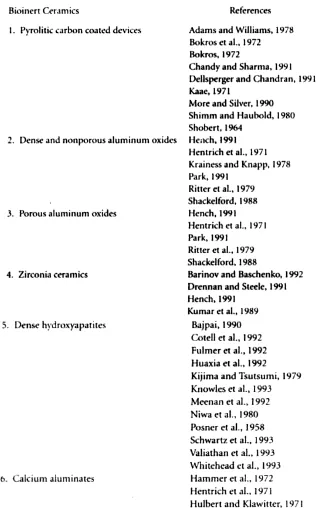
The poorly crystalline carbons are thought to contain unassociated or unoriented carbon atoms. The hexagonal layers are not perfectly arranged, as shown in Figure 39.2. Properties of individual crystallites seem to be highly anisotropic. However, if the crystallites are randomly dispersed, the aggregate becomes isotropic [Park and Lakes, 1992].
7/28
FIGURE 39.2 Schematic presentation of poorly crystalline carbon. (a) Single-layer plane. (b) parallel layers in a crystallite, (c) unassociated carbon, (d) an aggregate
of crystallites, single layers and unassociated carbon. [From Bokros, J.C. 1972. Chem. Phys. Carbon. 5:70-41, New York. Marcel Dekker. With permission.]
8/28
FIGURE 39.4 Elastic modulus vs. density for unalloyed pyrolitc carbons. [From Kaae, J.L 1971, J. Nucl. Mater. 38:42—50. With permission.]
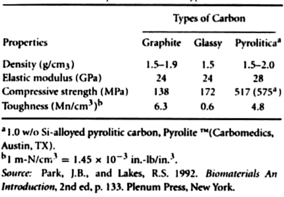
TABLE 39.6 Properties of Various Types of Carbon
Graphite and glassy carbon have a much lower mechanical strength than pyrolitic carbon (Table 39.6). However, the average modulus of elasticity is almost the saine for all carbons. The strength of pyrolitic carbon is quite high compared to graphite and glassy carbon. Again, this is due to the fewer number of flaws and unassociated carbons in the aggregate.
9/28
TABLE 39.7 Mechanical Properties of Carbon Fiber-Reinforced Carbon
Carbons exhibit excellent compatibility with tissue. Compatibility of pyrolitic carbon-coated devices with blood have resulted in extensive use of these devices for repairing diseased heart valves and blood vessels [Park and Lakes, 1992].
Pvrolitic carbons can be deposited onto finished implants from hydrocarbon gas in a fluidized bed at a controlled temperature and pressure. The anisotropy, density, crystallite size and structure of the deposited carbon can be controlled by temperature, composition of the fluidized gas, the bed geometry, and the residence time (velocity) of the gas molecules in the bed. The microstructure of deposited carbon should be highly controlled, since the formation of growth features associated with uneven crystallization can result in a weaker material (Figure 39.5). It is also possible to introduce various elements into the fluidized gas and co-deposit them with carbon. Usually silicon (10 to 20 w/o) is co-deposited (or alloyed) to increase hardness for applications requiring resistance to abrasion, such as heart valve discs.
Recently, success was achieved in depositing pyrolitic carbon onto the surfaces of blood vessel implants made of polymers. This type of carbon is called ultra low temperature isotropic (ULTI) carbon instead of low temperature isotropic (LTI) carbon. The deposited carbon has excellent compatibility with blood and is thin enough not to interfere with the flexibility of the grafts [Park and Lakes, 1992].
10/28
FIGURE 39.5 Microstructure of carbons deposited in a fluidized bed. (a) A granular carbon with distinct growth features. (b) An isotropic carbon without growth features. Both under
polarized light. 240x. (From Bokros, J.C.. LaGrange, LD., and Schoen, G.J. 1972. Chem. Phys. Carbon. 9: 103—171. New York, Marcel Dekker. With permission,)
39.3 BIODEGRADABLE OR RESORBABLE CERAMICS
11/28
TABLE 39.8 Examples of Biodegradable Bioceramics
12/28
39.3.1 Calcium PhosphateCalcium phosphate has been used in the form of artificial bone. This material has been synthesized and used for manufacturing various forms of implants, as well as for solid or porous coatings on other implants (Table 39.9).
Calcium phosphate can be crystallized into salts such as hvdroxyapatite and ß-whitlockite depending on the Ca:P ratio, presence of water, impurities, and temperature. In a wet environment and at lower temperatures (<900°C), it is more likely that hydroxyl- or hydroxyapatite will form, while in a dry atmosphere and at a higher temperature, ß-whitlockite will be formed [Park and Lakes 1992]. Both forms are very tissue compatible and are used as bone substitutes in a granular form or a solid block. The apatite form of calcium phosphate is considered to be closely related to the mineral phase of bone and teeth.
The mineral part of bone and teeth is made of a crystalline form of calcium phosphate similar to hydroxyapatite [Ca10(PO4)6(OH)2]. The apatite family of mineral [A10,(BO4)6X2]
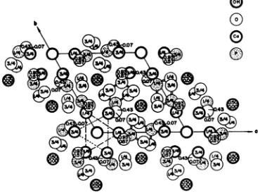
crystallizes into hexagonal rhombic prisms and has unit cell dimensions a = 9.432Å and c = 6.881 Å. The atomic structure of hydroxyapatite projected down the c-axis onto the basal plane is shown in Figure 39.6. Note that the hydroxyl ions lie on the corners of the projected basal plane and they occur at equidistant intervals (3.44 Å) along the columns perpendicular to the basal plane and parallel to the c-axis. Six of the ten calcium ions in the unit cell are associated with the hydroxyls in these columns, resulting in strong interactions among them [Park and Lakes, 1992].
FIGURE 39.6 Hydroxyapatite structure projected down the c-axis onto the basal plane (From Posner A.S., PerIotTA..and Diono A.OE ¡958. Acta. OysL 11:308—309.)
13/28
The mechanical properties of synthetic calcium phosphates vary considerably (Table 39.10). The wide variations in properties of polycrystalline calcium phosphates are due to the variations in the structure and manufacturing processes. Depending on the final firing conditions, the calcium phosphate can be calcium hydroxyapatite or ß-whitlockite. In many instances, both types of structures exist in the same final product [Park and Lakes, 1992].
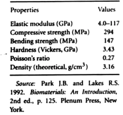
TABLE 39.10 Physical Properties of Calcium Phosphate
Polycrystalline hydroxyapatite has a high elastic modulus (40 to 117 GPa). Hard tissue such as bone, dentin, and dental enamel are natural composites which contain hydroxyapatite (or a similar mineral), as well as protein, other organic materials, and water. Enamel is the stiffest hard tissue, with an elastic modulus of 74 GPa, and contains the most mineral. Dentin (E = 21 GPa) and compact bone (E = 12 to 18 GPa) contain comparatively less mineral. The Poisson’s ratio for the mineral or synthetic hydroxyapat ite is about 0.27 which is close to that of bone (≈0.3) [Park and Lakes, 1992].
Hontsu et al. [1997] were able to deposit an amorphous HA film on Ti, α-Al2O3,
SiO//Si(100), and SrTiO3 using a pulsed ArF excimer laser. Upon heat treatment the
amorphous film was converted to the crystalline form of HA. The HA film’s electrical properties were measured for the first time (Table 39.14).
Among the most important properties of hydroxyapatite as a biomaterial is its excellent biocompatibility. Hydroxyapatite appears to form a direct chemical bond with hard tissues [Piattelli and Trisi, 1994]. On implantation of hydroxyapatite particles or porous blocks in bone, new lamellar cancellous bone forms within 4 to 8 weeks [Bajpai and Fuchs, 1985]. Scanning electron micrograph (500x) of a set and hardened hydroxyapatite-cysteine composite is shown in Figure 39.7. The composite sets and hardens on addition of water.
Many different methods have been developed to make precipitates of hydroxyapatite from an aqueous solution of Ca(NO3)2 and NaH2PO4. There has been successful use of
14/28
FIGURE 39.7 Scanning electron micrograph (x 500) of a set and hardened hydrozyaptite (HA)-cvsteine composite. The small white
cysteine particles can be seen on the larger HA particles.
39.3.2 Aluminum-Calcium-Phosphate (ALCAP) Ceramics
Initially we fabricated a calcium aluminate ceramic containing phosphorous pentoxide [Hentrich et al., 1969, 1971; Graves et al., 1972]. Aluminum-calcium-phosphorous oxide ceramic (ALCAP) was developed later [Bajpai and Graves, 1980]. ALCAP has insulating dielectric properties but no magnetic or piezoelectric properties [Allaire et al., 1989]. ALCAP ceramics are unique because they provide a multipurpose crystallographic system where one phase of the ceramic on implantation can be more rapidly resorbed than the others [Wyatt et al., 1976; Bajpai, 1983; Matie and Bajpai, 1988]. ALCAP is prepared from stock powders of aluminum oxide, calcium oxide, and phosphorous pentoxide. A ratio of 50:34:16 by weight of AlO2:CaO:P2O5 is used to
obtain the starting mixture for calcining at 1350°C in a high temperature furnace for 12 h. The calcined material is ground in a ball mill and sieved by an automatic siever to obtain
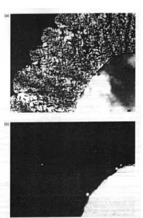
particles of the desired size. The particulate powder is then pressed into solid bbcks or hollow cylinders (green shape) and sintered at 1400°C for 36 h to increase the mechanical strength. ALCAP ceramic implants have given excellent results in terms of biocompatibility and gradual replacement of the ceramic material with endogenous bone [Bajpai, 1982; Mattie and Bajpai, 1988]. A scanning electron micrograph (l000x) of sintered porous ALCAP is shown in Figure 39.8.
15/28
39.3.3 CorallineCoral is a natural substance made by marine invertebrates. According to Holmes et al. [1984]. the marine invertebrates live in the limestone exostructure, or coral. The porous structure of the coral is unique for each species of marine invertebrate [Holmes et al., 1984]. Corals for use as bone implants are selected on the basis of structural similarity to bone [Holmes et al., 1984]. Coral provides an excellent structure for the ingrowth of bone, and the main component, calcium carbonate, is gradually resorbed by the body [Khavari and Bajpai, 1993]. Corals can also be converted to hydroxyapatite by a hydrothermal exchange process. Interpore 200, a coral hydroxyapatite, resembles cancellous bone [Sartoris et al., 1986]. Both pure coral (Biocoral) and coral transformed to hydroxyapatite are currently used to repair traumatized bone, replace diseased bone, and correct various bone defects.
Biocoral is composed of crystalline calcium carbonate or aragonite, the metastable form of calcium carbonate. The compressive strength of Biocoral varies from 26 (50% porous) to 395 MPa (dense) and depends on the porosity of the ceramic. Likewise, the modulus of elasticity (Young’s Modulus) of Biocoral varies from 8 (50% porous) to 100 GPa (dense) [Biocoral, 1989].
39.3.4 Tricalcium Phosphate (TCP) Ceramics
A multicrystalline porous form of β-tricalcium phosphate [β-Ca3(PO4)2] has been used
successfully to correct periodontal defects and augment bony contours [Metsger et al.. 1982]. X-ray diffraction of ß-tricalcium phosphate shows an average interconnected porosity of over 100 µm [Lemons et al., 1979]. Often tribasic calcium phosphate is mistaken for ß-tricalcium phosphate. According to Metsger et al. [1982], tribasic calcium phosphate is a nonstoichiometric compound often bearing the formula of hydroxyapatite [Ca10(PO4)6(OH)2].
ß-Tricalcium phosphate is prepared by a wet precipitation procedure from an aqueous solution of Ca(NO3)2 and NaH2PO4 [Bajpai et al., 1988]. The precipitate is calcined at
1150°C for 1 h, ground, and sieved to obtain the desired size particles for use as bone substitutes [Bajpai et al., 1988; Bajpai, 1990] and for making ceramic matrix drug delivery systems [Morris and Bajpai, 1989; Nagy and Bajpai, 1994; Moldovan and Bajpai, 1994]. These particles are used as such or pressed into cylindrical shapes and sintered at 1150 to 1200°C for 36 h to achieve the appropriate mechanical strength for use as drug delivery devices [Bajpai, 1989, 1992, 1994; Benghuzzi et al, 1991]. A scanning electron micrograph (500x) of a set and hardened TCP-cysteine composite is shown in Figure 39.9. The composite sets and hardens on the addition of water. TCP is usually more soluble than synthetic hydroxyapatite and, on implantation, allows good bone ingrowth and eventually is replaced by endogenous bone.
16/28
39.3.5 Zinc-Calcium--Phosphorous Oxide (ZCAP) Ceramics
Zinc is essential for human metabolism and is a component of at least 30 metalloenzymes [Pories and Strain, 1970]. In addition, zinc may also be involved in the process of wound healing [Pones and Strain 1970]. Thus zinc-calcium-phosphorous oxide polyphasic ceramics (ZCAP) were synthesized to repair bone defects and deliver drugs [Binzer and Bajpai, 1987; Bajpai, 1988, 1993; Arar and Bajpai. 1992]. ZCAP is prepared by a thermal mixing of zinc oxide, calcium oxide, and phosphorous pentoxide powders [Bajpai, 1988]. ZCAP, like ALCAP, has insulating dielectric properties but no magnetic or piezoelectric properties [Allaire et al., 1989]. Various ratios of these powders have been used to produce the desired material [Bajpai, 1988]. The oxide powders are mixed in a ball mill and subsequently calcinied at 800°C for 24 h. The calcined ceramic is then ground and sieved to obtain the desired size particles. Scanning electron micrograph (500 x) of a set and hardened ZCAP-cysteine composite is shown in Figure 39.10. The composite sets and hardens on addition of water. To date, ZCAP ceramics have been used to repair experimentally induced defects in bone and for delivering drugs [Binzer and Bajpai, 1987; Bajpai. 1993].
FIGURE 39.10 Scanning electron micrograph (x500) of a set and hardened ZCAP—cysteine composite. The small white cysteine particles have blended with the ZCAP particles.
39.3.6 Zinc-Sulfate-Calcium-Phosphate (ZSCAP) Ceramics
Zinc-sulfate-calcium-phosphate polyphasic ceramics (ZSCAP) are prepared from stock powders of zinc sulfate, zinc oxide, calcium oxide, and phosphorous pentoxide [Bajpai, 1988]. A ratio of 15:30:30:25 by weight of ZnSO4:ZnO:CaO:P2O5 is mixed in a crucible
and allowed to cool for 30 min after the exothermal reaction has subsided. The cooled mixture is calcined in a crucible at 650°C for 24 h. The calcined ceramic is ground in a ball mill and the particles of the desired size are separated by sieving in an automatic siever. Scanning electron micrograph (2000 x) of set and hardened ZSCAP particles (45 to 63 sim) is shown in Figure 39.11. ZSCAP sets and hardens on addition of water. ZSCAP particles, on implantation in bone, set and harden on contact with blood and have been used to repair experimentally induced defects in bone [Scheidler and Bajpai, 1992].
39.3.7 Ferric-Calcium-Phosphorous-Oxide (FECAP) Ceramics
17/28
by means of a hydraulic press and calcined at 1100°C for 12 h. The calcined ceramic blocks are crushed and ground in a ball mill. The calcined ceramic is ground in a ball mill and the particles of the desired size are separated by sieving in an automatic siever. A scanning electron micrograph (l000x) of a set and hardened FECAP-α ketoglutaric acid composite is shown in Figure 39.12. The composite sets and hardens on the addition of water. Studies conducted to date suggest complete resorption of FECAP particles implanted in bone within 60 days [Larrabee et al. 1993]. This particular ceramic could be used in patients suffering from anemia and similar diseases [Fuski et al., 1993].
FIGURE 39.11 Scanning electron micrograph (x2000) of a set and hardened ISCAP particles (45-63 µm). Sulfate is hardly visible between the cube shaped ZCAP particles.
FIGURE 39.12 Scanning electron micrograph (x1000) of a set and hardened FECAP-α- ketoglutaric acid composite.
39.4 BIOACTIVE OR SURFACE-REACTIVE CERAMICS
19/28
9.4.1 Glass CeramicsSeveral variations of Bioglass and Ceravital glass ceramics have been used by various workers within the last decade. Glass ceramics used for implantation are silicon oxide based systems with or without phosphorous pentoxide.
Glass ceramics are polycrystalline ceramics made by controlled crystallization of glasses developed by S.D. Stookey of Corning Glass Works in the early 1960s [Park and Lakes, 1992]. Glass ceramics were first utilized in photosensitive glasses, in which small amounts of copper, silver, and gold are precipitated by ultraviolet light irradiation. These metallic precipitates help to nucleate and crystallize the glass into a fine grained ceramic which possesses excellent mechanical and thermal properties. Both Bioglass and Ceravital glass ceramics have been used as implants [Yamamuro et al., 1990].
The formation of glass ceramics is influenced by the nucleation and growth of small (<1
µ m diameter) crystals as well as the size distribution of these crystals. It is estimated that about 1012 to 1015 nuclei/cm3 are required to achieve such small crystals. In addition
to the metallic agents already mentioned, Pt groups. TiO2, ZrO2, and P2O5 are widely
used for nucleation and crystallization. The nucleation of glass is carried out at temperatures much lower than the melting temperature. During processing the melt viscosity is kept in the range of 1011 and 1012 Poise for 1 to 2 h. In order to obtain a
larger fraction of the microcrystalline phase, the material is further heated to an appropriate temperature for maximum crystal growth. Deformation of the product, phase transformation within the crystalline phases, or redissolution of some of the phases should be avoided. The crystallization is usually more than 90% complete with grain sizes 0.1 to 1 µm. These grains are much smaller than those of conventional ceramics. Figure 39.13 shows a schematic representation of temperature-time cycle for a glass ceramic [Park and Lakes, 1992]. The glass ceramics developed for implantation are SiO2—CaO—Na2O—P2O5 and Li2O—ZnO—SiO2 systems. Two major groups are
experimenting with the SiO2—CaO--Na2O—P2O5 glass ceramic. One group varied the
compositions (except for P2O5) in order to obtain the best glass ceramic composition for
inducing direct bonding with bone (Table 39.13). The bonding to bone is related to the simultaneous formation of a calcium phosphate and SiO2-rich film byer on the surface,
as exhibited by the 46S5.2 type Bioglass. If a SiO2-rich layer forms first and a calcium
phosphate film develops later (46 to 55 mol % SiO2 samples) or no phosphate film is
formed (60 mol % SiO2) then direct bonding with bone does not occur [Park and Lakes,
1992]. The approximate region of the SiO2-CaO—Na2O system for the tissue—glass—
ceramic reaction is shown in Figure 39.14. As can be seen, the best region (region A) for good tissue bonding is the composition given for 46S5.2 type Bioglass (see Table 39.13) (Park and Lakes, 1992].
FIGURE 39.13 Temperature-time cycle for a glass-ceramic. (From Kingery W.D., Bowen H.K., and Uhlmann D.R. 1976. Introduction to Ceramics, 2nd ed., p. 368. New York, John
20/28
39.4.2 CeravitalThe composition of Ceravital is similar to that of Biogass in SiO2 content but differs
somewhat in other components (see Table 39.13). In order to control the dissolution rate, Al2O3, TíO2, and Ta2O5 are added in ceravital glass ceramic. The mixtures, after melting
in a platinum crucible at 1500°C for three h, are annealed and cooled. The nucleation and crystallization temperatures are 680 and 750°C, respectively, each for 24 h. When the size of crystallites reaches approximately 4 Å and the characteristic needle structure is not formed, the process is stopped to obtain a fine grain structured glass ceramic [Park and Lakes, 1993].
FIGURE 39.14 Approximate regions of the tissuc.giass.cerarnic bonding for the SiO3—CaO—
Na2O system. A: Bonding within 30 days. B: Nonbonding; reactivity is too low. D: Bonding
does not form glass. [From Hench LL and Ethridge E.C. 1982. Biormaterials: An Inserfacial Approach. p. 147. New York. Academic Press. With permission.)
Glass ceramics have several desirable properties compared to glasses and ceramics. The thermal coefficient of expansion is very low, typically 10−7 to l0−5 oC−1, and in some
21/28
A transmission electron micrograph of Bioglass glass ceramic implanted in the femur of rats for six weeks showed intimate contacts between the mineralized bone and the Bioglass (Figure 39.15). The mechanical strength of the interfacial bond between bone and Biogass ceramic is on the same order of magnitude as the strength of the bulk glass ceramic (850 kg/cm2 or 83.3 M Pa), which is about three-fourths that of the host bone strength [Park and Lakes, 1992]
.
FIGURE 39.15 Transmission electron micrograph of well-mineralized bone (b) juxtaposed to the glass-ceramic (c) which fractured during sectioning. x51,500. Insert a is the diffraction
pattern from ceramic area and b is from bone area. (From Beckham C.A., Greenlee T.K. Jr,and Crebo A.R. 1971. Calc. Tiss. Res. 8: 165-171.With permission.)
A negative characteristic of the glass ceramic is its brittleness. In addition, limitations on the compositions used for producing a biocompatibile (or osteoconductive) glass ceramic hinders the production of a glass ceramic which has substantially higher mechanical strength. Thus glass ceramics cannot be used for making major load-bearing implants such as joint implants. However, they can be used as fillers for bone cement, dental restorative composites, and coating material (see Table 39.12). A glass ceramic containing 36 wt% of magnetite in a ß-wollastonite- and CaOSiO2-based glassy matrix
has been synthesized for treating bone tumors by hyperthermia [Kokubo et al, 1992].
39.5 DETERIORATION OF CERAMICS
22/28
strength occurs if water pe1etrates the ceramic. Decrease in strength was not observed in samples which did not show water marks on the fractured surface (Figure 39.16). The presence of a small amount of silica in one sample lot may have contributed to the permeation of water molecules that is detrimental to the strength [Park and Lakes, 1992]. It is not clear whether the static fatigue mechanism operates in single crystal alumina. It is reasonable to assume, that static fatigue will occur if the ceramic contains flaws or impurities, because these will act as the source of crack initiation and growth under stress [Park and Lakes, 1992].
FIGURE 39.16 Flexural strength of dense alumina rods alter aging under stress in Ringer’s solution. Lot 1 and 2 are from different batches of production. (From Krainess FL and Knapp
WJ. 1978. J. Biomat. Mater. Res. 12: 245. With permission.)
FIGURE 39.17 Strain vs. number of cycles to failure (o = absence of fatigue cracks in carbon film; = fracture of carbon film due to fatigue failure of substrates; = data from substrate determined in single-cycle tensile test). (From Shimm HS. and Haubold AD. 1980.
Biomater. Med. Dev. Art. Org. 8:333—344. With permission.)
Studies of the fatigue behavior of vapor-deposited pyrolitic carbon fibers (4000 to 5000 Å thick) onto a stainless steel substrate showed that the film does not break unless the substrate undergoes plastic deformation at 1.3 x 10—2 strain and up to one million cycles
23/28
predicted by assuming that the fatigue fracture is due to the slow growth of preexisting flaws. Generally, the strength distribution, σi, of ceramics in an inert environment can be
correlated with the probability of failure F by the following equation:
Both m and s0 are constants in the equation. Figure 39.18 shows a good fit for Bioglass
coated alumina [Park and Lakes, 1992].
FIGURE 39.18 Plot of Ln Ln [1/(1 −F)] vs. Ln S for Bioglass-coated alumina in a tris-hydroxyaminomethane buffer and liquid nitrogen. F is the probability
of failure and S is strength. (From Ritter I.E. Jr. Greenspan D.C.. Palmer R.A..and Hench L.L 1979. J. Biomed. Mater. Res. 13: 260. With permission.) A minimum service life (tmin) of a specimen can be predicted by means of a proof test
wherein it is subjected to stresses that are greater than those expected in service. Proof tests also eliminate the weaker pieces. This minimum life can be predicted from the following equation:
Here σp is the proof test stress, σa is the applied stress, and B and N are constants.
Equation 39.2 after rearrangement reads as follows:
Figure 39.19 shows a plot of Equation 39.3 for alumina on a logarithmic scale [Park and Lakes, 1992].
39.6 BIOCERAMIC MANUFACTURING TECHNIQUES
24/28
device, whether it is for hard tissue replacement or the integration of the device within the surrounding tissue.
FIGURE 39.19 Plot of Equation 39.3 for alumina after proof testing. N = 43.85, m = 1321, and σ0 = 55728 psi. (From Ritter I.E. Jr. Greenspan D.C., Palmer R.A.,
and Hench L.L 1979. J. Biomed. Mater. Res. 13: 261. With permission.)
39.6.1 Hard Tissue Replacement
Hard tissue replacement implies that the bioceramic device will be used for load bearing applications. Although it is desirable to have a device with a sufficient porosity for the surrounding tissue to infiltrate and attach to the device, the most important and immediate property is the strength of the device. In order to accomplish this, one must manufacture a bioceramic implant with a density and strength sufficient to mimic that of bone. However, if the bioceramic part is significantly stronger than the surrounding bone, one runs mio the common problem seen with metals called stress shielding. The density of the biocerarnic greatly determines its overall strength. As the density increases so does the overall strength of the bioceramic. Some of the techniques used to manufacture dense bioceramics are injection molding, gel casting, bicontinuous microemulsion, inverse microemulsion, emulsion, and additives.
25/28
achieved 98% relative density with only 16% shrinkage using injection molding. The maximum flexural strength was 60 MPa for HA products sintered at 1473 K. However, this is still not strong enough for load bearing applications. They also observed that HA decomposed to α-TCP at temperatures greater than 1573 K (Cihlar and Trunec, 1996).
In gel casting, HA is formed using the standard chemical precipitation. The calcium phosphate precipitant (30% w/v) is then mixed with glycerol and filtered. The “gel cake” is sintered at 1200°C for 2 h. This yielded a density of >99% and a highly uniform microstructure [Varma and Sivakumar, 1996].
Bicontinuous microemulsion, inverse microemulsion, and emulsion are all wet chemistry based methods to produce nanometer size HA powders. All three methods yield >97% relative density upon sintering at 1200°C for 2 h. The biocontinuous and inverse microemulsion resulted in the two smallest HA particle sizes, 22 and 24 nm respectively [Lim et al., 1997].
Another strategy to increase the density of ceramics is to use additives or impurities in small weight percents during sintering. The main disadvantages to this technique include (1) the possible decomposition of the original pure bioceramic and (2) results may end in all or portions of the bioceramic being nonbiocompatible.
Suchanek et al. (1997) studied the addition of several different additives to HA in 5 wt% amounts. The additives studied were K2CO3, Na2CO3, H3BO3, KF, CaCl2, KCl,
KH2PO4, (KPO3)n, Na2Si2O5, Na2P2O7, Na3PO4, (NaPO3)n, Na5P3O10 , and ß-NaCaPO4.
HA has a fracture toughness of 1 MPa.m1/2, whereas human bone has a fracture
toughness of 2 to 12 MPa..m1/2. One of the ways to improve the mechanical properties is
to improve the densification of HA. Suchanek et al. [1997] found that the following additives (5 wt%) did not improve the densification of HA: H3BO3, CaCl2, KCl,
KH2PO4, (KPO3)n, and Na2Si2O5. The densification of HA was improved through the
addition (5 wt%) of K2CO3, Na2CO3, KF, Na2P2O7, Na3PO4, (NaPO3)n, Na3P3O10, and
ß-NaCaPO4. However, H3BO3, CaCl2, KH2PO4, (KPO3)n, Na2Si2O5, K2CO3, Na2CO3,
26/28
that improved densification, did not cause formation of ß-TCP or CaO, and provided a weak interface for HA was ß-NaCaPO4.
Another additive that has been investigated to improve the performance of HA is lithium (Li). The addition of lithium can increase the microhardness and produces a fine microstructure in HA. Fanovich et al. [1998] found that the addition of 0.2 wt% of Li to HA produced the maximum microhardness (5.9 GPa). However, the addition of high amounts of Li to HA results in abnormal grain growth and large pores. Furthermore, Li addition to HA results in the formation of ß-TCP upon sintering.
Zirconia has been used as an additive to HA in order to improve its mechanical strength. Kawashima et al. [1997] found that addition of partially stabilized zirconia (PSZ) to HA can be used to increase the fracture toughness to 2.8 MPa m.1/2. Bone has a fracture
toughness of 2-12 MPa m.1/2 [Suchanek
et al., l997]. PSZ was added to HA in different percentages (17,33,50 wt%) and it was found that 50 wt% PSZ had the highest fracture toughness. The surface energy of the PSZ-HA was not significantly different from HA alone. This suggests that the PSZ-HA composite could be biocompatible because of the similarity of the surface with HA [Kawashima et al., 1997].
39.6.2 Tissue Integration
The porosity is a critical factor for growth and integration of a tissue into the bioceramic implant. In particular the open porosity, that which is connected to the outside surface, is critical to the integration of tissue into the ceramic especially if the bioceramic is inert. Several methods have been developed to form porous ceramics, two of these are starch consolidation and drip casting.
In starch consolidation (Table 39.15), starch powders of a specific size are mixed with a bioceramic slurry at a predetermined weight percent. Upon heating, the starch will uptake water from the slurry mixture and swell. Upon sintering of the starch-bioceramjc mixture, the starch is burned out and the pores are left in their place. Starch consolidation has been used to form complex shapes in alumina with ultimate porosities between 23 and 70 vol %. By controlling the starch content, one can control the ultimate porosity and resulting pore sizes, large pores formed using starch consolidation in alumina were in the size range 10 to 80 µm whereas small pores varied between 0.5 and 9.5 µm [Lyckfeldt and Ferreira,1998].
Liu [1996] used a drip casting technique (Table 39.15) to form porous HA granules with pore sizes from 95to 400 µm. The granules had a total porosity from 24 to 76 vol %. The HA was made into a slurry using water and polv(vinyl butyral) powders. The slurry was then dripped onto a spherical mold surface. This technique is similar to that of drip casting by dripping an HA slurry into a liquid nitrogen bath. In both instances, calcining and sintenng procedures are used to produce the final product [Liu, 1996].
39.6.3 Hydroxyapatite Synthesis Method Prepare the following solutions:
Solution 1: Dissolve 157.6 g calcium nitrate letrahydrate [Ca(NO3)24H2O] in 500
ml DI water. Bring the solution to a pH of 11 by adding 70 ml ammonium hydroxide [NH4OH]. Bring the solution to 800 ml with DI water.
Solution 2: Dissolve 52.8 g ammonium phosphate dibasic [(NH4)2HPO4] in 500 ml
DI water. Bring the solution to a pH of 11 by adding 150 ml ammonium hydroxide [NH4OH]. Add Dl water until the precipitate is completely dissolved. 250-350 ml.
39.6.3.1 Special Note. If you use calcium nitrate [Ca(NO3)2.nH2O] instead of calcium
27/28
Solution 1 on the basis of the absence of the extra four waters. If you do not, you will have to add a large amount of ammonium hydroxide to pH the solution:
1. Add one-half of Solution 1 to a 2 l separatory funnel 2. Add one-half of Solution 2 to a 2 l separatory funnel
3. Titrate both solutions into a 4 l beaker with heat and constant stirring 4. Boil gently for 30 min
5. Repeat steps l - 4 for the rest of Solutions 1 and 2
6. Let cool completely allowing precipitate to settle to bottom of beaker 7. Pour contents of beaker into 250 ml polypropylene bottles
8. Centrifuge bottles for l0 min at (10,000 rpm) 16,000 g
9. Collect precipitate from the six bottles into two and resuspend with DI water
10. Fill the four empty bottles with the reaction mixture and centrifuge all six as before 11. Collect precipitant from the two bottles that were resuspended with DI water 12. Combine remaining four bottles into two bottles and resuspend with DI water 13. Repeat steps 10 - 12 as necessary
14. Dry precipitate for 24 - 48 h at 70°C
15. Calcine the precipitate for one hour at 1140°C 16. Grind and sieve product as desired
DEFINING TERMS
Alumina: Aluminum oxide (Al2O3) which is very hard (Moh’s hardness is 9) and
strong. Single crystals are called sapphire or ruby depending on color. Alumina is used to fabricate hip joint socket components or dental root implants.
Calcium phosphate: A family of calcium phosphate ceramics including aluminum calcium phosphate, ferric calcium phosphate, hydroxyapatite and tricalcium phosphate (TCP), and zinc calciumphosphate which are used to substitute or augment bony structures and deliver drugs.
Glass-ceramics: A glass crystallized by heat treatment. Some of those have the ability to form chemical bonds with hard and soft tissues. Bioglass and Ceravital are well known examples.
Hydroxyapatite: A calcium phosphate ceramic with a calcium to phosphorus ratio of 513 and nominal composition Ca10(PO4)6(OH)2. It has good mechanical properties and
excellent biocompatibility. Hydroxyapatite is the mineral constituent of bone.
LTI carbon: A silicon alloyed pyrolitic carbon deposited onto a substrate at low temperature with isotropic crystal morphology. It is highly compatible with blood and used for cardiovascular implant fabrication such as artificial heart valve.
Maximum radius ratio: The ratio of atomic radii computed by assuming the largest atom or ion which can be placed in a crystal’s unit cell structure without deforming the structure.
Mohs scale: A hardness scale in which 10 (diamond) is the hardest and 1 (talc) is the softest.
ACKNOWLEDGMENTS