INFECTION PROCESS OF
Fusarium oxysporum
FUNGUS: A CAUSE OF
DAMPING-OFF
ON
Acacia mangium
’
s
SEEDLINGS
S. M. Widyastuti1*), Susanti Tasik2) and Harjono1) 1)Faculty of Forestry, Gadjah Mada University Jl. Agro - Bulaksumur Yogyakarta 55281 Indonesia 2)Faculty of Forestry, The State University of Papua Jl. Gunung Salju Amban Manokwari Papua Indonesia
*) Corresponding author Phone:+62-274- 512102 E-mail: smwidyastuti@ugm.ac.id
Received: April 1, 2013/ Accepted: May 9, 2013
ABSTRACT
Fusarium oxysporum is the causal agent of damping-off disease. The fungus attacks seedlings of many plant species, including Acacia mangium. In order to effectively control the disease, detailed information on how the fungus infects seedlings of A. mangium and how the plant responds to the fungal infection is essentially needed. The objectives of this research were to investigate: (1) the infection process of F. oxysporumon seedlings of A. mangium, (2) the defence response of A. mangium seedling to infection by F. oxysporum. The fungal pathogen was identified, followed by performance of pathogenicity test. The infection process was followed by macroscopic observation as well as microscopic observation. The result indicated that fungal spore germination was observed at two-day post inoculation in planta. At four-daypost inoculation, hyphae of F. oxysporum had penetrated the collar root of A. mangium seedling via stomata aperture. In addition, fungal hyphae had grown intercellulary in to the vascular tissue. Correspondingly, hypersensitive response was also detected at the stomata aperture. However, this defence mechanism is not effective in stopping the fungus since F. oxysporum is a necrotropic pathogen. Moreover, accumulation of lignin, but not callose, was observed.
Keywords: Fusarium oxysporum, damping-off, Acacia mangium.
INTRODUCTION
Demand for wood material from industrial plantation forest has increased after its introduction in 1984. Hardiyanto and Arisman (2004) reported that as an important tree species in industrial plantation forest, Acacia mangium Will has some advantages, such as fast growth, suitable characteristic of its timber for pulp and paper raw material, construction material, light construction and coal. To support raw material availability, many healthyA. mangium seeds are required to achieve the expected maximal production target.
Acacia mangium seedling is susceptible to disease in nursery such as damping off. Fusarium oxysporum is a soil borne pathogen fungus, causing damping-off disease in A. mangium. The fungus is also reported to cause damping-off disease in forestry plant nurseries particularly in A. mangium seedling (Wardani, 2007), Pinus merkusii (Achmad et al., 2012), P. Pinea (Machon et al., 2009), Eucalyptus viminalis (Salerno et al., 2004). The objectives of this research were to investigate the infection process of F. oxysporum on seedlings of A. Mangium and the defence response of A. mangium seedling to infection by F. oxysporum
MATERIALS AND METHODS
Fusarium oxysporum Isolationes and Acacia mangium Seedling
Isolated F. oxysporum was obtained from two-week old, infected A. mangium seedling indicating damping-off symptom. The F. oxysporum from infected A. mangium seedling was isolated and grown in Potato Dextrose Agar
Accredited SK No.: 81/DIKTI/Kep/2011
(PDA) media. Fusarium oxysporum was identified when colony was 4 days old in PDA media. The identification process included size and shape of the observed spore as well as its color and the change of colony color (Leslie and Summerell, 2006). Acacia mangium seedling was obtained by planting A. mangium seed in sterile soil media.
Defense Response of Acacia mangium Seedling Against F. oxysporum
Infected A. mangium seedling was observed microscopically. The infected seedling was treated with chlorophyll removal by soaking the seedling using 96% ethanol solution, heated at 1000C for 10 minutes, and left for cooling for 1 hour at room temperature. Then, the seedling was treated using transparent tissue and soaked into 20 mL (2.5 g/mL) chloral hydrate solution (Merck) for 12 hours. Observation of the infection process of F. oxysporum in A. mangium seedling was conducted consecutively at 2, 4, 6, and 8 days after the suspended spore inoculation in the 1-week A. mangium. Infected A. mangium seedling was then stained with lactophenoltrypan blue (Merck). The modified Ruzin’s (1999) method was used for lignin accumulation. Tissue that was treated with transparent tissue was soaked in 20 mL phloroglucinol (Merck) solution for 30 minutes. Callose accumulation assay was done using the modified Ruzin’s (1999) method as well. Tissue treated using transparent tissueagent was soaked into 20 mL aniline blue (Merck) solution for 10 minutes. Then, microscopic observation was done using Axioskop 40 fluorescent microscope and was recorded with Canon Power Shot A 620 camera digital.
RESULTS AND DISCUSSION
Fusarium oxysporum Pathogenicity in Acacia mangium Seedling
Acacia mangium seedling in nurseries showed symptom of damping-off disease at two weeks after germination (Figure 1). Early symptom was indicated with wilted seedling, and the rot began from base up to whole seedling stem when seedling was 6 days old that caused damping off and death.
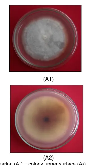
The fungus was isolated from Acacia mangium seedling showing the symptom and sign. The fungus was then sub cultured in PDA media at 27 0C and pure culture was obtained
(Figure 2). Observation of the fungal colony morphology showed that the pathogenic fungi grown in PDA media were thin and rare, white as cotton to pale violet. Another side of the colony was yellowish to purplish.
Figure 1.Eight-day infected Acacia mangium seedling by Fusarium oxysporum
(A1)
(A1)
(A2)
Remarks: (A1) = colony upper surface (A2)= colony lower surface
Remarks: (A) Macro conidia, (B) Micro conidia, (C) and (D) Chlamydospore (arrow sign) (H) F. oxysporumhypha (Bar = 10m)
Figure 3.
F. oxysporum
spore
Next identification indicated that pathogen fungi infecting A. mangium seedling had three spore types i.e.macroconidia, microconidia and chlamydospore (Figure 3). The spore size was various. Macroconidia had size ranging from (12-18) x (3-5)m, with 3 septa and no septa, the fusiform was slightly bent and pointed at two ends. Microconidia had 2-4 septa and no septa. Its shape was ovoid ellipse, and its size was between (4-8) x (1.5-2.5)m. Microconidia was smaller than macroconidia and chlamydospore. Chlamydospore presented at hypha end/ terminal and within hypha or intercalary, had smooth wall, round shape, in single and pairing configurations with diameter of 2.25-3.75 m. Based on macroscopic and microscopic observations, isolation of pathogenic fungi infecting A. mangium seedling was F. oxysporum Schlechtendahl emend. Snyder and Hansen (Leslie and Summerell, 2006).
Fusarium oxysporum Infection on Acacia mangium Seedling
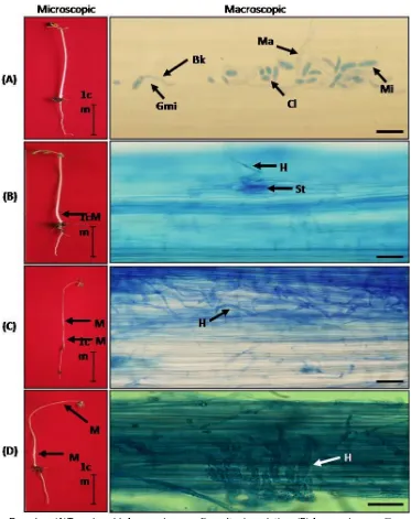
Remakrs: (A)Two-day-old A. mangium seedling after inoculation, (B)A. mangium seedling, 4 days after inoculation, (C)A. mangium seedling, 6 days after inoculation, (D)A. mangium
seedling, 8 days after inoculation, (H)F. oxysporum hypa, (Mi) Microconidia, (Gmi) Germinated microconidia, (Ma) Macroconidia, (Ch) Chlamydospore, (St) Stomata, (M) Mycelium F. oxysporum, (Bk) Shoot reed. (Bar = 50m)
Microscopic observation indicated that F. oxysporum hypha penetrated through stomata causing damping-off disease when A. mangium seedling occured at 2 and 4 days after inoculation (Figure 4). Penetration continued into plant tissue in intercellular way at phloem and xylem when seedling was at 6 and 8 days after inoculation resulting in seedling death. Vidhysekaran (2008) stated that F. oxysporum hypha performed direct penetration and colonized cortex. The colonization process caused fungi to be able to reach xylem vessel tissue and spread fast to stimulate wilt sign of the plant.
Saprophytic microorganism can turn pathogenic in plant through parasitic adaptation and had various levels from obligate parasite to facultative parasite (Widyastuti et al., 2005). F. oxysporum is one of saprophytic microorganism that can change into pathogenic microorganism that can cause damping-off on A. mangium seedling. Moreover, Widyastuti et al. (2005) stated that capacity of a microorganism to be a pathogen in plant was determined by its capacity to (1) penetrate into plant tissue, (2) break host defense, and (3) cause disease. Based on the current research result, soil-borne pathogen F. oxysporum was provento have the capacity to penetrate phloem and xylem vessel tissue of A. mangium seedling, which finally caused damping-off disease over A. mangium seedling.
Defense Response of Acacia mangium Seedling due to Fusarium oxysporum Infection
Defense response of A. mangium seedling due to F. oxysporum infection was marked with lignin accumulation (Figure 5). Lignin accumulation inA. mangium seedling stem was detected when the seedling was at 4 and 6 days after inoculation. It was marked with light red to violet colour in area around phloem and xylem vessel tissue of the infected A. mangium seedling after stained with Phloro-glucinol (Ruzin, 1999; Vidhyasekaran, 2008). Observation showed that lignin was also accumulated in infected phloem
and xylem tissue, but not found as many as it was in area around infected phloem and xylem vessel tissue (Figure 4). Lignin was also not detected in uninfected xylem vessel tissue. It is expected that lignin in A. mangium seedling not inoculated by F. oxysporum was still produced but it was not as much as an infected A. mangium seedling. Lignin as chemical component in cell wall of xylem vessel tissue functioning to strengthen mechanical defense of plants was also produced (Vanholme et al., 2008). Accumulated lignin was detected around stomata and xylem tissue in the inoculated A. mangium seedling stem, indicating response from the plants to strengthen mechanic system defense due to penetration of F. oxysporum hypha. Lignin was also one of polymers which is difficult to be degraded by pathogen (Nicholson and Hammerschmidt, 1992).
Figure 5. Lignin accumulation in Acacia mangium seedling due to Fusarium oxysporum infection. (A1) Stomata of healthy A. mangium, (A2) Stomata of A. mangium 4 days after inoculation, (B1) Xylem of healthyA. mangium, (B2) Xylem of A. mangium 6 days after infected, (La) Lignin accumulation, (H) Hypha F. oxysporum, (St) stomata, (Xy) Xylem, (Bar = 50m).
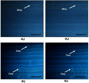
Result of A. mangium seedling defense response with histological stain Aniline blue indicated that there was callose accumulation but only in infected and non infected xylem (Figure 6). Callose was expected to accumulate and increase in area around xylem vessel tissue of an infected A. mangium. Microscopic examination indicated that seedling at 4 and 6 days after inoculation had no callose accumulation at site around the infected xylem vessel tissue. Interaction occurring betweenA. mangium seedling as the host and F. oxysporum as pathogen was expected to form a compatible interaction. The interaction resulted in A. mangium seedling vulnerable to F. oxysporum infection. It was evidenced with wilted seedling at 4 and 6 days after inoculation so the seedling may be classified as vulnerable to pathogen (Figure 4).
Figure 6. Callose accumulation in A. mangium seedling due to Fusarium oxysporum infection. (A1) Xylem of healthy A. mangium, (A2) Infected xylem of A. mangium 4 days after inoculation, (B1) Xylem of healthyA. mangium, (B2) Infected xylem of A. mangium 6 days after inoculation, (Caxy) Callose accumulation in xylem. (Bar = 50m)
Hypersensitive reaction was found when there was F. oxysporum infection on seedling stem surface at 4 days after inoculation (Figure 7). Based on the figure, there appeared difference in stomata colour between healthy tissue and tissue infected with F. oxysporum at the same age. In infected tissue, stomata was darker blue than that around healthy tissue (Figure 7A), while stomata in healthy tissue indicated the same colour as other surrounding tissues (Figure 7B). Staining with lactophenol trypan blue can be used to detect the existence of fungi structure or dead cell in plant, marked blue which is darker than other tissue. Similar finding was also presented by Feys et al. (2001) that staining with lactophenol trypan blue on Arabidopsis leaf infected with Pseudomonas parasitica could detect mycelium and might also detect hypersensitive reaction. The detected hypersensitive reaction was marked with darker
colour in leaf tissue experiencing more hyper-sensitive reaction than other surrounding tissues.
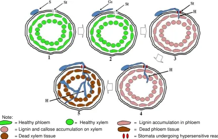
phloem vessel tissue to xylem with intercellular way through intercellular space. In the fifth steps, F. oxysporum hypha developed in A. mangium seedling tissue and the seedling died. Lignin
accumulation occurred in the fourth and fifth steps. Lignin accumulation was not detected in the last fifth step because the seedling had died.
Remarks: (A) Healthy stomata, (B) Infected stomata, (St) stomata, (g) Guard cell, (H) F. oxysporum hypha(Bar = 50
m)Figure 7. Hypersensitive reaction on Acacia mangium stomata due to Fusarium oxysporum infection 4 days after inoculation
Note:
= Healthy phloem = Healthy xylem = Lignin accumulation in phloem = Lignin and callose accumulation on xylem = Dead phloem tissue
= Dead xylem tissue = Stomata undergoing hypersensitive reaction
CONCLUSIONS
In conclusion F. oxysporum in vitro penetrated through stomata into A. mangium seedling stem base until phloem vessel tissue and xylem intercellular through intercellular space. Hypersensitive reaction and lignin accumulation occurred due to F. oxysporum infection.
ACKNOWLEDGEMENT
We thank to Tanoto Foundation for the financial support and toI. Riastiwifor preparing this manuscript.
REFERENCES
Achmad, S., S. Hadi, E.G. Harran, B. Sa'id, Satiawihardja and K. Kardin. 2012. Attack mechanism of damping off patho-gens of Pinus merkusii seedling. Jurnal Silvikultur Tropika. 3 (1):57-64. Abstract in English
Brockmann, B., R. Smit and P. Tudzynski P. 1992. Characterization of an extracellular b-1,3-glucanase of Claviceps purpurea. Physiology Molecular Plant Pathology 40: 191-201.
Feys, B.J., J.L. Moisan, N. Mari-Anne and E. J. Parker. 2001. Direct interaction between the arabidopsis disease resistance sig-naling proteins, EDS1 and PAD4. The EMBO Journal 20: 5400-5411,.
Hadi, S. 2001. Forest Pathology (in Indonesia). its development in Indonesia. Forestry Faculty. Bogor Agricultural University. Hardiyanto, E.B. and H. Arisman. 2004.
Esta-blishment of forest plantation Acacia mangium (in Indonesia). The experiment in PT. Musi Hutan Persada. South Sumatera
Kang, Z. and H. Buchenauer. 2000. Ultra-structural and immunocytochemical investigation of pathogen development and host responses in resistant and susceptible wheat spikes infected by Fusarium culmorum. Physiology Mole-cular Plant Pathology 57: 255-268. Leslie J. F. and B.A. Summerell. 2006. The
Fusarium Laboratory manual. Blackwell Publishing, Lowa, USA.
Machón P.J.A.P., J.J. Diez and F.M. Alves-Santos. 2009. Influence of the ecto-mycorrhizal fungus Laccaria laccata on pre-emergence, post-emergence and late damping-off by Fusarium oxy-sporum and F. verticillioides on stone pine seedlings. Symbiosis 49: 101-109. Nicholson R.L.and P.E. Hammerschmidt. 1992.
Phenolic compounds and their role in disease resistance. Annual Review Phytophatology 30: 369-389.
Ruzin S.E. 1999. Plant microtechnique and microscopy. Oxford University Press, New York, 322p.
Salerno M.L., S. Gianinazzi and V. Gianinazzi-Pearson. 2004. Cell Interaction between a non pathogenic Fusarium oxy-sporumstrain and root tissues of Eucalyptus viminalis. J.Gen.Plant Pathol 70:153-158
Southerton, S.G. and B.J. Deverall. 1990. Histochemical and chemical evidence for lignin acumulation during the expression of resistance to leaf rust fungi in wheat. physiology. Plant Patho-logy 36: 483-494.
Vanholme, R., M. Kris, R. John and W. Boerjan. 2008. Lignin engineering. Current Opinion in Plant Biology 11: 278–285. Vidhyasekaran, P. 2008. Fungal pathogenesis in
plants and crops: Molecular Biology and Host Defense Mechanism.
Wardani, B.A. 2007. Isolation and characteri-zation a cause damping-off Acacia mangium Willd seedling (Abstract in English). Forestry Faculty. University of Gadjah Mada
Widyastuti, S.M., Sumardi and Harjono. 2005. Forest Pathology (In Indonesia). Gadjah Mada University Press, Yogyakarta.