See discussions, stats, and author profiles for this publication at: https://www.researchgate.net/publication/47427410
Micro leakage of Posterior Composite Restorations
Lined with Self-adhesive Resin Cements
ARTICLE in OPERATIVE DENTISTRY · SEPTEMBER 2010
Impact Factor: 1.67 · DOI: 10.2341/09-189-L · Source: PubMed
CITATION
1
READS
49
4 AUTHORS, INCLUDING:
Mohammed Al-Saleh
University of Alberta
16PUBLICATIONS 60CITATIONS
SEE PROFILE
SUMMARY
Purpose: This study determined the microleak-age of Class II composite restorations lined with self-adhesive resin-cements as bonding agents. Methods: Forty-five caries-free extracted molars were sterilized, mounted in acrylic bases and divided into five equal groups according to the
adhesive used: RXU (RelyX-Unicem, adhe-sive resin-cement, 3M ESPE), BRZ (Breeze,
self-adhesive resin-cement, Pentron Clinical
Technologies), MON (Monocem, self-adhesive cement, Shofu), PAN (Panavia-F-2.0, resin-cement with self-etch primer, Kuraray) and SBMP (Scotchbond Multi-Purpose, total-etch three-step adhesive, 3M ESPE). Class II MOD cavities were prepared with gingival floors located on dentin at one side and on enamel on the other. The bonding agent SBMP, used accord-ing to the manufacturer’s directions, or a thin layer of resin cement, was applied on all cavity walls and cavosurface margins. Filtek Z250 (3M ESPE) was used to restore cavities in all groups. The specimens were subjected to 1,000 thermo-cycles between 5°C and 55°C. All tooth surfaces were sealed with nail-varnish to within 1 mm from the restoration margins. The specimens were immersed in 2% fuchsine red solution for 24 hours at 37°C. The teeth were then sectioned mesiodistally and dye penetration was assessed
Microleakage of
Posterior Composite Restorations
Lined with
Self-adhesive Resin Cements
M Al-Saleh • O El-Mowafy
L Tam • A Fenton
Clinical Relevance
Two self-adhesive resin cements resulted in minimal microleakage scores when used as liners in Class II composite restorations.
©Operative Dentistry, 2010
,
35-5, 556-563*Mohammed Al-Saleh, BDS, MSc, Department of Clinical Dental Sciences, Faculty of Dentistry, University of Toronto, Toronto, Canada
Omar El-Mowafy, BDS, PhD, FADM, professor, Restorative Dentistry, Department of Clinical Dental Sciences, Faculty of Dentistry, University of Toronto, Toronto, ON, Canada
Laura Tam, DDS, MSc, associate professor, Department of Clinical Dental Sciences, Faculty of Dentistry, University of Toronto, Toronto, ON, Canada
Aaron Fenton, DDS, MS, FRCD(C), professor of dentistry, direc-tor of Implant Unit and IDAPP, Faculty of Dentistry, University of Toronto, Toronto, ON, Canada
*Reprint request: 124 Edward St, Toronto, ON M5G 1G6, Canada; e-mail: [email protected]
557
according to a five-point scale. Data were statis-tically analyzed with the Chi-square test (p<0.05). Results: Microleakage scores revealed that, on enamel margins, the SBMP group had significantly less microleakage than the RXU and BRZ groups, which, in turn, had significant-ly less microleakage than the MON and PAN groups; whereas on dentin margins, the RXU
and BRZ groups had significantly less
microleakage than the SBMP, MON and PAN groups. Conclusions: Thisin-vitrostudy showed that, when two self-adhesive resin-cements (RXU & BRZ) were used as liners in Class II com-posite restorations, they resulted in low microleakage scores as compared to the two other cements at both the enamel and dentin cavosurface margins.
INTRODUCTION
One major cause of failure of Class II resin composite restorations is recurrent caries.1 Microgaps at the
tooth-restoration interface permit the ingress of fluids and bacteria, a phenomenon referred to as microleak-age, which contributes to the development of recurrent caries.2-4 Composite materials undergo a volumetric
polymerization shrinkage of about 2%.5Stresses
gen-erated by polymerization shrinkage can reach values ranging from 13 to 17 MPa.6-7Such high stresses can
cause bond failure, with the material pulling away from the cavity margins during polymerization, with subsequent gap formation.8 In the Class II situation,
bond strength at the gingival margin is typically less than ideal and the gingival margin is particularly sus-ceptible to marginal gap formation and microleakage.9
Several methods have been developed to counteract stress buildup along the tooth-restoration interfaces during the polymerization of Class II composite restorations. These methods utilize different light polymerization cycles and strategic incremental place-ment techniques in order to reduce the C-factor.7-12
More recently, the use of glass fiber inserts was sug-gested for reducing overall polymerization contraction at the gingival margins of Class II composite restora-tions.13
Self-adhesive resin cements have been developed for use without the need for a separate bonding agent. By eliminating the etching, rinsing, priming and adhesive application steps, usage is simpler and less prone to potential errors associated with these steps. While these materials are manufactured to be primarily used for the cementation of crowns, fixed partial dentures and inlays/onlays, their potential for use as cavity lin-ers has not as yet been explored.
Self-adhesive resin cements offer a simplified bond-ing procedure that is similar to self-etch bondbond-ing
agents. However, self-adhesive cements are partially filled and would be expected to have improved mechanical properties over self-etch bonding agents. Furthermore, a self-adhesive tooth-cement-composite joint would be thicker than a tooth-adhesive-composite and, therefore, may potentially better resist polymer-ization contraction forces by viscous flow prior to com-plete setting. This may result in improved stress dis-tribution as an intermediary layer between structures with greater elastic moduli, dentin and composite sub-strates.
The current study determined the microleakage of Class II resin composite restorations lined with self-etch or self-adhesive resin cements and compared them to conventionally-bonded resin composite restorations (total-etch three-step bonding). The null hypothesis being that there will be no significant dif-ference in microleakage scores among the different experimental groups.
METHODS AND MATERIALS
Forty-five freshly extracted intact molars were employed. The teeth were sterilized with gamma irra-diation (Gamma cell 220, Atomic Energy Ltd, Mississauga, Canada) and stored in water at 4°C. They were then cleaned with periodontal scalers and their apical foraminae were sealed with glass ionomer cement (GC Fuji I, GC Corporation, Tokyo, Japan). Two layers of nail varnish were applied onto the root surfaces to prevent dye penetration during microleak-age testing. The roots of the teeth were then embedded in acrylic resin bases (Ivolen, Ivoclar Vivadent, Schaan, Liechtenstein) up to 2 mm apical to the cemento-enamel junction (CEJ). The teeth were then pumiced and divided into five equal groups (n=9). Standardized Class II MOD cavities were prepared with gingival margins located in dentin at one side (1.0 mm below the CEJ), and enamel at the other (1.0 mm above CEJ). All line angles were prepared rounded with tungsten carbide burs #245 (SS White, Great White Series, Lakewood, NJ, USA) in a water-cooled air turbine handpiece. The cavities were 4.0 mm wide bucco-lingually, with a pulpal floor depth of 2 mm and a 1.5 mm axial depth. The dimensions of the prepara-tions were verified with a periodontal probe. A univer-sal metal matrix band/retainer was placed around each prepared tooth and supported externally by low-fusing compound to maintain adaptation of the band to the cavity margins. Five different materials were used for bonding composite restorations: RXU (RelyX-Unicem, self-adhesive resin cement, 3M ESPE, St Paul, MN, USA), BRZ (Breeze, self-adhesive resin cement, Pentron Clinical Technologies, Wallingford, CT, USA), MON (Monocem, self-adhesive resin cement, Shofu Inc, Kyoto, Japan), PAN (Panavia-F 2.0, resin cement with self-etch primer, Kuraray Dental
558 Operative Dentistry
Co, Okayama, Japan) and, as a control group, SBMP (Scotchbond Multi-Purpose, total-etch adhesive, 3M ESPE).
The materials were applied according to the manu-facturers’ instructions (Table 1). For the cement groups, a thin layer of mixed cement was carefully
applied to the entire cavity walls and floor and onto the cavosurface margins with a microbrush (Figure 1). Table 1 outlines the application procedures of the cements/bonding agent for each group. A Demi-LED light polymerization unit (Kerr Corporation, Middleton, WI, USA, mW/cm2) was used for photopoly-merization. Filtek Z250 universal composite restora-tive (3M ESPE) was used as the restorarestora-tive material. An approximately 1 mm-thick horizontal layer of the composite was carefully adapted onto the gingival floor and light-polymerized for 40 seconds before a second diagonal increment was added and similarly light-polymerized. Third, fourth and fifth increments, filling up the remainder of the prepared cavity, were placed and similarly light-polymerized. The occlusal surfaces of the restorations were contoured with football-shaped multi-fluted carbide burs in a high-speed handpiece with water-cooling. Polishing was followed with alu-minum oxide disks (Sof-Lex LX Pop-on, 3M ESPE). Excess proximal flash was removed with a sharp hand scaler. One investigator performed all the cavity prepa-rations and restoprepa-rations, while another examined the specimens to ensure that the cavities conformed to the dimensions and the restorations were free from defects. The restored teeth were stored in distilled water at 37°C for seven days. The specimens were then thermocycled between 5°C and 55°C (dwell time 30
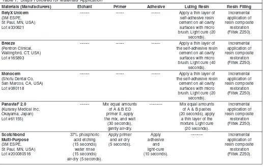
Materials (Manufacturers) Etchant Primer Adhesive Luting Resin Resin Filling
RelyX Unicem --- --- --- Apply a thin layer of Incremental
(3M ESPE, self-adhesive resin application of
St Paul, MN, USA) cement on all cavity resin composite
Lot #330621 surfaces with micro restoration
brush. Light cure (20 (Filtek Z250). seconds).
Breeze --- --- --- Apply a thin layer of Incremental
(Pentron Clinical, the self-adhesive resin application of
Wallingford, CT, USA) cement on all cavity resin composite
Lot #165893 surfaces with micro restoration
brush. Light cure (40 (Filtek Z250). seconds).
Monocem --- --- --- Apply a thin layer of Incremental
(Shofu Dental Co, the self-adhesive resin application of
San Marcos, CA, USA) cement on all cavity resin composite
Lot #080118 surfaces with micro restoration
brush. Light cure (20 (Filtek Z250). seconds).
PanaviaF 2.0 --- Mix equal amounts --- Mix equal amounts Incremental
(Kuraray Medical Inc, of A & B ED of A & B pastes application of
Okayama, Japan) primer II, apply (20 seconds), apply resin composite
Lot #61155) the mix, and wait a thin layer of the restoration
(30 seconds), mixture. Light cure (Filtek Z250).
gently air-dry. (20 seconds).
Scotchbond 37% phosphoric Apply primer Apply --- Incremental
Multi-Purpose acid etching and air dry adhesive application of
(3M ESPE, (15 seconds) (5 seconds). and resin composite
St Paul, MN, USA) water rinse light-cure restoration
Lot #200080516 (15 seconds) (10 seconds). (Filtek Z250).
air-dry (5 seconds). Table 1: Steps Followed for Materials Application
seconds in each water bath with a transfer time of 15 seconds between baths) for 1,000 cycles.
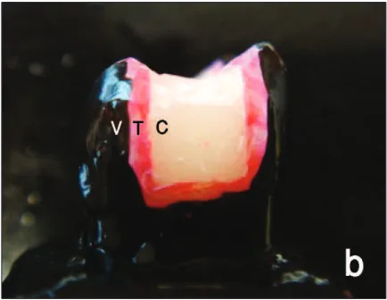
Two layers of nail varnish were applied to the coronal tooth surfaces to within 1 mm from the tooth-restora-tion margins (Figures 2a and 2b). The specimens were then immersed in a 2% fuchsine red solution for 24 hours at 37°C, then rinsed with tap water for five min-utes. Each tooth was then sectioned mesiodistally with a low-speed microslicing machine (Isomet, Buehler, Lake Buff, IL, USA) into two equal sections. All the sections were photographed with a high-resolution scanner (4800 x 9600 dpi, CanoScan 880F, Canon USA Inc, Melville, NY, USA) to produce digital images. The section with more dye penetration was selected to rep-resent the specimen. The extent of die penetration was scored according to a five-point scale: 0 = no leakage, 1 = leakage extending to the outer half of the gingival floor, 2 = leakage extending to the inner half of the gin-gival floor, 3 = leakage extending past the gingin-gival floor up to two-thirds of the axial wall, 4 = leakage extending through the axial wall up to the DEJ level. Two independent examiners evaluated the extent of dye penetration for each section. In case of disagree-ment, a third examiner was consulted to resolve the dispute. Chi square analysis (Procedure Frequency of SAS) was used to test the effect of the adhesive and tooth structure (enamel and dentin) on dye penetra-tion scores (p<0.05). Statistical analysis was carried out using an SAS program (SAS 1988, Release 6.03 ed, SAS Institute, Cary, NC, USA).
The interfacial cement thicknesses at the axial wall and pulpal and gingival floors were measured with a traveling spot insight camera (Model 3.2.0, Diagnostic Instruments Inc, Sterling Heights, MI, USA) under an optical microscope at 60x magnification (SMZ800, Nikon Instruments Inc, Melville, NY, USA), and the
range of the intermediate layer thickness was recorded.
RESULTS
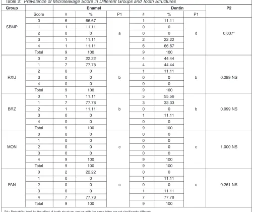
For the SBMP group only, microleakage scores were significantly higher at the dentin gingival margin than at enamel (p=0.037). For each of the four cement groups, no significant difference was found between the microleakage scores at dentin and the enamel gingival margins (p>0.05). However, the Chi-square test revealed a significant difference in microleakage scores at the dentin gingival margins among the groups, except between RXU and BRZ (p>0.05) and between MON and PAN (p>0.05). Similar results were reported in the enamel gingival margins for the four cement groups. The RXU and BRZ groups showed the lowest microleakage scores at the dentin side, followed by the control (SBMP) group. While at the enamel side, SBMP showed the lowest microleakage, followed by the RXU and BRZ groups. The highest microleakage scores were recorded with the PAN and MON groups. Distribution of the microleakage scores among all groups is given in Table 2. Figures 3-7 show images of representative tooth sections from each group.
The intermediate cement layer thickness ranges were: RXU (26-61 µm), BRZ (23-68 µm), MON (28-74 µm) and PAN (13–100 µm). The thickness of the inter-facial SBMP bonding agent could not be detected under light microscope.
DISCUSSION
Previous studies reported that composite restorations showed relatively greater microleakage at the gingival margins than at the occlusal margins.14-15The method used for the assessment of microleakage of Class II composite restorations in the current study has been
Al-Saleh & Others: Composite Microleakage with Resin Cement Liners 559
Figure 2a:Two layers of black nail varnish were applied all over exposed surfaces to within 1 mm of the tooth-restoration inter-face. (C) = composite, (T) = tooth structure, (V) = varnish seal.
Figure 2:Proximal views of representative restored specimens.
560 Operative Dentistry
previously reported.13 Sterilization of the extracted teeth with gamma irradiation was used in the current study, as it has been shown to be effective and has no adverse effects on dentin structure or its permeabili-ty.16 It was also reported that gamma irradiation nei-ther affects the bond strength to dentin nor alters the dentin surface morphology.17Although seven days stor-age in distilled water at room temperature is relative-ly short compared to the life expectancy of composite restorations, it was deemed to be appropriate in the current study, considering that cements are more sus-ceptible than restorative composites to dissolution dur-ing and immediately after their initial set.18
Since statistical analysis revealed variability in the mean microleakage scores among the different groups, the null hypothesis could not be substantiated. The
out-standing performance of self-adhesive resin cements with indirect restorations (inlays, onlays and crowns) was the main motivating factor for conducting the cur-rent study.19-20Under the indirect restoration technique, self-adhesive resin cements are subjected to hydraulic stresses transmitted by the hard restoration under the biting forces exerted by the patient during cementation. This might be helpful for some cements that produce a better seal, as it may assist in limiting the adverse effects of polymerization contraction. However, varying results were obtained among the groups of self-adhe-sive cements used in the current study with direct restoration. RXU and BRZ resulted in significantly lower microleakage scores and, therefore, can be con-sidered for use under direct composite restorations. In contrast, PAN and MON resulted in significantly
high-Group Enamel Dentin P2
Score # % P1 # % P1
0 6 66.67 1 11.11
SBMP 1 1 11.11 0 0
2 0 0 a 0 0 d 0.037*
3 1 11.11 2 22.22
4 1 11.11 6 66.67
Total 9 100 9 100
0 2 22.22 4 44.44
1 7 77.78 4 44.44
2 0 0 1 11.11
RXU 3 0 0 b 0 0 b 0.289 NS
4 0 0 0 0
Total 9 100 9 100
0 1 11.11 5 55.56
1 7 77.78 3 33.33
BRZ 2 1 11.11 b 0 0 b 0.099 NS
3 0 0 1 11.11
4 0 0 0 0
Total 9 100 9 100
0 0 0 0 0
1 0 0 0 0
MON 2 0 0 c 0 0 c 1.000 NS
3 0 0 0 0
4 9 100 9 100
Total 9 100 9 100
0 2 22.22 0 0
1 0 0 1 11.11
PAN 2 0 0 c 0 0 c 0.261 NS
3 0 0 1 11.11
4 7 77.78 7 77.78
Total 9 100 9 100
P1= Probability level for the effect of tooth structure, groups with the same letter are not significantly different. P2 = Probability level for the effect of tooth structure.
Means with the same letter within each column are not significantly different atp≤0.05. NS = Insignificant (p>0.05).
Al-Saleh & Others: Composite Microleakage with Resin Cement Liners
er microleakage scores at both the dentin and enamel margins and, therefore, should not be considered for such application. It is speculated that the different microleakage results of the self-adhesive resin cements can be explained partly by their different functional monomers and different chemical compositions. Differences in pH values may influence the ability of some of these cements to appropriately condition enam-el and dentin in the direct restoration technique, thus resulting in less than ideal adhesion with subsequent microleakage. RXU was shown to have a low initial pH that rapidly rises upon mixing.21 It seems, however, that this was sufficient to provide restorations with minimal microleakage.
Two studies reported low microleakage and good mar-ginal seal when PAN was used with metal and ceramic crowns.22-23 Other studies reported high bond strength with PAN when used with prepolymerized resin com-posite blocks.20,24-25 The less-than-ideal performance of 561
Figure 3: Representative photograph of microleakage for the RXU group. The enamel side (E) shows no microleakage at the tooth-composite interface (only the enamel tooth structure is stained with the dye). The dentin side (D) shows no microleak-age.
Figure 4:Representative photograph of microleakage for the BRZ group. The enamel (E) and dentin (D) sides show microleakage extending to the outer half of the gingival floor (score 1).
Figure 5:Representative photograph of microleakage for the MON group. Photograph shows microleakage extended through the axial wall up to the pulpal floor (score 4) at both the enamel side (E) and the dentin side (D).
Figure 6:Representative photograph for the PAN group. The photograph shows microleakage extended through the axial wall up to the pulpal floor (score 4) at both the enamel (E) and dentin side (D).
562 Operative Dentistry
PAN in the current study can be attributed to the direct (unpolymerized) nature of the restorations that were placed. Under the indirect restoration technique, the resin cement is sandwiched between the cavity surfaces and overlying prepolymerized restoration, both hard structures, thus, it is subjected to reduced polymeriza-tion contracpolymeriza-tion stresses. In contrast with direct restoration, the resin cement is subjected to contraction stresses of the overlaying resin composite increment.
Different thermal expansion coefficients of the restorative material and tooth structure may explain the high microleakage scores of PAN and MON. In the current study, all specimens were subjected to 1000 cycles between 5°C and 55°C, which is considered an appropriate artificial aging test according to ISO TR Standard 11405: 1994.26The coefficient of thermal expan-sion for Filtek Z250 was reported to be 41.5x 10⁻⁶/°C,27 while for enamel and dentin, the values were signifi-cantly lower at 11 x 10-6/°C and 17 x 10-6/°C, respective-ly.28 It has been reported that the warm water bath (55°C) may accelerate the hydrolysis of the component of the interfacial material (bonding agent) with subse-quent water absorption and leaching of the broken down collagen or poorly-polymerized resin oligomers.29 The mismatch in the coefficient of thermal expansion may also create stresses at the bonding interface, allow-ing water percolation and, thus, increased microleak-age. However, Crim and others found no difference in dye penetration during microleakage testing when specimens were thermocycled between 100 and 1500 cycles.30Therefore, one can only assume a limited coef-ficient of thermal expansion influence on the current study results.
The main purpose of applying a low-stiffness interme-diate material layer is to absorb part of the stresses generated by composite polymerization shrinkage. For this reason, thicker adhesive layers of unfilled adhe-sives, filled adhesives and flowable composites, have been proposed for the dentin-composite interface. Van Meerbeek and others confirmed the positive effect of the elastic and low viscosity intermediate layer on the marginal adaptation and retention of composite restorations.31Braga and others also suggested that an elastic intermediate layer will increase cavity wall com-pliance and, therefore, decrease the destructive con-traction stresses of direct composite restorations.7 However, the results of the current study did not con-firm the benefits of a thick intermediate layer at the tooth-composite interface. RXU and BRZ, which had an intermediate layer thickness range (23-68 µm), had low microleakage scores, while PAN and MON showed high microleakage results regardless of their thick interme-diate layer (13–100 µm). It is possible that other fac-tors, such as chemical composition or degree of poly-merization contraction, played a larger role in microleakage than did the cement thickness or elasticity.
CONCLUSIONS
Within the limitations of this in vitrostudy, it can be concluded that:
1. Minimal leakage was observed with RelyX-Unicem and Breeze cements at both the dentin and enamel margins.
2. Panavia F 2.0 and Monocem resulted in signifi-cantly higher microleakage scores at both the dentin and enamel margins.
Acknowledgements
The authors thank 3M/ESPE, Kuraray Dental Co, Shofu Inc, Pentron Clinical Technologies and Kerr Corporation for provid-ing materials/instruments for this study.
(Received 24 June 2009; Accepted 18 April 2010)
References
1. Mjör IA (1998) The location of clinically-diagnosed second-ary cariesQuintessence International29(5)313-317.
2. Kidd EA (1976) Microleakage in relation to amalgam and composite restorations—A laboratory studyBritish Dental Journal141(10)305-310.
3. Ciucchi B, Bouillaguet S & Holz J (1990) Proximal adapta-tion and marginal seal of posterior composite resin restora-tions placed with direct and indirect techniques Quintessence International21(8)663-669.
4. Hilton TJ, Schwartz RS & Ferracane JL (1997) Microleakage of four Class II resin composite insertion techniques at intraoral temperature Quintessence International28(2)135-144.
5. Coelho Santos MJM, Santos GC Jr, Nagem Filho H, Mondelli RFL & El-Mowafy OM (2004) Effect of light cur-ing method on volumetric polymerization shrinkage of resin compositesOperative Dentistry29(2)157-161.
6. Davidson CL, deGee AJ & Feilzer A (1984) The competition between the composite-dentin bond strength and the poly-merization contraction stressJournal of Dental Research
63(12)1396-1399.
7. Braga RR & Ferracane JL (2004) Alternatives in polymer-ization contraction stress managementCritical Reviews in Oral Biology and Medicine15(3)176-184.
8. Lutz F, Krejci I & Barbakow F (1991) Quality and durabil-ity of marginal adaptation in bonded composite restora-tionsDental Materials7(2)107-113.
9. Bertolotti RL (1991) Posterior composite technique utiliz-ing directed polymerization shrinkage and a novel matrix Practical Periodontics and Aesthetic Dentistry3(4)53-58.
10. Ericson D & Derand T (1991) Reduction of cervical gaps in Class II composite resin restorations The Journal of Prosthetic Dentistry65(1)33-37.
563
Al-Saleh & Others: Composite Microleakage with Resin Cement Liners
12. Miller MB, Castellanos IR, Vargas MA & Denehy GE (1996) Effect of restorative materials on microleakage of Class II compositesJournal of Esthetic Dentistry8(3) 107-113.
13. El-Mowafy O, El-Badrawy W, El-Tanty A, Abbasi K & Habib N (2007) Gingival microleakage of Class II resin composite restorations with fiber inserts Operative Dentistry32(3)298-305.
14. Hattab FN, Mok NY & Agnew EC (1989) Artificially formed caries like lesions around restorative materialsJournal of the American Dental Association118(2)193-197.
15. Alani AH & Toh CG (1997) Detection of microleakage around dental restorations: A review Operative Dentistry
22(4)173-185.
16. Sperandio M, Souza JB & Oliveira DT (2001) Effect of gamma radiation on dentin bond strength and morphology Brazilian Dental Journal12(3)205-208.
17. White JM, Goodis HE, Marshall SJ & Marshall GW (1994) Sterilization of teeth by gamma radiation Journal of Dental Research73(9)1560-1567.
18. Swartz ML, Sears C & Phillips RW (1971) Solubility of cement as related to time of exposure in waterThe Journal of Prosthetic Dentistry26(5)501-505.
19. Piwowarczyk A, Lauer HC & Sorensen JA (2004)In vitro shear bond strength of cementing agents to fixed prostho-dontic restorative materials The Journal of Prosthetic Dentistry92(3)265-273.
20. Hikita K, Van Meerbeek B, De Munck J, Ikeda T, Van Landuyt K, Maida T, Lambrechts P & Peumans M (2007) Bonding effectiveness of adhesive luting agents to enamel and dentinDental Materials23(1)71-80.
21. Saskalauskaite E, Tam LE & McComb D (2008) Flexural strength, elastic modulus, and pH profile of self-etch resin luting cementsJournal of Prosthodontics17(4)262-268. 22. Piwowarczyk A, Lauer HC & Sorensen JA (2005)
Microleakage of various cementing agents for full cast crownsDental Materials21(5)445-453.
23. Schenke F, Hiller KA, Schmalz G & Federlin M (2008) Marginal integrity of partial ceramic crowns within dentin with different luting techniques and materials Operative Dentistry33(5)516-525.
24. De Munck J, Vargas M, Van Landuyt K, Hikita K, Lambrechts P & Van Meerbeek B (2004) Bonding of an auto-adhesive luting material to enamel and dentinDental Materials20(10)963-971.
25. Piwowarczyk A, Bender RL, Peter OT & Hans L (2007) Long-term bond between dual-polymerization cementing agents and human hard dental tissue Dental Materials
23(2)211-217.
26. ISO-Standards (1994) ISO TR 11405 Dental materi-als–guidance on testing of adhesion to tooth structure, In anonymous Genève: International Organization for Standardization1stedition 15-25.
27. Kwon YH, Jeon GH, Jang CM, Seol HJ & Kim HI (2006) Evaluation of polymerization of light-curing hybrid com-posite resins Journal of Biomedical Materials Research Part B, Applied Biomaterials76(1)106-113.
28. Xu HC, Liu WY & Wang T (1989) Measurement of thermal expansion coefficient of human teeth Australian Dental Journal34(6)530-535.
29. Hashimoto M, Ohno H, Kaga M, Endo K, Sano H & Oguchi H (2000) In vivo degradation of resin-dentin bonds in humans over 1 to 3 yearsJournal of Dental Research79(6)
1385-1391.
30. Crim GA & García-Godoy F (1987) Microleakage: The effect of storage and cycling durationThe Journal of Prosthetic Dentistry57(5)574-576.
31. Van Meerbeek B, Willems G, Celis JP, Roos JR, Braem M, Lambrechts P & Vanherle G (1993) Assessment by nano-indentation of the hardness and elasticity of the resin-dentin bonding area Journal of Dental Research 72(10)