INFLUENCE OF GAP SHAPE ON BIOMECHANICAL
PROPERTIES OF EXTRA-ARTICULAR DISTAL
HUMERAL FRACTURE – A FINITE ELEMENT STUDY
Srećko Sabalić1, Hrvoje Maričić2, Zvonimir Tomičević2 and Janoš Kodvanj2 1Sestre milosrdnice University Hospital Center, University Hospital for Traumatology; 2Faculty of Mechanical Engineering and Naval Architecture, University of Zagreb, Zagreb, Croatia
SUMMARY – Th e aim of the study was to assess the infl uence of gap shape on biomechanical results in extra-articular distal humeral fracture: with contact on the posterior part (by anterior gap) and contact on ulnar column (by radial gap). Th e goal was to examine if and to what extent did displacements decrease in comparison with previously examined parallel gap without bony contact. Th e fi nite element analysis on the three diff erent plate constructs was performed, i.e. parallel, perpen-dicular and newly designed Y shape plate were considered. Displacements were measured on articular surface and gap point. Th e most visible decrease of maximum displacements in the distal part of the model was detected in the Y plate model with axial loading: in case of anterior gap 58.5% and espe-cially at radially formed gap 60.9%. Similarly, at axial loading, displacement at the analyzed point on fracture gap most signifi cantly decreased in Y plate model (by 49.4%) at posterior bony contact. More-over, the latter showed displacement decrease by 68.5% at ulnar bone contact. Furthermore, if a longer radial plate than the ulnar one was used, varus stress could have been avoided. Study results suggested that suffi cient stability could be ensured with the newly designed Y shape plate.
Key words: Humeral fractures – surgery; Fracture fi xation, internal; Biomechanical phenomena; Finite element analysis; Bone plates
Correspondence to: Srećko Sabalić, MD, PhD, Medveščak 85, HR-10000 Zagreb, Croatia
E-mail: [email protected]
Received March 15, 2016, accepted September 2, 2016
Introduction
Fractures of distal humerus in adults are often challenging in operative treatment. In practice, it has been shown that 16% of humeral shaft and 10% of distal humerus fractures in adults are distal humeral shaft and extra-articular supracondylar humerus frac-tures1. Th e focus of this article is comminuted
extra-articular distal humeral fracture. Th is type of fracture often results from a gunshot wound (Fig. 1) or motor vehicle injuries in the younger population2. Such
inju-ries can also result from a simple fall in the elderly population3. Although Y shape plate4,5 has been used
for years in the treatment of intra-articular distal
hu-meral fracture, with a few modifi cations with two dor-sal plates6 or distal part of Y shape plate7 (Lambda®
plate, Zimmer, Étupes, France), it is not frequently used in the treatment of extra-articular distal humeral fracture and distal shaft fracture.
Th e aim of this study was to preconfi gure the old Y reconstructive plate intended primarily for the treat-ment of intra-articular fractures of distal humerus and to convert it for extra-articular fractures of distal hu-merus and distal humeral diaphysis, as well as to exam-ine its biomechanical performance comparing it to the existing osteosynthesis methods with two reconstruc-tion plates in perpendicular and parallel posireconstruc-tion using the fi nite element method.
Th e principal objective of treating extra-articular distal humeral fractures is restoring alignment and achieving stable fi xation aimed at facilitating early el-bow range of motion, essential for good functional
outcome. It is often diffi cult to obtain rigid fi xation in distal fractures of humeral diaphysis without compro-mising the elbow8. However, fi xation of these fractures
remains a challenge due to the restricted space for in-strumentation at the distal segment and the need to maintain repair integrity under a large range of motion and low to moderate loading9.
Th rough many stages of development, from con-servative to operative treatment, open reduction and internal fi xation with dual plating systems are the gold standard for fi xation of distal humerus fractures2,3,6,8-17.
Double-plating techniques using two 3.5 mm recon-struction plates or LCP plates in dorsal plating, 90-90°
or 180-180° (Fig. 2) pattern are generally accepted for
both intra-articular3,8,10-16,18-23 and extra-articular
frac-tures1-3,9,10,17. Previous studies have shown that surgery
surpasses the results of conservative treatment3,8.
Sco-laro et al.24 in a recent biomechanical study suggest
that single posterolateral column fi xation of extra-ar-ticular humerus fractures is appropriate for more prox-imal fractures. However, it has been reported that the treatment with dual plate fi xation is more suitable for distal fractures.
Even if the above mentioned techniques are ap-plied, this does not exclude non-union as a complica-tion of distal humerus fractures, with a reported inci-dence of 8% to 25%14. Poor initial fi xation, which is not
easily manageable in the presence of extensive com-minution and osteopenia, can be the main factor for hardware failure25.
Migration of the plate and screws or non-union with cubitus varus deformity (gunstock deformity) can occur when applied to inadequate osteosynthesis with one plate (Fig. 3).
Fig. 1. Gunshot fracture of distal humerus: (left) treated with an external fi xator; and (right) later treated with two plates in perpendicular position (90-90º).
Fig. 2. Double-plating technique using two 3.5-mm reconstruction plates in parallel (180-180º)
confi guration.
Fig. 3. Varus deformity after non-union of the extra-articular distal humeral fracture after osteosynthesis: (left) with one plate; and (right) with two plates.
In a previous study, by using the fi nite element cal-culations, attempts were made to fi nd the optimal plate confi guration which would improve its biome-chanical properties, taking into account the specifi c anatomy of distal humerus26.
Th e article presents the eff ect of the gap shape in case of extra-articular distal humerus fracture on bio-mechanical properties. Th e fi nite element calculations are used to simulate these phenomena. In the fi rst study, models with parallel gap26 were investigated,
while the goal of this study was to examine whether contact in the posterior part (by anterior gap) and con-tact on ulnar column (by radial gap) infl uenced biome-chanical properties.
Materials and Methods
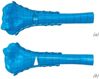
Th e fi nite element analysis was performed on the three diff erent plate constructs. Two diff erent gaps placed 25 mm above the olecranon fossa were taken into account. First, anterior gap and bony contact in posterior segment were investigated, while the second one dealt with radial gap and bony contact in ulnar segment (Fig. 4). Th e creation of numerical models and the prescribed boundary, as well as the loading conditions are described in detail in Sabalic et al.26.
Th erefore, this article only briefl y depicts them. Th e fi nite element models of distal humerus with the newly designed Y plate and two 3.5 reconstruction plates in parallel and perpendicular confi guration were created from 3D optical scans (Atos III Triple Scan, GOM mbH, Germany). Each model was 140 mm long and constrained with respect to the proximal end. Th e loading conditions of the models were the same as those previously used26. Nonlinear computational
sim-ulations under axial (200 N), lateral and bending (30 N) loading regimes were conducted using the fi nite element software Abaqus 6.10-1 (Dassault Systèmes, France). In all fi nite element models, contact interac-tions were applied as surface to surface fi nite sliding with a coeffi cient of friction of 0.3. Th e contact is de-fi ned between the bone and the plates and the connec-tion of osteotomy interfaces. Tied constraints were ap-plied between the screws and plates, as well as between the screws and the surrounding bone in all constructs. All models were meshed using ten node quadratic tet-rahedral elements. Th e number of elements in the pro-(a)
(b) Fig. 4. Computational model: (a) with anterior gap; and (b) with radial gap.
Fig. 5. Maximum displacements on the distal end of the
models. 0 1 2 3 4 5 Parallel gap Anterior gap Radial gap Parallel gap Anterior gap Radial gap Parallel gap Anterior gap Radial gap
Axial compression Posterior deflection Varus loading
Displacement,
mm
Loading
posed models altered from 796100 to 884200 elements depending on the plate type and number of screws.
Th e chosen material was defi ned as linear elastic, homogeneous and isotropic. Th e Poisson’s ratio for os-teoporotic bone was taken to be 0.4. Young’s modules applied for the cortical bone in axial compression, pos-terior defl ection and varus loading were 3400 MPa, 1150 MPa and 660 MPa, respectively. Both stainless steel and titanium alloys were assigned with a Poisson’s ratio of 0.3, while the Young’s module for steel equals 200 GPa and for titanium 110 GPa.
Results
In the following section, the calculation results for the three diff erent plate designs are reported. Figure 5 shows the maximum computed displacements on the distal end for the investigated plate models subjected under three diff erent loading directions (i.e. axial,
bending and lateral direction) when the gaps were po-sitioned 25 mm above the olecranon fossa and defi ned as parallel, radial and anterior.
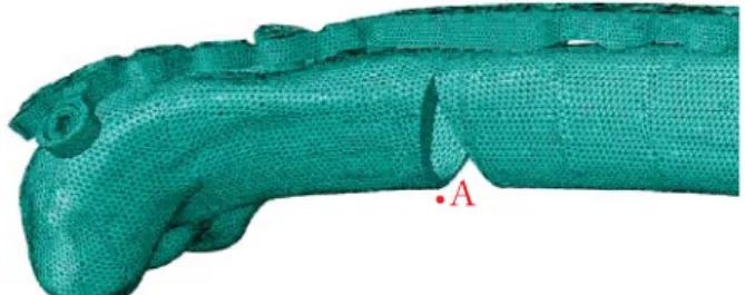
Displacements on the fracture gap for all models are observed at point A (Fig. 6), due to the reasons mentioned earlier26. Maximum displacements at point
A on the distal edge of the fracture gap for all the tests are reported in Figure 7. Estimated maximum stress levels in the bones and plates for the three investigated plate designs subjected under various loading regimes and gap defi nitions are shown in Table 1.
Discussion
By forming a gap in the segment of distal humerus with cortical bony contact using computational simu-lations we wanted to examine the infl uence of the gap shape on the biomechanical properties of the fracture in the segment of distal humerus in three methods of osteosynthesis. For this purpose, models from the pre-vious study were used26. Th e aim was to examine if and
to what extent the displacements decreased in com-parison with previously examined parallel gap without bony contact.
Th e most visible decrease of maximum displace-ments in the distal part of the model was detected in the Y plate model with anterior gap at axial loading (58.5%) and especially at radially formed gap (60.9%). Th is was not detected in the models with parallel and perpendicular plate confi guration. In all three models, Fig. 6. Point A on the distal edge of the fracture gap.
Fig. 7. Displacements in point A on the distal edge of the fracture gap. 0.0 0.5 1.0 1.5 2.0 2.5 Displacement, mm Loading
Y plate Parallel plate Perpendicular plate
Parallel gap Anterior gap Radial gap Parallel gap Anterior gap Radial gap Parallel gap Anterior gap Radial gap
Axial compression Posterior deflection Varus loading
such visible changes were not detected at bending and lateral loading when compared to radial and anterior gap.
Similarly, at axial loading, the displacement at ana-lyzed point A on fracture gap most signifi cantly de-creased in Y plate model (49.4%) at dorsal bony contact, and by 68.5% at ulnar bone contact. In general, the shape of fracture gap, that is bony contact, did not have signifi cant infl uence on displacements at bending and lateral loading of radial condyle, as in axial loading.
Signifi cant stress changes were also detected in axial loading in terms of decreasing the stress in the plate and increasing the stress in the bone, in all three models. Stress decrease was most evident at axial loading of Y plate, specifi cally in radial gap (354%), with increase in the stress in bone (97%). Such visible stress changes were not detected at bending, where stresses in Y plate in the models with anterior and radial gap were even greater than in the model with parallel gap. At lateral loading of Y plate model the stresses in the plate de-creased (55.7%), while bone stresses inde-creased (36.7%).
Th e current standard of treatment of the extra-ar-ticular distal humerus fracture is osteosynthesis with two plates in posteromedial and posterolateral posi-tion. Biomechanical stability on the transition of the distal humerus diaphysis into the distal segment of the humerus has been the object of several studies.
Th e incidence of distal humeral fracture is relative-ly low, with a large number of fracture subtypes. Clini-cal studies are often functionally insuffi cient because of the limited number of patients. Th ere are no pub-lished prospective randomized studies, the majority of the studies were retrospective and carried out on a small number of samples. Th erefore, on the basis of these clinical studies, it is not possible to draw conclu-sions about the recommendable implant confi guration
in case of fractures of distal humerus. A biomechanical study is therefore required26.
Th e majority of non-unions happen at the supra-condylar level, while healing of the articular compo-nents may occur in their reduced position. Nonethe-less, the stability of the construct requires adequate bony contact with interfragmentary compression. In case of a distal humeral fracture, by far the greatest number of fi xation failures occurs at the supracondylar level, while typically the articular fragments unite, and, with time, fracture union occurs at this level.
Maximizing stability between the distal fragments and the shaft of the humerus at the metaphyseal level should be the focus of the fi xation strategy7,10.
O’Driscoll10 lists the technical principles to apply in
order to achieve stable internal fi xation of distal intra-articular humeral fractures.
From the previous research10 concerning the plates
used for fi xation, it has been reported that the latter should be applied in such a manner as to achieve com-pression at the supracondylar level for both columns. However, at the same time, the plates used must be strong and stiff enough to resist breaking or bending before union occurs at the supracondylar level.
Th e practical application of these principles in-volves ‘parallel’ plates that permit a total of at least 4 to 6 long screws to be placed in the distal fragments, from one side across to the other. Th e plates are placed with a slight off set, posteromedially and posterolaterally18.
Pajarinen et al.27 conclude that satisfactory results
can be obtained when the stability of the humeral col-umns is achieved and the articular platform recon-structed.
O’Driscoll10 points out that the literature on
frac-tures of distal humerus pays far too little attention to the reason why the failure of the fi xation generally be-Table 1. Maximum von Mises stresses in the bones and plates
Axial compression Posterior defl ection Varus loading
Parallel gap Anterior gap Radial gap Parallel gap Anterior gap Radial gap Parallel gap Anterior gap Radial gap
Parallel plate Bone 10.86 14.42 22.46 13.98 15.02 15.09 14.12 20.51 23.64
Plate 309.40 173.30 152.40 143.10 119.60 140.30 160.80 153.60 153.60 Perpendicular plate Bone 18.48 25.06 46.29 14.95 15.01 15.12 15.19 15.47 18.45 Plate 280.40 255.30 205.30 152.40 137.20 149.70 193.80 165.80 165.90 Y plate Bone 25.01 32.56 48.49 14.87 15.37 15.42 16.54 22.58 22.67 Plate 567.40 276.80 160.30 189.00 221.10 220.90 189.00 83.66 83.68
gins in the lateral column. Th e force of gravity acting on the long lever arm (the forearm), while the elbow is fl exed and extended during apparently minimal-use activities leads to repetitive varus stresses across the el-bow. Th is can be typically seen in the action of a person reaching out to grab something, a glass of water for instance, followed by bringing the hand to the mouth. Th is causes varus torque across the elbow, distracting the lateral column away from the fi xation placed along its posterior surface10.
Th e load transfer in the elbow joint can be de-scribed by a two column model1,28. Th e medial ulnar
column and the lateral radial column form the articu-lar block. Th e lateral column shares 60% and the me-dial column 40% of the load1,22,28. Th is two-column
model is the basic principle of double plating osteo-synthesis of C-type fractures of distal humerus1,10,16.
In recent publications, a higher stiff ness and strength of osteosynthesis in the parallel plating technique were compared to the perpendicular technique with diff erent plate designs. Th e mechanical advantages of a parallel plate confi guration have been demonstrated for conven-tional reconstruction plate design29, as well as for
lock-ing plate construct23. In other studies, no signifi cant
dif-ferences were found1,9. As noted in previous studies,
pseudoarthrosis of distal humerus usually occurs in the region at the metaphyseal and supracondylar level of ra-dial columns due to varus stresses.
So far, there have been two biomechanical studies on the supracondylar metaphyseal level on the border of the distal humerus diaphysis. Except for these two24,26, previous studies examined intra-articular or
low supracondylar fractures20-23,29.
In the proposed research, it is believed that frac-tures in this part of the humerus have diff erent biome-chanical demands than intra-articular fractures of dis-tal humerus. Th e previously described new design of Y shape plate, which supports both columns and is at the same time longer in the radial, more loaded column, could be at least equal in strength as the two plates.
To prove the hypothesis, the following tests should be performed: biomechanical studies at the supracondy-lar metaphyseal level on the border of the distal humer-us diaphysis humer-using the fi nite element method, where the gap would be done at the level of distal humerus 25 mm above the fossa olecrani (Fig. 4). Th e gap is shown in the fi gure as an interruption of the bone continuity.
Th e biomechanical study should be made to the axial load in a position of fl exion of 5°, the bending
load in a position of fl exion of 75°and the lateral load
on the radial condyle. After applying the fi nite ele-ment computations in order to choose the optimal po-sition of the plates, a biomechanical study of the syn-thetic or cadaver humerus would be undertaken. Th is study would compare the stiff ness of the Y-shaped, parallel and perpendicular plate confi guration by ap-plying the same loads as those applied in the fi nite ele-ment analysis.
In their publication Penzkofer et al.1 state the
fol-lowing: “... the system stiff ness is infl uenced by two kinds of factors: factors which cannot be infl uenced by the surgeon and factors which allow the individual ad-justment of an osteosynthesis. Th e initial situation is the fracture pattern and fracture geometry with the number and shape of the fragments. Th is initial frac-ture situation strongly dictates the options for plate positioning. On the other hand, the overall construct stability can largely be infl uenced by placing the plates at diff erent anatomical positions”. Th is study did not take into account the lateral load, as there is already a load in the position of fl exion and extension.
Furthermore, Zalavras et al.23 found that parallel
plate designs had signifi cantly higher stiff ness than the perpendicular ones during cyclic varus loading. Th e measurement of displacement was not made by precise measuring instruments at the gap. In this article, loos-ening of the implants was defi ned as gross displacement (backing-out) of the screws during cyclic loading of the specimens. When done, varus loading to failure resulted in ligamentous disruption in all specimens, which oc-curred prior to any catastrophic failure of fi xation19. In
this way, they could not see displacements in the gap and assess the mechanical stability of the specimens.
Recent biomechanical studies have considered loads on the distal segment of the humerus in a posi-tion of fl exion of 75° 22,23 or 50°24 to the longitudinal
axis of the humerus, or a position of fl exion of 5°23 or
15° 22. However, Zalavras et al.24 conducted
experi-ments with radial varus loads.
In clinical studies, signifi cantly better results were achieved with surgical than with conservative treatment of extra-articular distal humeral fracture3,11. Shin et al.17
compared clinical outcomes in patients with intra-artic-ular distal humerus fractures and concluded that both parallel and orthogonal plate positioning could provide adequate stability and anatomic reconstruction of the distal humerus fractures, while Sanchez-Sotelo et al.16,18
Prasarn et al.8 have recently reported their good
clin-ical experience with the use of the locking compression plate for extra-articular fractures of distal humerus, add-ing two additional reconstructive plates to the radial column. Consequently, Scolaro et al.24 support this good
clinical experience with relevant biomechanical study. As noted in previous studies, pseudarthrosis of dis-tal humerus usually occurs in the region of the me-taphyseal and supracondylar level of the radial columns due to various stresses.
Previous studies have been diff erently designed with diff erent directions and types of loads, i.e. various static and dynamic forms were applied with diff erent cycles on a wide range of diff erent samples. Furthermore, mea-suring instruments and their degree of precision in measuring displacements and deformations are diff er-ent and hardly comparable. Th erefore, it is diffi cult to compare the results of biomechanical studies. Also, so far, displacements in diff erent directions when loads are signifi cantly diff erent have not been taken into account. Th erefore, we believe that the direction of the load which causes greater displacements has a greater impact on the overall evaluation of the stability of the implant. Consequently, displacements caused by bending and lateral loads compared to the radial condyle are consid-erably larger than the axial load, and thus with a greater signifi cance in the overall assessment of the structural stability of implants. Th erefore, the role of the implant is to minimize the forces that cause greater displacements in the area. Likewise, in the case of osteoporotic frac-tures, we should know that osteoporosis is more pro-nounced in the posterolateral part of the radial con-dyle26, and that the area of the lateral columns,
espe-cially the capitulum and the distal part of the lateral column, has very thin cortices31.
A disadvantage of biomechanical studies of this kind is the inability to take into account all factors that infl uence treatment outcome. Among them, dynamic loads that occur during everyday activities have an im-portant place.
Th e well known issue of anatomical variations in distal humerus requires making plates of diff erent size with the ability of remodeling according to anatomical diff erences.
Conclusion
Th e biomechanical study proposed in this work provides a proof of the hypothesis that the two plates
model where the plate at the radial side is longer re-sults in better biomechanical stability in comparison with two plates of equal length in fractures of distal humerus diaphysis at the turn of the distal humerus. Furthermore, it has been shown that a newly designed Y shape plate, as described previously, provides biome-chanical stability as the two plate model.
In order to avoid varus stress, radial plate should be longer than the ulnar plate. Suffi cient stability can be ensured with the newly designed Y shape plate with the longer radial arm. Improving biomechanical stabil-ity would avoid complications such as pseudarthrosis or malunion.
Th e newly designed Y plate for extra-articular frac-tures of distal humerus is a possible alternative to the usual osteosynthesis method with two plates. A de-fi nitive conclusion would require biomechanical stud-ies, either with a synthetic or cadaveric model.
References
1. Penzkofer R, Hungerer S, Wipf F, von Oldenburg G, Augat P. Anatomical plate confi guration aff ects mechanical perfor-mance in distal humerus fractures. Clin Biomech (Bristol, Avon). 2010 Dec;25(10):972-8. http://dx.doi.10.1016/j.clin-biomech.2010.07.005
2. Tejwani NC, Murthy A, Park J, McLaurin TM, Egol KA, Kummer FJ. Fixation of extraarticular distal humerus fractures using one locking plate versus two reconstruction plates: a labo-ratory study. J Trauma. 2009 Mar;66(3):795-9. http://dx. doi: 10.1097/TA.0b013e318181e53c
3. Robinson CM, Hill RM, Jacobs N, Dall G, Court-Brown CM. Adult distal humeral metaphyseal fractures: epidemiology and results of treatment. J Orthop Trauma. 2003 Jan;17(1):38-47. http://dx doi:10.1097/00005131-200301000-00006
4. Smrkolj V, Korošec B. Intra-articular fractures of the distal hu-merus. Acta Chir Iugosl. 1991;38(1):69-75.
5. Luegmair M, Timofi ev E, Chirpaz-Cerbat JM. Surgical treat-ment of AO type C distal humeral fractures: internal fi xation with a Y-shaped reconstruction (Lambda) plate. J Shoulder Elbow Surg. 2008;17:113-20. http://dx,. doi: 10.1016/j.jse. 2007.04.007
6. Bogataj M, Kosel F, Norris R, Krkovic M, Brojan M. Biome-chanical study of diff erent plate confi gurations for distal hu-merus osteosynthesis. Med Biol Eng Comput. 2015 May; 53(5):381-92. http://dx. doi: 10.1007/s11517-015-1247-1 7. Saragaglia D, Rouchy RC, Mercier N. Fractures of the distal
humerus operated on using the Lambda® plate: report of 75 cases at 9.5 year follow-up. Orthop Traumatol Surg Res. 2013 Oct;99(6):707-12. http://dx. doi: 10.1016/j.otsr.2013.04.007 8. Prasarn ML, Ahn J, Paul O, Morris EM, Kalandiak SP, Helfet
the humeral shaft. J Orthop Trauma. 2011 Jan;25(1):57-63. http://dx. doi: 10.1097/BOT.0b013e3181df96a7
9. Schwartz A, Oka R, Odell T, Mahar A. Biomechanical com-parison of two diff erent periarticular plating systems for stabi-lization of complex distal humerus fractures. Clin Biomech (Bristol, Avon). 2006 Nov;21(9):950-5. http://dx.doi.org/10. 1016/j.clinbiomech.2006.04.018
10. O’Driscoll SW. Optimizing stability in distal humeral fracture fixation. J Shoulder Elbow Surg. 2005;14:186S-94. http:// dx.doi.org/10.1016/j.jse.2004.09.033
11. Jawa A, McCarty P, Doornberg J, Harris M, Ring D. Extra-articular distal-third diaphyseal fractures of the humerus. A comparison of functional bracing and plate fi xation. J Bone Joint Surg Am. 2006 Nov;88(11):2343-7. http://dx.doi: 10.2106/JBJS.F.00334
12. Schatzker J. Fractures of the distal End of the humerus (13-A, B and C). In: Schatzker J, Tile M (Eds). Th e Rationale of Op-erative Fracture Care. Springer, Berlin-Heidelberg-New York, 2005;103-121.
13. McKee MD. Fractures of the shaft of the humerus. In: Bucholz RW, Heckman JD, Court-Brown CM (Eds). Rockwood and Green’s Fractures in Adults. Lippincott Williams & Wilkins, Philadelphia; 2006;1117-59.
14. Becker EH, Stein J. Advancements in the treatment of distal humeral fractures. Curr Orthop Pract. 2009 Aug;20(4):345-8. http://dx doi: 10.1097/BCO.0b013e3181a65514
15. DeLuise A, Voloshin I. Current management of distal humerus fractures. Curr Opin Orthop. 2006;(17):340-7. http://dx. doi: 10.1097/01.bco.0000233730.49010.c5
16. Sanchez-Sotelo J, Torchia ME, O’Driscoll SW. Complex distal humeral fractures: internal fixation with a principle-based par-allel-plate technique. J Bone Joint Surg Am. 2007;89:961-9. http://dx.doiI: 10.2106/JBJS.E.01311
17. Shin SJ, Sohn HS, Do NH. A clinical comparison of two dif-ferent double plating methods for intraarticular distal humerus fractures. J Shoulder Elbow Surg. 2010 Jan;19(1):2-9. http:// dx.doi: 10.1016/j.jse.2009.05.003
18. Sanchez-Sotelo J, Torchia ME, O’Driscoll SW. Principle-based internal fi xation of distal humerus fractures. Tech Hand Up Extrem Surg. 2001 Dec;5(4):179-87. http://dx.doi: 00130911-200112000-00001
19. Th eivendran K, Duggan PJ, Deshmukh SC. Surgical treatment of complex distal humeral fractures: functional outcome after internal fi xation using precontoured anatomic plates. J Shoul-der Elbow Surg. 2010 Jun;19(4):524-32. http://dx.doi: 10.1016/j.jse.2009.09.011
20. Korner J, Diederichs G, Arzdorf M, et al. A biomechanical evaluation of methods of distal humerus fracture fi xation using locking compression plates versus conventional reconstruction plates. J Orthop Trauma. 2004;18:286-93. http://dx.doi: 10.1097/00005131-200405000-00004
21. Schuster I, Korner J, Arzdorf M, Schwieger K, Diederichs G, Linke B. Mechanical comparison in cadaver specimens of three diff erent 90-degree double-plate osteosyntheses for simulated C2-type distal humerus fractures with varying bone densities. J Orthop Trauma. 2008;22:113-20. http://dx.doi:10.1097/ BOT.0b013e3181632cf8
22. Windolf M, Maza ER, Gueorguiev B, Braunstein V, Schwieger K. Treatment of distal humeral fractures using conventional implants. Biomechanical evaluation of a new implant confi -guration. BMC Musculoskelet Disord. 2010 Aug 4;11:172. https://dx.doi.org/10.1186/1471-2474-11-172
23. Zalavras CG, Vercillo MT, Jun BJ, Otarodifard K, Itamura JM, Lee TQ. Biomechanical evaluation of parallel versus orthogo-nal plate fi xation of intra-articular distal humerus fractures. J Shoulder Elbow Surg. 2011 Jan;20(1):12-20. http://dx.doi: 10.1016/j.jse.2010.08.005
24. Scolaro JA, Hsu JE, Svach DJ, Mehta S. Plate selection for fi xa-tion of extra-articular distal humerus fractures: a biomechanical comparison of three diff erent implants. Injury. 2014 Dec;45 (12):2040-4. http://dx.doi: 10.1016/j.injury.2014.08.036 25. Sommer C, Babst R, Müller M, Hanson B. Locking
compres-sion plate loosening and plate breakage: a report of four cases. J Orthop Trauma. 2004 Sep;18(8):571-7. http://dx.doi: 10.1097/ 00005131-200409000-00016
26. Sabalic S, Kodvanj J, Pavic A. Comparative study of three mod-els of extra-articular distal humerus fracture osteosynthesis us-ing the fi nite element method on an osteoporotic computa-tional model. Injury. 2013 Sep;44 Suppl 3:S56-61. http://dx. doi:10.1016/S0020-1383(13)70200
27. Pajarinen J, Bjorkenheim JM. Operative treatment of type C intercondylar fractures of the distal humerus: results after a mean follow-up of 2 years in a series of 18 patients. J Shoulder Elbow Surg. 2002;11:48-52. https://dx.doi.org/10.1067/ mse.2002.119390
28. Halls AA, Travill A. Transmission of pressures across the elbow joint. Anat Rec. 1964;150:243-7.
29. Arnander MW, Reeves A, MacLeod IA, Pinto TM, Khaleel A. A biomechanical comparison of plate confi guration in distal humerus fractures. J Orthop Trauma. 2008 May-Jun;22 (5):332-6. http://dx. https://dx.doi.org/10.1097/BOT.0b013e 31816edbce
30. Park SH, Kim SJ, Park BC, Suh KJ, Lee JY, Park CW, Shin IH, Jeon IH. Th ree-dimensional osseous micro-architecture of the distal humerus: implications for internal fi xation of osteopo-rotic fracture. J Shoulder Elbow Surg. 2010 Mar;19(2):244-50. http://dx.doi: 10.1016/j.jse.2009.08.005
31. Diederichs G, Issever AS, Greiner S, Linke B, Korner J. Th ree-dimensional distribution of trabecular bone density and corti-cal thickness in the distal humerus. J Shoulder Elbow Surg. 2009 May-Jun;18(3):399-407. https://dx.doi.org/10.1016/j. jse.2008.11.001
Sažetak
UTJECAJ OBLIKA PUKOTINE NA BIOMEHANIČKA SVOJSTVA EKSTRAARTIKULARNIH PRIJELOMA DISTALNOG HUMERUSA – STUDIJA METODOM KONAČNIH ELEMENATA
S. Sabalić, H. Maričić, Z. Tomičević i J. Kodvanj
Cilj ovoga rada bio je procijeniti utjecaj oblika pukotine na biomehaničke rezultate kod extraartikularnih prijeloma distalnog humerusa s kontaktom na stražnjem dijelu (procijepom u prednjem dijelu) i s kontaktom na ulnarnoj kolumni (procijepom na radijalnom dijelu). Cilj je bio ispitati smanjuju li se i u kojoj mjeri pomaci u usporedbi s prethodno ispitanim paralelnim procijepom bez koštanog kontakta. Provedene su računalne simulacije metodom konačnih elemenata na mode-lima s paralelnom i perpendikularnom konfi guracijom rekonstrukcijskih pločica i s novodizajniranom Y pločicom. Mjerenja su izvršena na zglobnoj plohi i u mjernoj točki na distalnom rubu pukotine. Najizraženije smanjenje maksimalnih pomaka bilo je kod Y ploče pri aksijalnom opterećenju s procijepom u prednjem dijelu (58,5%), a osobito s radijalnim procijepom (60,9%). Slično, pri aksijalnom opterećenju smanjenje pomaka u analiziranoj točki na frakturnoj pukotini bilo je najizraže nije kod Y pločice (49,4%) s dorzalnim kontaktom i ulnarnim koštanim kontaktom (68,5%). Kako bi se izbjegla varusna napre-zanja, radijalna ploča treba biti duža od ulnarne. Studija ukazuje na to da se dostatna stabilnost može osigurati novodizajni-ranom Y pločicom.
Ključne riječi: Humerus, frakture – kirurgija; Frakture, fi ksacija; Biomehanički fenomeni; Metoda konačnih elemenata; Kost, fi ksacija pločicom