www.elsevier.nlrlocateraqua-online
Approaches to improve cultured pearl formation in
Pinctada margaritifera through use of relaxation,
antiseptic application and incision closure during
bead insertion
John H. Norton
a,), John S. Lucas
b, Ian Turner
c,
Robert J. Mayer
a, Raymond Newnham
da
Queensland Department of Primary Industries, Oonoonba Veterinary Laboratory, PO Box 1085, TownsÕille,
Queensland 4810, Australia
b
Aquaculture, School of Marine Biology and Aquaculture, James Cook UniÕersity, TownsÕille,
Queensland 4811, Australia
c
55 Robert Street, Henley Brook 6005, Western Australia
d
Ministry of Marine Resources, GoÕernment of the Cook Islands, PO Box 85, AÕarua, Rarotonga,
Cook Islands
Accepted 8 September 1999
Abstract
Three treatments were tested on blacklip pearl oysters, Pinctada margaritifera, during the bead insertion process as part of research to improve the efficiency of round pearl culture. The oysters
Ž100 to 150 mm shell height were maintained on a commercial farm at Manihiki atoll, Cook. Ž .
Islands. The three treatments were: 1 immersion of the oysters in 2 mlrl propylene phenoxetol
Ž .
for 15 min to relax them before operating; 2 disinfection of the operation site with an antiseptic
Ž1:50 aqueous dilution of 10% povidone iodine solution ; and 3 closure of the surgical incision. Ž .
with a flexible cyanoacrylate adhesive. The three treatments were applied or not applied to oysters in a 3-factor arrangement to give eight treatment combinations. The treatment combinations were
Ž
applied to two groups of oysters: Group I, consisting of 96 oysters 12 sets=8 treatment
. Ž
combinations , and Group II, consisting of 768 oysters 12 sets=8 treatment combinations=8
.
oysters per set . After 6 weeks, all oysters were inspected for mortality and bead rejection, and Group I oysters were sacrificed. Group I oysters were necropsied and checked for the presence of pearl sacs. After 18 months, the pearls from Group II oysters were harvested and graded for
) C o rresp o n d in g au th o r. T el.: q6 1 -7 -4 7 2 2 2 -6 0 7 ; fax : q6 1 -7 -4 7 7 8 4 -3 0 7 ; e-m ail:
0044-8486r00r$ - see front matterq2000 Elsevier Science B.V. All rights reserved. Ž .
quality; scoring for lustre, surface perfection, main colour, tint, shape, diameter, length and weight. Inserted beads ranged from 6.3–7.8 mm diameter and their size was not correlated significantly with shell size. Relaxant use was associated with significantly higher mortality in
Ž .
both Groups of oysters and total failures deathsqbead rejections in Group II oysters compared with controls. Relaxant use was also associated with pearls of significantly lighter weight, i.e., with lower nacre secretion, further suggesting that there was an adverse effect from this treatment.
Ž .
Adhesive use was associated with more deaths significant in Group II and with more bead
Ž .
rejections significant in Group I . Adhesive use, however, had a positive effect on pearl quality through improved shape. Seventy-six of the 78 Group I oysters that retained beads for 6 weeks
Ž .
had formed pearl sacs, but 55 of the beads 71% already had imperfections in the form of projecting calcified ‘tails’ from the bead. Histological examination showed that the ‘tails’ enclosed accumulations of inflammatory cells in the incision pathway. Adhesive use significantly reduced the prevalence of beads with ‘tails’, apparently by better closing the incision pathway. The reduction in ‘tails’ at 6 weeks was reflected in a significant reduction in the mean lengthrdiameter ratio of pearls at harvest, i.e., a higher level of symmetry. This was the only significant, positive effect of the three treatments on pearl quality parameters. Antiseptic applica-tion to the operaapplica-tion site had no significant effect on mortality, bead rejecapplica-tion rate or ‘tail’ formation. However, antiseptic was the only treatment that resulted in a lower percentage of total failures compared to controls. Inflammatory cells, arising from suspected bacterial infections, were associated with failures to produce a pearl sac and the ‘tails’ imperfection. Better hygiene with beads, instruments, cloths, cutting boards and graft tissue should control infection levels. The additional times taken to apply the three treatments of this study were not excessive and encourage further innovative approaches to the bead insertion process. q2000 Elsevier Science B.V. All
rights reserved.
Keywords: Pearl; Bivalve; Pinctada; Technique; Relaxation; Antisepsis; Incision
1. Introduction
Ž .
Cultured black pearls from Pinctada margaritifera Linnaeus are a significant industry for peoples of the Southern Pacific. Currently, French Polynesia produces in excess of US$117 million annually, while the Cook Islands produce some US$2.6
Ž .
million Anon, 1996 . Other Pacific countries have become interested in pearl produc-tion. In countries that are remote from major air routes, cultured pearls offer a high value, non-perishable and small volume and weight commodity that is suited for culture in many of their coral lagoons.
The basic technique for round pearl formation in Pinctada species was developed by Ž
Japanese biologists many years ago and is used by most pearl technicians Anon, 1991; .
Gervis and Sims, 1992 . Briefly, it may include a period during which the oyster is ‘conditioned’ to weaken it prior to the operation. The oyster is then forced open and an incision is made in the gonad. A small piece of mantle from another oyster together with
Ž .
a bead or nucleus of shell is inserted through the incision. The oyster is then returned to the sea. There it is suspended from long-lines or rafts or rested on the substrate for up to 2 years, during which time the pearl forms.
epithelium spreads around the nucleus until it encloses the nucleus. This has been
Ž .
reported to take from 3 days Wada, 1968 in the Japanese pearl oyster P. imbricata Žrefer Shirai, 1994 to 23 days Scoones, 1996 in P. maxima. An organic periostracal. Ž .
Ž .
layer sconchiolin and a prismatic layer of calcite crystals are then laid down. After Ž
about 40 days, the nacreous layer of aragonite crystals begins to be secreted Kawakami, .
1952 . However, this process of deposition may become defective and result in
Ž .
abnormal pearls Scoones, 1996 . Details of the three main types of pearl sac epithelium and the pearl layers produced by these sacs have been documented for P. imbricata ŽAoki, 1966 and P. maxima Dix, 1972 . The three layers correspond to the three layers. Ž . of pearl oyster shell, secreted by the mantle.
However, despite the relative success of this pearl formation method, substantial failures continue to occur, including mortalities from the surgery, bead rejection and
Ž .
poor quality pearls Cabral, 1990; Scoones, 1990; Wada, 1991; Scoones, 1996 . Virtu-ally no studies of factors influencing the outcome of the insertion operation in pearl
Ž .
oysters have been published. Meng and Xing 1991 is one example of a preliminary
Ž .
study of factors influencing cultured pearls in P. margaritifera. Scoones 1996 also studied the development of the pearl sac in P. maxima with reference to pearl defects. This study is part of a broader investigation into the application of surgical techniques used in modern veterinary science to improve the quality and yield of cultured pearls. In particular, this experiment examined the effects of relaxation, antiseptic application and incision closure on the round pearl formation technique, when these treatments were used in association with the bead insertion procedure in P. margaritifera.
2. Materials and methods
2.1. Oysters
The blacklip pearl oysters, Pinctada margaritifera, used in this study had been
Ž X
collected as spat on commercial spat collectors in the lagoon of Manihiki atoll 11824 S,
X
.
161801 W , one of the northern Cook Islands in the southern Pacific Ocean. They were reared on subsurface long-lines in the lagoon, some 3 to 5 m below the surface, prior to use in the experiments. Most were about 2 years old at the time of the experiment.
Oysters were collected from the long-lines in early morning on the day of bead insertion. They were transported a short distance to the work area, Takaniko Islet, at the southern end of the atoll, where they were held in shallow water. The oysters were scrubbed and washed clean of any loose debris, algae, etc. Just before operating on each group of eight oysters, each oyster was forced open about 5 to 10 mm with a pair of pearl oyster openers and then chocked with a plastic wedge. They were held in the air in the shade prior to the various treatment combinations and the pearl seeding operation. The oysters were not ‘conditioned’ or weakened prior to the seeding operation.
2.2. Treatment combinations
Ž . Ž . Ž . Ž . relaxant, 2 relaxant and antiseptic, 3 relaxant and adhesive, 4 antiseptic, 5
Ž . Ž . Ž .
antiseptic and adhesive, 6 adhesive, 7 relaxant and antiseptic and adhesive, and 8
Ž . Ž .
no treatment control . The bead-insertion technician Ian Turner selected from a range of bead diameters based on his subjective assessment of oyster and gonad size. After
Ž .
being operated on, each oyster was enclosed in a mesh bag a ‘ vomit’ bag with mesh fine enough to catch the bead if it was subsequently rejected by the oyster.
For the relaxant treatment, groups of eight oysters were placed in 30 L of seawater in a large plastic bin. Aeration was used to circulate the water containing propylene
Ž .
phenoxetol at 2 mlrl Norton et al., 1996 . The solution was renewed after every 32 to 48 oysters. Oysters were held in the solution for approximately 15 min. Water temperature ranged from 25 to 288C. The antiseptic, a 1:50 aqueous dilution of 10%
Ž
povidone iodine solution Betadine, Faulding Pharmaceuticals, Salisbury, South Aus-.
tralia , was applied to the incision site using a soft artist’s brush for a minimum of 30 s prior to surgery. Incisions were closed using a small amount of flexible cyanoacrylate
Ž .
adhesive Loctite 454, Loctite Australia, Carringbah, New South Wales, Australia on
Ž .
the end of a thin metal spatula. Excess fluid mainly haemolymph and gonad products were removed from the incision site with a sterile gauze swab before application of the adhesive. More than one application of adhesive was often needed to seal the incision. Often an oyster with its opening clamp in place had to be removed from the operating stand and inverted so that excess haemolymph could be drained from the incision site before swabbing.
The commercial beads inserted into the oyster’s gonad were produced from
freshwa-Ž . Ž
ter mussel shell and ranged in size from 2.1 to 2.6 bu 6.3–7.8 mm diameter . The .
Japanese unit of measurement, ‘bu’, is equivalent to 3 mm . Each oyster was tagged
Ž .
with a length of coloured plastic tubing 5 mm diameter , with eight colours to identify the eight different treatment combinations. Each tag was tied to the oyster with monofilament fishing line inserted through a hole drilled in the posterior-dorsal corner of the shell. A second hole was drilled in this region through which a piece of stainless steel wire was inserted. This wire was used to attach each oyster to an approximately 2 m long vertical rope, with eight oysters per rope at about 20 cm intervals. Each vertical
Ž .
rope drop line was individually identified with a large plastic tag and number, and it was attached to a subsurface long-line with a stainless steel clip. The oysters were thus suspended about 3 to 5 m below the surface of the lagoon.
Ž .
The times taken to insert the beads were recorded for a run of eight oysters on 5 randomly chosen occasions for each treatment combination. However, since the time to relax the oysters was not part of the actual insertion process, only four treatment combinations were recorded: control, antiseptic, adhesive, and antiseptic plus adhesive.
2.3. Groups of oysters
Group I consisted of 96 oysters, derived from the eight treatment combinations imposed in random order on eight oysters and repeated 12 times to form a randomised block design. The oysters were hung on 12 vertical ropes, each with eight oysters, one of each treatment per rope in random order from top to bottom. Each oyster was measured
Ž .
Group II consisted of 768 oysters. The treatment combinations were imposed in 12 blocks of 64 oysters. Groups of 8 oysters were given the same treatment, and the treatment combinations were applied in a random order within each block of 64 oysters.
Ž .
This occurred over a period of 4 days Sept. 27 to Oct. 1, 1996 . The 64 oysters of each block were then hung on 8 vertical ropes with 8 oysters of the same treatment on each rope. The oysters on 22 randomly selected vertical ropes were measured for size.
Six weeks after the beads were implanted, both groups of oysters were examined for
Ž .
deaths and bead rejections bead in mesh bag . The treatment combination, vertical rope
Ž .
number and depth oyster’s position on each vertical rope, from tops1 to bottoms8 were recorded for each dead oyster or bead rejection. All surviving Group I oysters were sacrificed. Their gonad tissue was removed and fixed in 10% seawater formalin. The Group II oysters were inspected for deaths and rejected beads. Then, eighteen months after the pearl operation, the pearls from the Group II oysters were harvested, counted
Ž .
and graded for quality April 1998 . The numbers of deaths and bead rejections were also recorded at this time.
2.4. Pearl classification
The basis of pearl classification is to assess their market value. Some pearl attributes, such as diameter, are quantitative and readily measured; others are more qualitative and not as readily measured. In seeking to try to quantify those more qualitative attributes of pearls that affect their value, e.g., lustre, main colour, tint, surface perfection and shape ŽRambaud, 1991 , a classification was developed scoring each pearl for these attributes.
Ž . Ž .
on a 5sbest to 1spoorest scale Table 1 . As well as these, diameter mm , length Žslongest dimension, mm and weight g of each pearl were measured. Pearls are. Ž . graded according to the combination of their attributes; and, to produce something equivalent to a commercial assessment, the scores for each pearl for lustre, surface perfection, main colour, tint and shape were pooled. Size is an important factor, so a
Ž
Numerical grading of cultured pearls from P. margaritifera with scores of 5 best to 1 poorest for various attributes
Pearl parameter Score
5 4 3 2 1
Lustre Very good Good Fair Poor Nil
Surface No more Several Numerous Ringed No clear
perfection than 1 or 2 scattered or small or face
small, close, grouped extensive
defects defects defects
Main Black White, yellow Grey Silver Other
colour or blue
Tint Green Purple Blackrblue Red Nil
Shape Completely Near spherical Symmetrical Symmetrical, Baroque
2.5. Histopathology
The preserved gonads from the Group I oysters were examined histologically to identify any variations in the pearl sacs or their process of formation that may have resulted from the different treatments. Where the shell bead had been retained for 6 weeks, an incision was made partially around the circumference of the sac that enclosed the bead. The incision was large enough to allow the bead to be removed. The spherical cavity was filled with warm 1% agarose agar so as to exclude air bubbles. After the agar had set, the oyster tissue was returned to 10% seawater formalin for 24 h. The original incision in the pearl sac was continued so as to divide the sac into two hemispheres that were placed cut-side down, processed and sectioned. This produced two circles of pearl sac tissue per oyster. The heights of the pearl sac epithelium of both circles were measured at the 08, 908, 1808and 2708positions to give eight readings.
The ‘tails’ from six of the beads that had large ‘tails’ were removed, decalcified and processed by routine procedures for histopathology. Haematoxylin and eosin slides of the ‘tail’ tissue were examined.
2.6. Statistical analyses
Group I and II experiments were arranged in separate randomised block designs with the three treatments in factorial combinations. Hence data from each experiment were analysed by standard factorial analysis of variance, estimating and testing the individual factors and all interactions. For percentage response data arcsine transformations were applied prior to analysis, and equivalent means, with indications of statistical differ-ences, presented in result tables.
3. Results
3.1. Oyster and bead sizes
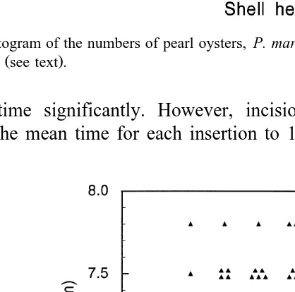
The mean shell height of the Group I oysters, used in the 6-week experiment, was
Ž . Ž .
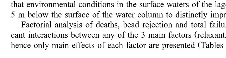
114.6"8.5 mm "S.D. and it was 119.3"8.2 mm for the Group II oysters Fig. 1 . An analysis of the bead diameter chosen for insertion into the 96 Group I oysters showed no significant difference in mean diameters chosen for the different treatment
Ž .
combinations range in mean diameter from 6.81 to 7.29 mm , nor between the
Ž .
successive replicates. There was no significant correlation rs0.19, P)0.05 between
Ž .
bead diameter and oyster shell height for these oysters Fig. 2 . This lack of correlation between bead diameter and oyster shell height suggests that the bead diameter selected for a particular oyster was based more on gonad size than on shell size.
3.2. Time taken to apply treatment combinations
Mean time for bead and mantle insertion was 1.22 min, i.e., about 50 insertions per
Ž .
Fig. 1. A histogram of the numbers of pearl oysters, P. margaritifera, in shell height size classes for Group 1
Ž .
and Group II see text .
insertion time significantly. However, incision closure with adhesive significantly extended the mean time for each insertion to 1.91 min. The combination of antiseptic
Ž
Fig. 2. Diameters of beads inserted into pearl oysters, P. margaritifera, vs. shell heights of the oysters Group
.
Table 2
Ž .
Mean times for simple bead insertion control into pearl oysters P. margaritifera and bead insertions together with additional treatments
Values followed by the same letter do not differ significantly P)0.05 .
and adhesive treatments further extended the mean insertion time to 2.30 min, almost twice the mean time for simple bead and mantle insertions.
Part of the extra time in applying the adhesive was often taken up in ridding the incision site of haemolymph. Many oysters bled profusely from the incision and it was necessary to remove this haemolymph before the adhesive would adhere to the incision site. As noted in Section 2, oysters often had to be removed from the operating clamp and inverted, causing haemolymph to pour copiously from the oyster.
3.3. Treatment effects
The depths at which the oysters were suspended, i.e., positions on the vertical ropes, had no significant effect on mortality or bead rejection rates in the data collected at 6 weeks either as a main effect or in interactions with the treatment combinations. The lack of any depth effect on mortality or bead rejection is not surprising. It is unlikely that environmental conditions in the surface waters of the lagoon vary enough over 3 to 5 m below the surface of the water column to distinctly impact the oysters.
Factorial analysis of deaths, bead rejection and total failure data showed no
signifi-Ž .
cant interactions between any of the 3 main factors relaxant, antiseptic, adhesive , and
Ž .
hence only main effects of each factor are presented Tables 3 and 4 .
Table 3
Ž .
Percentage mortalities, bead rejections, total failures deaths and bead rejections and beads with ‘tails’ in two
Ž . Ž .
groups of P. margaritifera, Group I 96 oysters and Group II 768 oysters , 6 weeks after the pearl operation.
ŽArcsine transformations were used in statistical analyses of percentage data.
Treatment % Deaths % Bead rejection % Total failure % With tails
Gp I Gp II Gp I Gp II Gp I Gp II Gp I
UU UU U UU
Relaxant 22 18 3 8 26 30 64
Non-relaxant 0 1 12 14 12 18 81
Antiseptic 5 7 5 9 16 21 81
Non-antiseptic 6 7 9 13 21 26 65
UU U U
Adhesive 8 11 12 12 23 28 53
Non-adhesive 4 4 3 10 14 20 89
U
Significantly different from no treatment at P-0.05. UU
Table 4
Ž .
Percentage mortalities, bead rejections, total failures deaths and bead rejections in pearl oysters, P.
Ž . Ž
margaritifera, at harvest 18 months after the pearl operation Group II oysters . Arcsine transformations were
.
used in statistical analyses of percentage data
Treatment % Deaths % Bead rejection % Total failure % Pearls
UU UU UU UU
Relaxant 24 6 39 58
No relaxant 2 16 21 74
Antiseptic 10 9 27 69
No antiseptic 12 12 32 63
UU
Adhesive 15 10 33 61
No adhesive 7 10 26 71
U
Significantly different from no treatment at P-0.05. UU
Significantly different from no treatment at P-0.01.
The use of the relaxant, propylene phenoxetol, significantly increased mortality in both groups of oysters. However, it resulted in a lower bead rejection rate, which was
Ž .
significant P-0.05 for the Group I oysters at 6 weeks. Taking account of total failure
Ž .
rates deaths and bead rejections , the higher mortality rates outweighed the lower bead rejection rates and the net effect of propylene phenoxetol was adverse. This greater percentage of total failures was significant for the Group II oysters both at 6 weeks and
Ž .
at 18 months P-0.01 . Following from the high percentage of total failures, the percentage of oysters yielding pearls was significantly lower for those treated with the
Ž . Ž .
relaxant P-0.01 Table 4 .
Application of an antiseptic, povidone iodine solution, had no significant effects on
Ž .
mortality, bead rejection or total failure rates in either group of oysters Tables 3 and 4 . Interestingly, however, antiseptic was the only treatment that resulted in lower percent-age total failures compared to no treatment both at 6 weeks and at 18 months. There is a suggestion of marginal improvement in total failure rate with application of this antiseptic.
The use of an adhesive, cyanoacrylate adhesive, to close the incision resulted in
Ž .
increased mortality, which was significant P-0.01 for Group II oysters at 6 weeks
Ž .
and at 18 months Tables 3 and 4 . Bead rejections were also higher for oysters treated
Ž .
with adhesive significant, P-0.05, for Group 1 oysters at 6 weeks . Total failures were thus greater for adhesive treatment than for no treatment, although the levels did
Ž .
not differ significantly Tables 3 and 4 .
3.4. Pearl sac and secretions
ŽCVs39% . Hence, the differences between the 8 treatment combinations mean cell. Ž .
heights ranging from 7.7 to 12.9mm were not significant. Of the 78 beads retrieved, the
Ž . Ž .
predominant colour was brown 74% , a combination of brown and silverrgrey 4%
Ž . Ž .
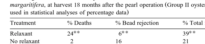
Fig. 3. A Pearl sac epithelium E of P. margaritifera. This was associated with a bead that was
Ž . Ž .
predominantly brown in colour. The scale bar is 22mm. B Pearl sac epithelium E P. margaritifera. This
Ž .
was associated with a bead that was predominantly silverrgray in colour. The scale bar is 22mm. C A longitudinal section of a conical ‘tail’ structure associated with an inserted shell bead in the pearl oyster, P.
Ž .
margaritifera. The ‘tail’ is encased by a calcified layer CL and encloses a large number of degenerating
Ž .
Ž .
and silverrgrey 22% . The brown beads had a mean pearl sac epithelial height of
Ž .
10.23"4.94mm "S.D. , while the silverrgrey beads had a mean pearl sac epithelial
Ž .
height of 6.75"1.31 mm. These mean heights were significantly different P-0.01 . In addition, the variation between the eight measurements made on each pearl sac epithelium was greater for the brown than for the silverrgrey beads. The standard deviation was 4.03 compared to 2.31. The brown colour is presumably the periostracal layer, while the silverrgrey is presumably the prismatic andror early nacreous layers. The appearance of the pearl sac epithelium associated with the brown and the silverrgrey beads is presented in Fig. 3A and B, respectively.
Many of the beads had ‘tails’, cone-shaped extensions of the initial secreted layers that extend into the passageway down which the shell bead had been inserted during the
Ž .
pearl operation. Of the 78 beads recovered, 55 71% had ‘tails’ that were equal to or greater than 0.1 mm long. There were differences in percentages of ‘tails’ in treated vs. non-treated oysters, with relaxant and adhesive treatments reducing the percentage of oysters with tailed beads, while antiseptic increased the percentage with tailed beads ŽTable 3 . However, only the adhesive treatment resulted in a significant difference. ŽP-0.01 compared to non-treatment. There were large numbers of dead inflammatory.
Ž .
cells within the ‘tail’ structure of all six ‘tails’ examined Fig. 3C .
The use of the relaxant significantly reduced the pearls harvested from 74% to 58% ŽTable 4 . However, use of the antiseptic or the adhesive had no significant effect on. pearls harvested.
3.5. Pearl quality
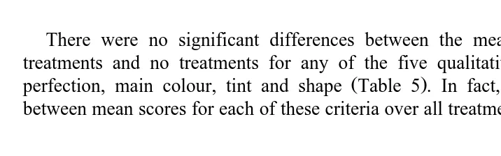
There were no significant differences between the mean scores for pearls from treatments and no treatments for any of the five qualitative criteria: lustre, surface
Ž .
perfection, main colour, tint and shape Table 5 . In fact, there was little variation between mean scores for each of these criteria over all treatments and no treatments. The
Table 5
Mean classification scores and dimensions of pearls harvested from pearl oysters, P. margaritifera, 18 months
Ž .
after the pearl operation Group II oysters Treatment Pearl classification
Lustre Surface Main Tint Shape Diameter Length Lengthr Weight
Ž . Ž . Ž .
score score colour score score mm mm diameter g
score ratio
U
Relaxant 2.97 2.56 3.71 3.42 2.60 10.59 12.53 1.184 1.72
No relaxant 3.01 2.70 3.88 3.44 2.74 10.72 12.72 1.187 1.83
Antiseptic 3.00 2.56 3.72 3.50 2.67 10.65 12.73 1.195 1.78
No antiseptic 2.98 2.70 3.87 3.36 2.68 10.66 12.52 1.175 1.77
U UU
Adhesive 2.99 2.70 3.74 3.50 2.70 10.68 12.34 1.156 1.76
No adhesive 2.99 2.56 3.85 3.36 2.64 10.63 12.91 1.214 1.79
U
Significantly different from no treatment at P-0.05. UU
mean scores for lustre, surface perfection, main colour, tint and shape were about 3.0, 2.6, 3.8, 3.4 and 2.6 out of 5, respectively, in all cases. Thus, there were no effects of
Ž
relaxation, antiseptic nor adhesive on these five criteria of pearl quality. The pearls were not polished, by the usual procedure of rubbing in salt, at the time of grading and the mean lustre values would be improved by this procedure. The absence of polishing
. is, however, a uniform effect.
In terms of quantitative criteria, pearl diameter, length, lengthrdiameter ratio and weight, there were significant differences. Use of propylene phenoxetol was associated with the smallest diameter pearls, although not significantly smaller than pearls from oysters not treated with the relaxant. Pearl weight, which is proportional to diameter3,
Ž .
was significantly lighter in oysters that had been treated with relaxant P-0.05 . The other significant results on quantitative criteria from a treatment were the effects
Ž .
of adhesive use in reducing the mean length of pearls P-0.05 and the mean ratio of
Ž . Ž .
pearl lengthrdiameter P-0.01 Table 5 . The pearls from oysters that were treated with adhesive tended to be less elongate.
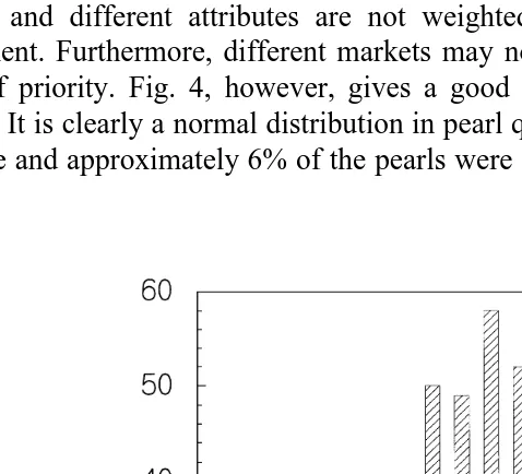
The scores for the pearls harvested from the experiment are presented in Fig. 4 as a frequency vs. pearl quality score histogram. Commercial grading of pearls is a complex process and different attributes are not weighted equally as has been done in this assessment. Furthermore, different markets may not rank the pearl colours in the same order of priority. Fig. 4, however, gives a good indication of the nature of the pearl harvest. It is clearly a normal distribution in pearl quality. No pearls achieved the perfect 30 score and approximately 6% of the pearls were in the top 20% of scores, 24qout of 30.
4. Discussion
4.1. Pearl quality
The overall quality of this pearl crop was low. This small percentage of gem quality
Ž .
pearls is similar to that reported by Cabral 1990 in P. margaritifera and by Scoones Ž1990 in P. maxima. Furthermore, pearl quality Fig. 4 had a normal distribution with. Ž . the mode around the score of 19. Ideally, the distribution needs to be skewed towards the maximum score of 30 so that a larger proportion of pearls are in the upper, gem-quality range. The lack of effect of the treatments on the qualitative aspects of pearl quality suggests the need for reevaluation of what factors might be influencing pearl quality.
4.2. Pearl sac formation
As the mantle tissue implanted into the operated oysters grew to form a pearl sac
Ž .
around 76 of the 78 beads recovered Group I, 6 weeks , it is unlikely that any significant host vs. non-host tissue reaction had occurred. If implanted tissue is to be
Ž .
rejected, this rejection occurs within 27 to 40 days, as demonstrated by Wada 1989 using freshwater mussels.
Ž .
In pearl sac formation in P. imbricata, Aoki 1966 found the mean heights of periostracal, prismatic and nacreous epitheliums to be 55.8"21.4, 8.1"9.0 and 3.4"2.1mm, respectively. The pearl sac epithelium thins progressively as it develops.
Ž .
Kawakami 1952 also working with P. imbricata, reported that the nacreous layer began to be secreted about 40 days after transplantation when the epithelial cells of the
Ž .
pearl sac became flat. Aoki 1966 stated that it is difficult to distinguish pearl sac epithelium which is producing a nacreous layer from that which is producing a prismatic layer on the basis of morphological features of these cells. In contrast, epithelial cells
Ž .
producing periostracum sconchiolin are quite distinct since they are extremely tall, cylindrical and ciliated and contain numerous acidophilic small granules in the cyto-plasm. It seems that more than 60%, but not all, of cultured pearls in P. imbricata have
Ž . Ž .
an organic layer of periostracum Tsujii, 1968 . Furthermore, Wada 1957 noted that periostracum does not always cover the whole surface of the bead. As the epithelium of
Ž .
the pearl sac is very sensitive to both internal and external stimuli Tsujii, 1968 , one Ž .
wonders what factor s control whether these cells secrete an organic layer of perios-tracum.
In this study of P. margaritifera, where 78% of beads were wholly or partially brown in colour 42 days post-operation, it seems that at least these beads had received a layer of periostracum. This does not preclude that all beads received a periostracal layer: it may be obscured by further layers in the other beads. The total or partial silverrgrey colour of 26% of the beads suggests that these were at the prismatic or nacreous stages. However, the mean epithelial heights of the pearl sacs, 7.7 to 12.9mm, suggest that the pearl sacs were at about the prismatic stage; that is, if the data for the pearl sac
Ž .
thinned to the mean height of nacreous epithelium in P. imbricata. There is thus some conflict between the observed colour of a substantial proportion of beads in P. margaritifera and the presumed stage of the pearl sac. If colour is a reliable indicator of the onset of the nacreous layer, then the age of these beads, 42 day post-operation, is
Ž .
comparable to the 40 days observed by Kawakami 1952 for P. imbricata. If epithelium height, based on P. imbricata, is a better indicator of pearl sac development, then P. margaritifera develops more slowly than the former species.
Ž .
The ‘tails’ attached to many of the beads Table 3 occurred in the ‘dead’ space above the shell bead. This ‘dead’ space was the persisting passageway made by the pearl knife and along which the shell bead was directed until it reached its final resting
Ž .
position adjacent to the mantle graft. Aoki 1961 observed that to produce superior pearls, the operation should be made without leaving a space between the bead and adjacent tissues. It would appear that this ‘dead’ space adjacent to the shell bead is ideal for the accumulation of haemolymph, bacteria and inflammatory cells. There would have been bacteria on the operating instruments, bead and mantle graft, as the pearl surgery technique used here was the usual industry technique, which was not aseptic. The pearl sac grew around this accumulation of dead inflammatory cells which had accumulated in the tail, as well as around the bead, producing a calcified ‘tail’ attached
Ž .
to the bead Fig. 3C . Similar haemocyte accumulations were reported by Scoones Ž1996 in 38% of 59 pearl sacs examined by him in P. maxima pearl oysters. He. concluded that the cells were a response to infection or tissue damage and that they contributed to the formation of low-value baroque pearls.
The significantly lower number of ‘tails’ on beads from oysters on which adhesive
Ž .
was used Table 3 suggests that adhesive closure of the incision helped to reduce or eliminate the ‘dead’ space adjacent to the shell bead. This was followed by a significant reduction in the mean length of harvested pearls and the mean proportion of lengthr di-ameter from oysters treated with adhesive compared to those without this treatment. It appears that reducing the development of ‘tails’ during early pearl formation continues into the subsequent pattern of nacre deposition, so that the pearls have significantly lower lengthrdiameter ratios.
4.3. Antiseptic use
The lack of any significant effect on post-operation mortality, bead rejection or pearl
Ž .
‘tail’ formation from the antiseptic treatment of the incision site Tables 3 and 4 accords with observations of the likely sources of bacteria in the bead and mantle graft
Ž .
to lower the frequency of these infections. Similar conclusions on the role of bacterial
Ž .
infections were made by Scoones 1996 when working with P. maxima pearl sacs.
4.4. Oyster mortality
Ž .
The high mortality rates in relaxed oysters Tables 3 and 4 contrast with previous Ž
work in the laboratory on small numbers of pearl oysters P. albina and P.
margari-. Ž . Ž
tifera Norton et al., 1996 and in the field on large numbers of farmed pearl oysters P.
. Ž .
maxima Mills et al., 1997 . Oyster survival was very high in both cases, e.g. with losses of 1% or less. Several factors may have contributed to the high mortality with relaxant treatment. Immersion of each oyster in the relaxant bath for 15 min should not
Ž .
have been a factor since Norton et al. 1996 immersed oysters for up to 60 min without mortalities. However, these latter oysters were not subjected to multiple stresses, including forced opening, haemolymph haemorrhaging and prolonged emersion. The oysters in this current study were held in the air for periods of 30 min to 1 h before and after the bead insertion operation. Water drained off or evaporated from the delicate oyster tissues during the period of emersion and some drying may have occurred.
The large loss of haemolymph during the pearl operation is also of concern. As much as one-third of the circulating haemolymph volume can be lost in this way from
Ž .
non-relaxed P. margaritifera Lintilhac, 1987 . This volume may be greater from
Ž .
relaxed oysters with collapsed mantles. Harley and Harley 1973 performed surgical operations on the gastropod mollusc, Aplysia californica, and found that if there was a
Ž .
weight loss of more than 15% during surgery mainly due to loss of haemolymph , a significant proportion of the animals died.
Further support for added stress arising from use of relaxant is the finding that the pearls from this treatment were significantly smaller than those from untreated oysters ŽTable 5 . Pearl diameter and weight are related to bead size and to thickness of nacre on. the bead. It is conceivable that significantly smaller beads were inserted into the oysters that were treated with relaxant and the final size effect is related to bead size. However, this seems unlikely. There was no significant difference between treatments in bead size and there was nothing to suggest any reason for such a bias during the course of the bead insertion program. The most likely explanation is that the nacre layer is thinner in pearls from relaxant-treated oysters. This suggests that the process of nacre secretion was delayed or inhibited, probably in the period after the stress of the bead insertion procedure.
Ž .
The increased mortality of oysters receiving the adhesive Tables 3 and 4 may be related to the extent of inflammation. The adhesive causes inflammation of the tissues to
Ž .
which it adheres Norton, unpubl. data . If a large quantity of adhesive is applied, the resulting large area of inflammation may lead to death.
4.5. Bead rejection
Ž .
tissues, allowing the bead to escape. The use of a smaller quantity of adhesive may help to reduce these losses or another method needs to be developed to close the incision, e.g., sutures. However, suturing is potentially unacceptable in terms of the time required,
Ž
especially when used at the current incision site for bead insertion Norton, unpubl. .
data . For this reason, suturing in combination with a more accessible incision site for bead insertion e.g., the gonad surface, is being investigated. Such a site also has the
Ž .
advantage of reducing tissue damage and haemorrhage see above .
5. Conclusions
Ž
There was a lack of positive effects of these treatments on percent failures mortality .
and bead rejections , percent yield of pearls and pearl quality, apart from the positive effect of adhesive on pearl symmetry. This suggests that reducing mortality and bead rejection rates are needed before expecting substantial improvements in pearl quality. Improving the level of hygiene for all aspects of the pearl surgery will have greater impact on reducing infections than treating the surface of the incision site only. This should reduce the prevalence of pearl sacs with inflammatory cell infiltrations and thence of baroque pearls.
Mortality associated with adhesive use may be reduced by using less adhesive or using adhesive in association with other material, e.g., cotton strips.
The increased times taken for additional procedures to the pearl formation operation
Ž .
were not excessive Table 2 . This was in spite of inexperience with the techniques and constant changing from one treatment to another as part of the experimental protocol. The times were undoubtedly longer than if the same treatment was routinely followed. This suggests that additional time for the operation should not be an obstacle to further innovative approaches to the bead insertion process. The cost of slower operation times could be far outweighed by greater proportions of high-quality pearls.
Acknowledgements
This research was funded by the Australian Centre for International Agricultural Research, Canberra, Project No. 9131. The assistance of Kora Kora and other family members and employees of Terone Pearl, Manihiki, is greatly appreciated. The authors are also very indebted to Dr. Miles Anderson of the Lagoon Ecology Monitoring and Management Project for collection of specimens and data at 6 weeks.
References
Anon, 1991. Pearl oyster Farming and Pearl Culture, Training Manual No. 8, Central marine Fisheries Research Institute, Tuticorin, India, 103 pp.
Ž
Aoki, S., 1961. Some experiments on the nuclear insertion in the pearl-culture of the oyster Pinctada
.
martensii : V. Pearl-sac formation in the material with a space between the inserted nucleus and adjacent tissue. Bull. Natl. Pearl Res. Lab. 6, 647–656.
Aoki, S., 1966. Comparative histological observations on the pearl-sac tissues forming nacreous, prismatic and periostracal pearls. Bulletin of the Japanese Society of Scientific Fisheries 32, 1–10.
Ž .
Cabral, P., 1990. Some aspects of the abnormal mortalities of the pearl oyster, Pinctada margaritifera L in
Ž .
the Tuamotu Archipelago French Polynesia . In: Advances in Tropical Aquaculture, Tahiti Feb. 20–March 4 1989. Aquacop Ifremer, Actes de colloque 9, pp. 217–226.
Ž .
Dix, T.G., 1972. Histology of the mantle and pearl sac of the pearl oyster Pinctada maxima Lamellibranchia . J. Malac. Soc. Aust. 2, 365–375.
Ž .
Gervis, M.H., Sims, N.A., 1992. The biology and culture of pearl oysters Bivalvia: Pteriidae . ICLARM Stud. Rev. 21, 49 p.
Harley, P., Harley, E., 1973. A technique for cutting or accessing nerves and connectives in Aplysia californica. Comp. Biochem. Physiol. 45A, 389–392.
Kawakami, K., 1952. Studies on pearl-sac formation: 1. On the regeneration and transplantation of the mantle
Ž . Ž .
piece in the pearl oyster. Mem. Fac. Sci. Kyushu Univ. Ser E 1 2 , 83–89.
Lintilhac, J., 1987. Black pearls of Tahiti; Royal Tahitian Pearl Book, Papeete, Tahiti, p. 53.
Meng, Z., Xing, K., 1991. The effects of various factors on the nucleus-insertion of the black lipped pearl oysters Pinctada margaritifera Linnaeus. Oceanologia et Limnologia Sinica 22, 8–13.
Mills, D., Thili, A., Norton, J., 1997. Large scale anaesthesia of the silver-lip pearl oyster, Pinctada maxima. J. Shellfish Res. 16, 573–574.
Norton, J.H., Dashorst, M., Lansky, T.M., Mayer, R.J., 1996. An evaluation of some relaxants for use with pearl oysters. Aquaculture 144, 39–52.
Rambaud, H., 1991. Tahiti Cultured Pearl: Grading Guide. Rambaudqpublication, Tahiti, French Polynesia, 12 pp.
Scoones, R.J.S., 1990. Research on practices in the Western Australia cultured pearl industry, Fishing Industry Research and Development Council, Project BP12, Final Report, Fisheries Research and Development, Canberra, Australia.
Ž .
Scoones, R.J.S., 1996. The development of the pearl sac in Pinctada margaritifera Jamieson 1901
ŽLamellibranchia: Pteriidae and the implications for the quality of cultured pearls. MSc Thesis, University.
of Western Australia, Nedlands, Western Australia.
Shirai, S., 1994. Pearls and Pearl Oysters of the World, Marine Planning, Okinawa, Japan.
Tsujii, T., 1968. Studies on the mechanism of shell and pearl formation. XI. The submicroscopical observations on the mechanism of formation of abnormal pearls and abnormal shell. Rep. Fac. Fish. Pref. Univ. Mie. 6, 59–66.
Wada, K., 1957. Microscopic observations of cultured pearls at their early formation. Bull. Nat. Pearl. Res. Lab. 31, 167–174.
Ž
Wada, K., 1989. Allograph and xenograph mantle transplantation in freshwater mussels. Venus Jap. J.
.
Malacol 48, 174–190.
Ž . Ž . Ž .