Introduction
Biotechnology is widely used in plant breeding to alter the genetic composition of plants to improve crop yields, alter cosmetic features, or facilitate transport. The alteration of genetic traits is performed by inserting genes from other plants, or even from other organisms, into the genome of the plant that is to be improved. In this way, new emerging genetic and physiological traits arise and these characters are passed to successive generations (Yuwono, 2006).
Changes in the properties of the transformant plant can be observed from its phenotype, but these may not be detectable for some time, or even until the plant reaches maturity. Selecting transformant plants could be made more efficient if the transformant plant could be detected earlier in its development. Thus, we have designed a system that relies on the plasmidbased genomic insertion of a fluorescent reporter gene to indicate that transformation has taken place. Our system spesifically reports the expression of flowering genes before the flowering phenotype can be observed. A number of systems were considered, including screenable markers such as β glucuronidase (GUS) assay, and luciferase. However, as enzymatic assays both luciferase and GUS assays require the
Early Detection of the Orchid Flowering Gene
PaFT1
in Tobacco
Cells Using a GFP Reporter
Sri Wahyuningsih
1,2§, Muhammad Dylan Lawrie
1§, Budi Setiadi
Daryono
1,3, Sukarti Moeljopawiro
1, Soenghoe Jang
4, Endang Semiarti
1,3,*1Graduate Study Program of Biology, Faculty of Biology, Universitas Gadjah Mada,Yogyakarta, Indonesia
2Department of Biology, University of Lampung, Lampung, Sumatera, Indonesia 3Department of Tropical Biology, Faculty of Biology, Universitas Gadjah Mada,
Yogyakarta, Indonesia
4Biotechnology Center in Southern Taiwan (BCST) of Agricultural Biotechnology Research Center (ABRC), Academia Sinica, Taiwan
§The two authors contributed equally in this paper
Abstract
Here we describe a novel method of usinggreen fluorescence protein(GFP) as a reporter gene for early detection of an integrated TDNA containing the orchid flowering gene,PaFT1(Phalaenopsis aphrodite Flowering locus T1) in the tobacco genome. Functional assays that report the presence of exogenous DNA early in development are especially useful in plants where the desired phenotype is only apparent after long periods of vegetative growth. The objective of this study is to establish a method for detecting an insertedPhalaenopsis orchid flowering gene and examining its function in tobacco. The p35S::PaFT1 35S::GFP construct was introduced intoAgrobacterium tumefaciensstrain EHA101. Transformed tobacco leaves were cultured on MS medium with addition of 1 mgL1NAA+3 mgL1BAP+50 mgL1Kanamycin+300 mgL1timentin for selection. Results showed bright green GFP fluorescent signals in 11 out of 15 (73%) tobacco leaf cells at a 2month time point after transformation. GFP and PaFT1 fragments were amplified in genomic PCR using GFP and PaFT1 specific primers. The accumulated PaFT1 transcripts were observed in 3 monthold transgenic tobacco plants containing p35S::PaFT135S::GFP. Green florescence was observed only in the transgenic plants at the 5 monthold stage but not in the wild type controls.
Keywords: Early flowering,GFP,PaFT1, reporter gene, tobacco
*Corresponding author:
Endang Semiarti
addition of an enzyme substrate to visually detect the result. Therefore we chose an approach based on Green Fluorescent Protein (GFP).GFPis a 27kDa protein (Xia
et al., 2002), which emits bright light when exposed to blue light or ultraviolet radiation (UV) (Britoet al., 2013).GFPis widely used because it does not require substrates or exogenous cofactors for fluorescence (Stewart, 2005; Puchta, 2003) and does not destroy the tissues of transformants (Hraska et al.2006). For these reasons, GFP
was selected as the reporter gene to improve early detection of flowering genes.
In this research, the construction of a plasmid based on pGAS101 was carried out by insertingGFPas a reporter gene. The main gene on pGAS101 plasmid is PaFT1.PaFT1
gene is one ofFThomologous genes isolated fromP. aphrodite(Janget al.2015). Analogous to Arabidopsis as the model plant, over expression of thePaFT1gene is expected to accelerate flowering in plants.GFPwas used as an evaluation tool of transformation parameters. Cells and tissues expressingGFP
could be distinguished easily from those that do not expressGFP without destroying the tissue sample, and can facilitate separating transformants and nontransformants (Hraska
et al., 2006). GFPexpression is indicated by green fluorescence, while the non transformant retains its natural red autofluorescence after being exposed under light with a wavelength of 480 nm (Ghorbel
et al., 1999).
Plasmid pGAS101 also includes the selection marker genes Neomycin Phosphotransferase (NPT) and tetracycline
resistance (tetR) genes. The selection marker genes are important for the development of plant transformation technology, because it allows the identification and isolation of cells expressing the cloned DNA and for monitoring and selecting offspring of the transformants (Miki and McHugh, 2004).
A number of transformation methods, which could be used to transfer foreign genes include particle bombardment,
Agrobacterium tumefaciensmediated, electroporation with protoplasts/protocorm,
seed imbibition and transformation through pollen tube (Chai and Yu, 2007). Transformation usingAgrobacteriumis an indirect transformation technique, which is the most frequently used. This technique has several advantages including transformation efficiency with higher copy numbers of single genes and ease of use with simple laboratory equipment. The
Agrobacteriummediated transformation method is commonly used to create transgenic plants, because it has more advantages compared to direct gene transfer methods such as particle bombardment, electroporation and silicon carbide fibers. The advantages include (1) stable gene expression; (2) a low number of transgene copies, (3) large DNA segments are able to be transferred (Ko and Korban, 2004).
Ti plasmid in a cell ofA. tumefaciens
contains a DNA fragment called TDNA (sized 10–30 kb), which can integrate into the DNA of the plant cell nucleus. The Ti plasmid in A. tumefacienscell is currently developed as a vector to insert foreign DNA into plant cells (Yuwono, 2006). This process requires functional transgenic constructs, including the desired main gene, promoter guiding the expression, marker genes, the transgene insertion into the Ti plasmid and the transformation of TDNA contained in the plasmid into Agrobacterium cells (Mohammed and Abalaka, 2011).
In this study, a binary vector containing p35S::PaFT135S::GFP construct was then transferred into A. tumefaciens
strain EHA101 and tobacco leaves. The detection of GFP at the beginning of plant growth indicates thatPaFT1has integrated into plant genome. The p35S::PaFT1 35S::GFP/A. tumefaciensstrain EHA101 is an efficient system for early detection and functional analysis of Flowering GenePaFT1
Materials and Methods
Plant materials and bacterial strains
Plant materials used as explants in this study were leaf dishes from two months old tobacco (Nicotiana tabacumL. var. Kemloko 1). Thein vitroseedlings were cultivated on Murashige and Skoog (MS) medium, under white continuous light, at (25±2)°C room temperature. The leaf dishes were used as explants forAgrobacteriummediated genetic transformation of p35S::PaFT135S::GFP /EHA101 construct. For bacterial strains,
Eschericia coli strain DH5α, A. tumefaciens
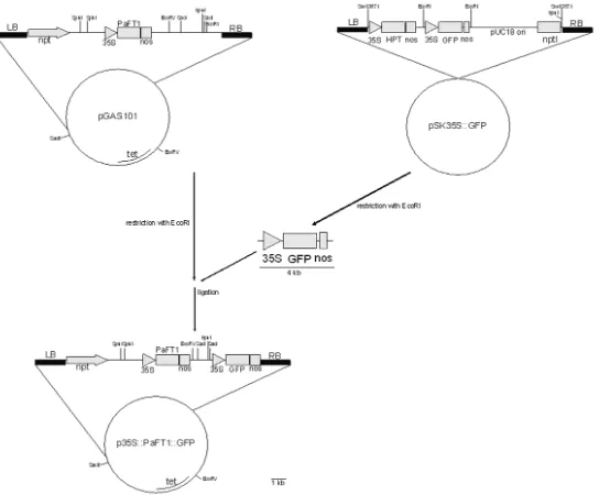
strain EHA101, and E.coli containing pSK35S::GFP, pGAS101 plasmid were used to construct35S::PaFT1::GFP. The pGAS101 plasmid (12.2 kb in length) was constructed previously to express PaFT1 gene using
Cauliflower Mosaic Virus (CaMV) promoter
35S, it containedNeomycin Phosphotransferase II(NPT II) gene, which encodes a resistance character against the antibiotic kanamycin as a selection marker. The pSK35S::GFP(15.8 kb in lengths) plasmid was obtained from Prof. Chiyoko Machida (Chubu University, College of Bioscience and Biotechnology, Japan) that was constructed based on the pSK1 plasmid (Kojima et al., 1999). The plasmid carriesGFP
gene betweenXbaIandNotIrestriction sites, which ws used as reporter in this research.
Insertion of 35S::GFP fragment into pGAS101 plasmid
The plasmid pGAS101 and pSK35S::GFP were isolated fromE. coliusing alkaline lysis method following the instruction manual of plasmid isolation kits (Qiagen, UK). The isolated pSK35S::GFP plasmids were cut using
EcoRI restriction enzymes, and then separated using electrophoreses and visualized with a UV Transilluminator. The obtained p35S::GFP fragment bands were cut from the gel and extracted using JETQUICK Gel Extraction Spin Kit following the manual instruction (Genomed, The Netherlands). The gel extraction product was checked by PCR using 2 pairs of specific primers: the sGFPFw (5' ACGAGAAGCGCGATCACAT3') – Nos Rev (5'GTATAATTGCGGGACTCTAATCA3')
primer pair. The pGAS101 plasmid containing
PaFT1gene, which was previously cut using
EcoRI restriction enzyme, and the p35S::GFP fragment was ligated to pGAS101 using Ligation HighDNA ligase at 16°C for 30 minutes. The presence of ligation products was checked using PCR with sGFPFw and Nos Rev primers as before. The p35S::PaFT1 35S::GFP plasmid (16.2 kb in length)was constructed as shown in Figure 1.
Transformation of p35S::PaFT135S::GFP plasmid into E. coli strain DH5α
Before the transformation, competent cells of E. coli strain DH5α were prepared according to the methods described in Sambrook and Russell (2001). Competent cells were subjected to transformation through the heat shock method. A volume of 800 µL of competent cells was divided into 8 microtubes with 100 µL competent cells each. A mixture of competent cells and p35S::PaFT135S::GFP plasmid were soaked in liquid SOC medium in a microtube and the obtained mixture was incubated for 45 minutes in water bath at 37°C. A 100 µL culture was taken from SOC medium, then inoculated it onto SOB solid medium plate containing 20 mM MgSO4and 50 mgL1 of kanamycin antibiotic using the
Bacterial transformation by insertion of
p35S::PaFT1::GFP plasmid into A.
tumefaciens strain EHA101
The p35S::PaFT135S::GFP plasmids were transferred intoA. tumefacienscompetent cells, following procedures described by Widyasari and Suhandono (2007) with modification.A. tumefacienscompetent cells in a volume of 100 µL in a 1.5 ml Eppendorf tube were mixed with 1 µL plasmid in cold condition with liquid nitrogen for 5 minutes. The mixture was then incubated at 37°C for 25 min. A volume of 1 mL liquid LB medium was added into the tube, and then the bacteria were grown at room temperature (28°C) for 3 hours with shaking at 50 rpm. Bacterial cells were later harvested by centrifugation at 12,000 rpm for 30 sec. Pellets were suspended with 1 mL LB. After that, 200 µL cell suspension was spread into the selection media (LB + 5 mgL1 tetracycline antibiotics + 50 mgL1
kanamycin), and then incubated at 28oC for
two days to obtain transformed colonies. The transformation results were checked by PCR with primer pair of Deg PaFT F2 and R1 to
detect the presence ofPaFT1, and sGFP Fw and Nos Rev to detectGFP, in the same way of that used in E.coli. The recombinant p35S::PaFT135S::GFP /A. tumefaciensstrain EHA101 was then ready to be used for genetic transformation in tobacco leaf explants.
Transfer of TDNA containing p35S::PaFT1 35S::GFP fragment into tobacco leaves
A single colony ofA. tumefaciensstrain EHA101 containing p35S::PaFT135S::GFP was cultured in a 15 mL corning tube containing 2 mL LB liquid medium with 50 mgL1kanamycin and 5 mgL1 tetracycline
antibiotics, incubated at 28oC for 48 h on a
rotary shaker. A volume of 100 µl Agrobacterium culture was added to 10 mL LB medium with 50 mgL1kanamycin and 5
mgL1tetracycline antibiotics, and incubated
at 28oC for 48 h on a rotary shaker. Bacteria
were then harvested by centrifugation at 6,000 rpm for 3 min. Bacterial cells pellet obtained was suspended in 10 mL liquid MS medium without sugar. After that, 2 mL bacterial culture was poured into 8 mL liquid MS
medium without sugar, and then homogenized. Next, thein vitroleaf explants (1 cm2 in size) were soaked into bacterial
suspension for 30 min. Leaf explants were later drained on sterile filter papers, then planted on MS medium with addition of 1 mgL1 NAA, 3 mgL1 BAP, and 100 mgL1
acetosyringone, then it were incubated in the dark room for 3 days. The transformed explants were then washed using 1,000 mgL1timentin
for 5 minutes, then rinsed its with liquid MS medium without sugar twice for 3 min each. Finally, the explants were dried up with sterile filter papers and transferred onto MS solid medium with addition of 1 mgL1 NAA, 3
mgL1 BAP, 50 mgL1 kanamycin, and 300
mgL1timentin for maintenance to form buds.
About 2 months afterAgrobacteriuminfection, some leaf pieces were analyzed for GFP
expression, leaf pieces were placed on object glass, dropped with glycerin, covered with a cover glass, and then was observed with a fluorescent microscope under UV light, and photographed using Canon IXUS digital camera (Japan).
Analysis of PaFT1 expression in transgenic tobacco plants
Detection of gene expression was performed by isolating mRNA from young leaves of 3 months old of p35S::PaFT1 35S::GFP containing tobacco transformant plants, followed with cDNA synthesis from the mRNA. Total RNA was isolated using
Total RNA Mini Kit Plant (Geneaid) and cDNA was synthesized from mRNA using
Thermo ScientificTM RevertAidTM First Strand cDNA Synthesis Kit(Thermo Fisher Scientific Inc.). To detect the expression ofPaFT1gene in putative transgenic, RTPCR was performed. The cDNA was used as template for RTPCR analysis using the primers specific for thePaFT1gene (deg PaFT F2 and R1) in putativePaFT1transgenic plants. RT PCR was also conducted forGFPgene using a pair of sGFP Fw and Nos Rev primers.
Phenotypic analysis of transgenic tobacco plants containing p35S::PaFT135S::GFP
Two monthold regenerated plants from leaf explants cultivated on MS medium with
addition of 3 mg.L1BAP + 1 mg.L1NAA and
50 mg.L1 Kanamycin+300 mg.L1 timentin
were transferred into MS medium for rooting. The plants were transferred into soil in pots and maintained in a greenhouse with natural environmental conditions and light photoperiods. The number of leaves, length of stem and time until detection of fluorescence were measured.
Results and Discussion
Construction of p35S::PaFT135S::GFP
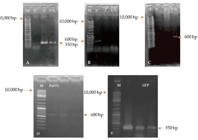
The 35S::GFP that originally came from pSK35S::GFP plasmid was successfully inserted into pGAS101 (Figure 2). Figure 2A shows isolated 12.2 kb circular plasmid DNA of pGAS101 DNA and pSK35S::GFP (12 kb). EcoRIdigested plasmid showed a single linear plasmid of pGAS101. On the other hand, pSK35S::GFP was split into two bands: 8 kb and 4 kb, indicating that pGAS101 has only oneEcoR1 restriction site, and pSK35S::GFP has 2 EcoR1 restriction sites (Figure 2 B). The presence of GFP in these two plasmids was confirmed by PCR analysis using sGFP Forward primers and Noster primers, that resulted in 350 bp amplified DNA fragments.
The linear plasmid DNA pGAS101 was ready to be inserted withGFPgene. A 35S::GFP DNA fragment of about 4 kb in length was gel extracted with theJet quick gel extraction kit(Genomed).
DNA bands resulting from amplified DNA pSK 35S::GFP using PCR with primers of sGFP Fw and Nos Rev were evaluated by gel extraction for the presenceGFPgene. As a negative control experiment, the detection ofHPTgene was checked using a pair of HPT primers (HPT Fw and HPT Rev). PCR products were then checked using electrophoresis and visualized using a UV Transilluminator (Figure 3). The presence ofGFPwas detected. This data confirmed that theGFPhad been amplified.
at temperature of 37°C for 30 mins. Products of the ligation process were used to transform theE.colistrain DH5α competent cells using the heat shock method. The heat shock method takes advantage of the membrane permeability created when cells undergo sudden temperature changes in a short period. Competent cells were diluted in CaCl2(0.1M)
cold solution (Sambrook and Russell, 2001) to induce competence so the cells could accept foreign DNA molecules and to undergo transformation (Madiganet al., 2012). The
E.coli competent cells were obtained by inducing the bacterial cells with CaCl2 treatment. The treatment resulted in cells that could be transformed by allowing DNA to
Figure 2. Confirmation of pGAS101 and pSK35S::GFP plasmids. (A) Isolated plasmid DNA of pGAS101 and pSK35S::GFP plasmids; (B)EcoRI restriction enzymedigested pGAS101 and pSK35S::GFP plasmids; (C) Amplified DNA from pSK35S::GFP [Lane 1. Markerλ/styI DNA, lane 2. pGAS101 plasmid DNA, lane 3. pSK35S::GFP plasmid DNA, lane 4. Marker of 1kb DNA ladder, lanes (1, 4 and 7).EcoRI restriction enzyme digested pGAS101 (5) and pSK35S::GFP plasmid (6); GFP amplified DNA from pSK35S::GFP (8 and 9)].
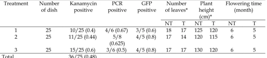
*5 months old; NT: nontransformant, T: transformant
Table 1. The initiation of flowering in transgenic tobacco containing p35S::PaFT135S::GFP.
Figure 4.p35S::PaFT135S::GFP plasmid/A. tumefaciensEHA101. (A) Colonies ofA. tumefaciensbacterial culture; (B) Isolated plasmid DNA of p35S::PaFT135S::GFP /A. tumefaciensEHA101, M=Marker (λ/styI).
Figure 5.Expression ofGFPin leaves of transgenic tobacco containing p35S::PaFT135S::GFP. (A) The 1.5 months old leaf culture of transgenic tobacco containing p35S::PaFT135S::GFP on MS medium + 1 mgL1NAA, 3 mgL1
BAP, 50 mgL1kanamycin, and 300 mgL1timentin; (F) The callus explants exposed under UV light illumination
showed green fluorescence; (B) A 2 months old plantlet of Wild type/non transformed on MS medium + 1 mgL1
NAA, 3 mgL1BAP; (C) Leaf of (B); (D) 2 months old Wild type/non transformed leaves examined under bright
light, and under UV light (E); (G) A 2 monthold plantlet of transgenic tobacco containing p35S::PaFT135S::GFP on MS medium + 1 mgL1NAA, 3 mgL1BAP, 50 mgL1kanamycin, and 300 mgL1timentin; (H) Leaf of (G); (I) the leaf
pass through theE.colicell wall and membrane, with Ca2+ ions neutralizing the negative
charges in DNA molecules so the DNA could more readily enter the cells followingheat shocktreatment (Casali, 2003). At the same time multiplication of pGAS101::GFP recombinant plasmids was occurred.
Checking whether bacteria carry pGAS101, pSK35S::GFP, and p35S::PaFT1 35S::GFP plasmids was performed by colony PCR to amplify DNA fragments using two pairs of primers, the DegPaFT F1 R1 and sGFP Fw Nos Rev. Amplified DNA fragments matching the sizes ofGFPandPaFT1genes, respectively were detected (Figure 3D and E).
The p35S::PaFT135S::GFP recombinant plasmid was isolated from E. coli strain DH5α. The plasmid was then transformed into competent cells of A. tumefaciens strain EHA101. Procedures of plasmid transformation into competent cells of A. tumefaciens was adopted from a method described by Widyasari and Suhandono (2007). The p35S::PaFT135S::GFP recombinant plasmid was isolated from A. tumefaciens strain EHA101, and then it was checked by electrophoresis and visualized with UV Transilluminator (Figure 4).
The p35S::PaFT135S::GFP/A. tumefaciens
EHA101 plasmid obtained as transformation products were checked using PCR with 2 pairs of primers, the degPaFT F2 deg PaFT
R1 and sGFPFw Nos Rev. The PCR products showed the presence ofPaFT1andGFPgenes (Figure 3).
Thus, the recombinant p35S::PaFT1 35S::GFP/A. tumefaciensstrain EHA101 was ready to be used for genetic tobacco transformation to accelerate flowering. Examination of the tobacco leaves under UV light showed the bright green fluorescence of GFP in the leaves of 2.5 months old transformants. (Figure 5, Table 1).
To examine gene expression in transformant plants, we used RTPCR using genespecific primers. This method was also used by Semiartiet al. (2010) to detectGFP
gene insertion inP. amabilisorchid protocorms, to evaluate transgenic plants carrying a copper inducible reporter gene expression system in
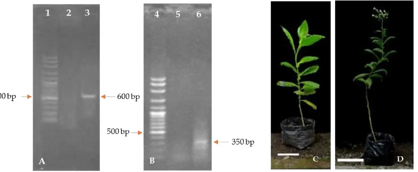
Arabidopsis thalianaby Saijo and Nagasawa (2015), to evaluate the efficiency of TDNA delivery in transformants of Phalaenopsis violaceaby Zulkifleet al.(2010) and to analyze FT expression in plant by Xuet al.(2012). In this study,PaFT1andGFPtranscripts were detected in putative p35S::PaFT135S::GFP transgenic tobacco plants (Figure 6) indicating that the TDNA of p35S::PaFT135S::GFP was integrated into the tobacco genome and functionally active. Transgenic tobacco plants with p35S::PaFT135S::GFP construct showed accelerated flowering with 14–17 leaves and 115–120 cm height at 5 months whereas wild
type (nontransformant) plants flowered around one month later than transgenic tobacco plants with more leaves (Table 1, Figure 6C and 6D).
These results indicate thatPaFT1 can mildly reduce the vegetative growth period and accelerate flowering in tobacco. The use of GFP for efficient and early detection of transgenes was very helpful for early judgement of positive transformants.
Conclusions
The use ofGFPfor p35S::PaFT135S::GFP
construct creates an efficient system for
Agrobacteriummediated plant transformation in plant. The recombinant plasmid, p35S::PaFT135S::GFP is a potential tool for genetic transformation in plants to accelerate flowering.
Acknowledgements
The authors thank the Directorate General of Higher Education (DGHE), Ministry of Education and Culture of RI for the Scholarship of National Graduate Study Program and for Doctor Research Grant to S.W, and for STRANAS research grant for 3 years (2012–2014) to E.S., PUPT UGM and PMDSU Research Grants (20162017) to ES and MDL. We wish also to thank Saka Wijaya for help to set the picture.
References
Brito, R.C.H., Carvalho, J.L., Zonari, A.A.C., Breyner, N.M., Carvalho, L., Gomes, D.A., and Goes, A.M. 2013. Genotype and expression of the enhanced green fluorescent protein in LEWTg (EGFP) F455.5/Rrrc rats. Braz. J. Vet. Res. Anim. Sci., 50(2), 8797.
Casali, N. (2003)Escherichia colihost strains. InE. coliPlasmid Vectors Methods and Applications (N. Casali and A. Preston, eds.) p 27. Humana Press, Inc.
Chai, D., and Yu, H. 2007. Recent Advances in Transgenic Orchid Production. Orchid Sci. Biotechnol., 1(2), 3439. Ghorbel, R., Juarez, J., Navarro, L., and Pena,
L. 1999. Green fluorescent protein as a screenable marker to increase the efficiency of generating transgenic
woody fruit plants. Theor. Appl. Genet., 99, 350358.
Hraska, M., Rakousky, S., and Curn, V. 2006. Green fluorescent protein as a vital marker for nondestructive detection of transformation events in transgenic plants. Plant Cell Tissue Organ Cult., 86, 303–318.
Jang, S., Choi, SC, Li, HY., An, G., Schmelzer, E., 2015. Functional Characterization of Phalaenopsis aphrodite Flowering GenesPaFT1and PaFD. PLoS ONE, 10(8), e0134987. doi:10.1371/journal pone.0134987 Julkiflie, A.L., Rathinam, X., Sinniah, U.R.,
and Subramaniam, S. 2010. Optimisation of Transient Green Fluorescent Protein (GFP) Gene Expression in Phalaenopsis Violacea Orchid Mediated by Agrobacterium
Tumefaciensmediated Transformation System. Aust. J. Basic & Appl. Sci., 4(8), 34243432.
Ko, T., and Korban, S. 2004. Enhancing the frequency of somatic embyogenesis following Agrobacteriummediated transformation of immature cotyledons of soybean (Glycine max (L.) Merrill). In Vitro Cell Dev. BiolPlant., 40, 552 558
Kojima, S., Banno, H., Yoshioka, Y., Oka, A., Machida, C., and Machida, Y. 1999. A binary vector plasmid for gene expression in plant cells that is stably maintained inAgrobacteriumcells. DNA Res., 6, 407410.
Madigan, M.T., Martinko, J.M., Stahl, D.A., and Clark, D.P. (2012) Brock Biology of Microorganisms. San Francisco: Pearson Education, Inc. p 160.
Miki, B., and McHugh, S. 2004. Selectable marker genes in transgenic plants: applications, alternatives and biosafety. J. Biotechnol., 107, 193–232.
Mohammed, A., and Abalaka, M.E. 2011.
Agrobacteriumtransformation: A boost to agricultural Biotechnology. J. Med. Genet. Genomics, 3(8), 126 – 130. Puchta, H. 2003. Markerfree transgenic
Cult., 74, 123–134.
Saijo, T. and Nagasawa, A. 2015. A new detection tool for bioavailable copper utilizing transgenic plants carrying recombinant yeast ACE1 transcription factor andGFPreporter genes. Soil Sci. Plant Nutr., 61, 281–286
Sambrook, J., and Russell, D.W. (2001) Molecular Cloning. New York: Cold Spring Harbor Laboratory Press. p 8.18. Semiarti, E., Indrianto, A., Purwantoro, A., Martiwi, I.N.A., Feroniasanti, Y.M.L., Nadifah, F., Mercuriani, I.S., Dwiyani, R., Iwakawa, H., Yoshioka, Y., Machida, Y., and Machida, C. (2010) High frequency genetic transformation of
Phalaenopsis amabilis orchid using tomato extractenriched medium for the preculture of protocorms. J. Hortic. Sci. Biotechnol., 85(3), 205210
Stewart, C.N. Jr. 2005. Monitoring the presence and expression of transgenes in living plants. Trends Plant Sci., 10(8), 390396.
Widyasari, W. B, and Suhandono, S. 2007. Konstruksi vektor biner untuk ekspresi gen dipp22 (yang diisolasi dari tebu varietas M 44251) pada tanaman. Berk. Penel. Hayati, 13, 1526.
Xia, N., Luo,W., Zhang, J., Xie, X., Yang, H., Li, S., Chen, M., and Ng, M. 2002. Bioluminescence of Aequorea macrodactyla, a Common Jellyfish Species in the East China Sea. Mar. Biotechnol., 4, 155–162.
Xu, F., Rong, X., Huang, X., and Cheng, S. 2012. Recent Advances of Flowering Locus T Gene in Higher Plants. Int. J. Mol. Sci., 13, 37733781.