VOL. 4, NO. 1, pp. 19-25, January, 2014
JTLS | J. Trop. Life. Science 19 Volume 4 | Number 1 | January | 2014
Inhibition of Bacterial Adhesion on Mice Enterocyte by the
Hemagglutinin Pili Protein 12,8 kDa Klebsiella Pneumoniae Antibody
Dini Agustina1*, Sumarno Retoprawiro2, Noorhamdani AS2
1
Department of Microbiology, Faculty of Medicine, University of Jember, Jember, Indonesia
2
Department of Microbiology, Faculty of Medicine, Brawijaya University, Malang, Indonesia
ABSTRACT
Klebsiella pneumoniaeas one of the most common causes of Ventilator Associated Pneumoniae is also the second most common cause of both community and hospital acquired gram negative bloodstream infections. The process of bacterial infection begins with bacterial adhesion to the host cell mediated by pili or outer membrane protein. There has not been any reported research on the hemagglutinin pili protein of K. pneumoniae as adhesion factors in VAP cases. This study was conducted in order to determine the hemagglutinin pili protein of K. pneumoniae, polyclonal antibody produced from pili protein immunization, and its ability to inhibit K. pneumoniae adhesion in mice enterocytes in VAP cases. Adhesion inhibition test used HA antibody with the implementation of dose dilutions of 1/100, 1/200, 1/400, 1/800, 1/1600, 1/3200 and 0 (control); while the immunocytochemistry test used HA pili protein with the implementation of dose dilutions of 1/10000, 1/20000, 1/40000, 1/80000, 1/160000, 1/320000 and 0 (control). Hemagglutinin pili protein was found in K. pneumoniaehaving MW 12.8 kDa. Pearson correlation analysis of the adhesion test showed that there was a significant correlation between antibody dilution titer with bacterial adhesion (p= 0.032, R= -0.797). Furthermore, Anova analysis of IT showed that there were significant differences between the various dilution titer with antigen-antibody reaction (p= 0.000). Antibody of hemagglutinin pili protein with MW 12.8 kDa of K. pneumoniaecan inhibit the adhesion of K. pneumoniaeto the enterocytes of mice in VAP cases.
Keywords:adhesion molecule, antibody, adhesion test, immunocytochemistry, K. pneumoniae
INTRODUCTION
Klebsiella pneumoniae is a member of the family of Enterobacteriaceae bacteria which is a gram negative, rod-shaped, non-motile, capsulated bacteria and facultative anaerobes [1]. K.pneumoniae
can cause a variety of infections that usually attack the respiratory system and the urinary tract such as pneumonia and UTI [2]. In addition, it is also the second most common cause of community and hospital acquired gram negative bloodstream infection, 27% is noso-comial, 43% is healthcare associated community onset, and 30% is community acquired. The fatality case rate is 20%, and the annual population mortality rate is 1.3 per 100,000 and as one of the most common cau
*
Corresponding author: Dini Agustina
Department of Microbiology, Faculty of Medicine, University of Jember, Jl. Kalimantan No.37 Kampus Tegalboto, Jember 68121, East Java, Indonesia
Email: dini.agustina83@gmail.com
-ses of VAP [3,4]. K.pneumoniae is widely reported to have antibiotic resistance, so the treatment for infection by these bacteria is very limited [5].
There are three pathogenicity factors of
K.pneumoniae including the polysaccharide cap-sule, adhesion factors and lipopolysaccharide (LPS) [6]. The process of infection caused by direct contact with infectious agents begins with the host cell adhesion process either by pili or by afimbria adhesin (AFA) [7]. Gram negative bacterial adhesin played by a protein that is able to agglutinate erythrocytes of mammals known as hemagglutinin protein. Some examples of gram negative adhesin protein that has been found is the adhesin protein 36 kDa and 48 kDa on S. typhi, 16 kDa protein in A. baumanni, Outer Membrane Protein (OMP) on P. mirabilis 35 kDa, 38.19 kDa protein pili on P. aeruginosa, pili protein of 37.8 kDa on V. cholera, and OMP (Outer Membrane Protein) with MW 20kDa of
JTLS |J. Trop. Life. Science 20 Volume 4 | Number 1 |January | 2014
of pili K. pneumoniaeas an adhesion factor in VAP cases. Therefore, this study was conducted in order to determine the molecular weight of the hemagglutinin pili pro-tein of K. pneumoniae, poly-clonal antibody produced from pili protein immunization, and its ability to inhibit K.pneumoniae
adhesion on mice enterocytes in VAP cases.
MATERIALS AND METHODS
This experiment used some methods, including: bacterial identification and culture, isolation of bacterial pili, isolation of bacterial hemaglutinin pili protein, hemagglutination test, and production of polyclonal antibody, serum collection of mice, adhesion inhibition test, and immunocytochemistry test. These methods were used to determine which hemagglutinin pili protein that would be used in adhesion inhibition test and immunocytochemistry test.
Culture of Klebsiella pneumoniae
Samples were taken from bronchial aspirate specimens of VAP patients admitted to the In-tensive Care Unit during the period of January to June 2013 from Clinical Microbiology laboratory of Saiful Anwar Public Hospital, Malang. Iden-tification of the bacteria was performed accor-ding to the instructions by Ling et al. and Sikarwar & Batra. Bacteria were grown in TCG medium. The growing culture was collected by scraped, previously poured sufficient sterile PBS pH 7.4. The bacterial suspension was put in a bottle containing of 1000 ml of Brain Heart Infusion Broth (BHI). The suspension was shaken for 30 minutes on a powerful water bath at 370C. Bacterial suspension was taken as much as 10 ml and put in TCG medium and incubated for 48 hours at 370C [15-17].
Isolation of K.pneumoniae Pili
This method refers to the isolation of bacterial pili by Ehara. Bacteria were harvested from bacterial cultures and were added Tri Chloroacetic Acid (TCA) to a concentration of 3% and homogenized. Suspension was then stored at room temperature for one hour, follow -ed by centrifugation at 6,000 rpm for 30 min at 40C. Pellets were taken and re-suspended in PBS pH 7.4 with liquid ratio 1:10 between pellets and liquids. Bacteria were then sheared using a mixer (pili cuter) designed specifically to shear the pili in Biomedical Laboratory of the Faculty of Medi cine, University of Brawijaya. Bacteria were shaved at 5,000 rpm for 30 second; the process was repeated up to four times with one minute
rest period. The results obtained from the shearing of pili bacteria were then centrifuged for 30 minutes, 12,000 rpm, at 40C. The supernatants containing the bacterial pili were stored at -200C [17].
Isolation of K.pneumoniae hemaglutinin pili protein (SDS-PAGE)
Determination of protein’s molecular weight was done by using SDS-PAGE. Protein samples were heated at 100°C for 5 min in a buffer solution containing 5 mM Tris HCl pH 6.8, 2-mercaptoethanol 5%, Sodium Dodecyl Sulfate 2.5% w/v, glycerol 10% v/v with color tracer Bromophenol blue. Separating gel concentration chosen was 12.5% mini slab gel with a 3% stacking gel. 120 mV and 400 mA voltage was used, with a running time of over 90 minutes. Commassie Brilliant Blue R-250 was used as a dye and as a pre-stained broad range protein marker [18].
Hemagglutination test
Hemagglutination test was performed to determine which pili cut had the highest hemagglutination titer to be used in the next protocol. Hemagglutination test was done accor-ding to the instructions of Li. Serial dilutions of samples were made for each dilution and the volume of each micro plate well was 50 µl. A suspension of red blood cells of mice with a concentration of 0.5% was added to each sample with the same volume as much as 50 µl. Then it was shaken with rotator plate for 1 minute, and then subsequently placed in room temperature for 1 hour. The magnitude of the titer was determined by observation on the presence of agglutination of red blood cells on the lowest dilution [19].
Production of polyclonal antibodies
Mice used in this study was strain BALB/C female mice, aged 6-8 weeks. Antigens used were previously selected hemagglutinin pili protein of K. pneumoniae. The antigen in the syringe was emulsified with Complete Freud's Adjuvant (CFA). Mice were injected intraperitoneally with a dose of 50 µg antigen diluted in normal saline solution. Booster injection was performed in week two, three and four by using antigen emulsified with Incomplete Freud's Adjuvant (IFA) with the same dose. Serum will be taken ten days after the last booster [20-22].
Serum collection method
JTLS | J. Trop. Life. Science 21 Volume 4 | Number 1 | January | 2014
incubator with temperature of 370C in a tilted position for 30 minutes. Then it was stored in a refrigerator with the temperature of 40C for 10 min and then centrifuged at 10,000 rpm for 5 minutes. Supernatants were taken and put in sterile tubes and stored at -200C [23].
Isolation of mice enterocyte cells
Mice enterocyte were used as a model of bacterial adhesion on host cell. Isolation of enterocytes cells of mice was performed according to the Weisser method. Mice used were healthy mice with approximate weight of 25 grams. Mice were anesthetized using chloroform and then parts of the small intestine were cut and taken. The small intestine was washed with PBS pH 7.4 containing 1 mM dithiothretiol at 40C until it looked clean. Parts of small intestine then put in the fluid containing of 1.5mM KCl, 9.6 mM NaCl, 27 mM NA citrate, 8 mM KH2SO4 and 5.6 mM Na2HPO4 with pH 7.4, then incubated in shaking incubator for 15 minutes, with a temperature of 370C. Supernatant was discarded and the tissue was transferred to a liquid containing 1.5 mM EDTA and 0.5 mM dithiothretiol. It was then shaken strongly for 15 minutes at 370C, the supernatant was discarded. The tissue was washed with PBS and centrifuged for 5 minutes at 1,000 rpm, and the process was repeated three times. Enterocytes isolated by the suspension of tissues by using sterile PBS were subsequently analyzed with a spectrophotometer at a wavelength of 560 nm to a concentration of 106 cells/ml. Enterocytes were ready to be used in adhesion test [24].
Adhesion inhibition test
The adhesion test used a modified method of Nagayama et al. K.pneumoniae cultured in lactose broth at 370C. Furthermore, the bacteria were harvested using centrifugation of 6,000 rpm for 10 min at 40C. The precipitate was suspended with PBS and bacteria content were made 108cells/ml using a spectrophotometer at a wavelength of 600 nm. Dilution titer antibody preparations were made of each 1/100, 1/200, 1/400, 1/800, 1/1600, 1/3200. At each dose of the suspension 300 µL of enterocytes was added and it was shaken gently in shaking water bath at 370C, for 30 minutes [24].
Gram staining
Staining was performed to gain main description of the morphology of enterocytes and bacterial adhesion of K.pneumoniae on enterocytes. Slide was protected using the crystal
violet for 1 min and rinsed with water. Later, lugol was dropped for 1 minute followed with 96% ethyl alcohol washing. Furthermore, safranin was dropped for 30 seconds and the slide was rinsed with water. The observation was done under the microscope with a magnification of 1000x.
Immunocytochemistry test
Prior to the immunocytochemistry procedure, sample preparations of hemagglutinin pili protein on mice enterocytes were performed as pre-viously described in the methods. Samples were then fixed with methanol and washed 3 times with PBS pH 7.4 for 5 minutes, then given a 3% H2O2for 15 min and washed again with PBS pH 7.4 3x5 minutes. Blocking was done with triton X-100 (0.25%) in BSA blocking buffer for 1 h incubation at room temperature, washed with PBS pH 7.4 3x5 minutes, subsequently incubated for 24 h at 40C with primary antibodies (poly-clonal antibody hemagglutinin pili protein) 1:100 with BSA blocking buffer, washed with PBS pH 7.4 3x5 minutes. The next step was incubation with secondary antibody anti-mouse IgG (1:200) for 1 h, washed again with PBS pH 7.4 3x5 minutes. Then the SA-HRP (1:500) was dropped in each preparation, incubated for 40 min, washed with PBS pH 7.4 for 3x5 minutes and distilled water for 2x5 minutes. After that, it was incubated with di-aminobenzine (DAB) for 20 min, washed again with PBS pH 7.4 for 3x5minutes and distilled water for 2x5 minutes. Mayer hematoxylin was last given for 10 minutes, covered with a cover glass and spilled entelan. The results was ready to be observed under 400-1000x [9,25] microscope magnification.
RESULTS AND DISCUSSION
Hemagglutinin protein is a protein that is able to agglutinate mammalian erythrocytes. The existence of the hemagglutinin protein in bacteria is a marker that the bacteria have the ability to perform the process of adhesion, an important process in the initiation and pro-gression of clinical symptoms of a disease. The sample used in this study had the highest hemagglutination titer (data not shown) [7,26].
JTLS |J. Trop. Life. Science 20 Volume 4 | Number 1 |January | 2014
cells but not in the bare cell. It means that those seven bands were presented only in pili (Figure 1).
Figure 1. Profile of subunit pili protein of K.pneumoniae
(SDS-PAGE 12.5%). 1: Marker, 2: Pili I, 3: Pili II, 4: Pili III, 5: Pili IV, 6: Bare cell (20x dilution), 7: Bare cell (50x dilution), 8: Whole cell (20x dilution). There were seven bands presented only in pili (91.2 kDa, 42.4 kDa, 36 kDa, 27.4 kDa, 20.9 kDa, 12.8 kDa, and 10.8 kDa)
Four pili cutting were then performed the hemagglutination test to determine which pili cutting to be used in the next step. The result showed that Pili I had the highest hem-agglutination titer (1/256 titer) (data not shown). Seven bands in Pili I were then performed the hemagglutination test. The results can be seen in Figure 2.
Figure 2. Profile of hemagglutination test of subunit pili protein of K.pneumoniae. 1: 91.2 kDa, 2: 42.4 kDa, 3: 36kDa, 4: 27.4 kDa, 5: 20.9kDa, 6:12.8 kDa, 7: 10.8 kDa, and K: control. Subunit pili protein with MW 12.8 kDa (row no.6) showed the highest HA titer (1/256 titer).
The result were then confirmed by spectrophotometer and showed the same result, in which subunit pili protein with MW 12.8 kDa had the highest concentration at 1.0779 mg/ml (data not shown). Thus, the most likely factor in
bacterial adhesion was played by pili protein of 12.8 kDa.
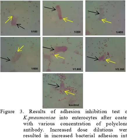
Based on the result, subunit pili protein 12.8 kDa were selected to produce polyclonal antibody in mice. The antibody obtained from immunization of mice was then used in adhesion inhibition test. This test used various concen-tration of antibody which were 1/100, 1/200, 1/400, 1/800, 1/1600, 1/3200 and 0 (control) (p<0.05) (Figure 3).
Figure 3. Results of adhesion inhibition test of
K.pneumoniae into enterocytes after coated with various concentration of polyclonal antibody. Increased dose dilutions were resulted in increased bacterial adhesion into enterocytes cells.
Based on the result showed in Figure 3, the amount of bacteria attached into enterocytes could be calculated. The adhesion index (AI) was calculated by counting the number of bacteria attached to 100 enterocytes (Table 1). Pearson correlation analysis was performed based on the observation of AI. The result is shown in Table 1.
Table 1. The correlation between various concentration of polyclonal antibody with Adhesion Index.
group N Mean ± SD p-value Pearson
correlation Titer 1/100 3 116 ± 14.42
0.032 -0.797 Titer 1/200 3 113.33 ±
20.01 Titer 1/400 3 135.67 ±
10.02 Titer 1/800 3 163.67 ±
8.50 Titer
1/1600
3 280 ± 18.68
Titer 1/3200
3 289 ± 30.55
Control 3 293.33 ±
27.43
JTLS | J. Trop. Life. Science 21 Volume 4 | Number 1 | January | 2014
The ability of polyclonal antibodies (immuno globulin G) against pili protein MW 12.8 kDa to inhibit bacterial adhesion was then tested by the adhesion inhibition test. The results indicate that the adhesion of K. pneumoniae bacteria was inhibited by polyclonal immuno-globulin G resulting from the pili protein immunization (Figure 3). As seen in Figure 3, the lowest dilution (1/10.000 titer) resulted in the smallest amount of bacteria that attached to the enterocytes cells of mice, then in the next dilution the number of bacteria attached were increasing. This is caused by the ability of the circulating antibodies to pili bacteria to bind actively to bacterial pili so the bacteria cannot attach to the enterocytes [27,28]. Based on the result of adhesion inhibition test using an antibody of subunit pili protein K. pneumoniae
MW 12.8 kDa, it can be concluded that the polyclonal antibody is an antibody that can fight against an adhesion molecule. Similar results are obtained in a study conducted by Agustina et al. proving that the polyclonal antibodies obtained from the injection of 49.8 kDa subunit pili of S. dysentriae in mice acts as an antibody against adhesion molecules [29].
Based on the result of Pearson correlation test, there was a significant correlation between antibody dilution titer with adhesion index (p= 0.032) with a correlation coefficient of -0.797 (Table.1). This means that the dose of antibodies affect adhesion index as much as 79.7 %. Decreased antibodies concentration would results in increased bacterial adhesion index. These results are similar to the results of the study conducted by Hidayati revealing that in the adhesion of
Pseudomonas aeruginosa pili, decreased antibodies concentration will increase the bacterial adhesion index significantly [31]. Similar results have also been obtained by other studies [9,27,30].
Of the three bacterial attachment patterns (local, aggregative, and diffuse), the pattern of bacterial attachment of K. pneumoniae in this study could be concluded to be more likely to the diffuse type. This can be seen by looking at the results of the adhesion test in Figure 3. It appears that the attachment of K. pneumoniae bacteria was evenly distributed on the enterocytes cells. This attachment is mediated by the hemagglutinin pili protein MW 12.8 kDa of K. pneumoniae. Previous research conducted by Di Martino et al. proves the role of nonfimbrial protein K. pneumoniae with MW 29 kDa as an intermediary diffuse type of bacterial adhesion to Caco-2 cells [31].
The second method used to ensure that the subunit pili protein MW 12.8 kDa of K.pneumoniae
is an adhesion molecule was immunocytochemistry
test. Adhesion molecules can attach to receptor molecules and the receptors are part of the cell [29]. In an immunocytochemistry test, antigen and antibody reactions can be evaluated from the color that appears. Brown color indicates that there is conformity antigen-antibody binding, while the blue color indicates a discrepancy between the two [9].
The concentration of antibody used in this test was 1/100, based on the strongest antigen-antibody reaction by Dot Blot method (data not shown). The effect of various concentration of subunit pili protein with MW 12.8 kDa of
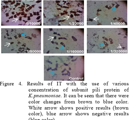
K.pneumoniae in immunocytochemistry test (IT) will produce color in enterocyte. The result showed the brown and blue color (Figure 4).
Figure 4. Results of IT with the use of various concentration of subunit pili protein of
K.pneumoniae.It can be seen that there were color changes from brown to blue color. White arrow shows positive results (brown color), blue arrow shows negative results (blue color).
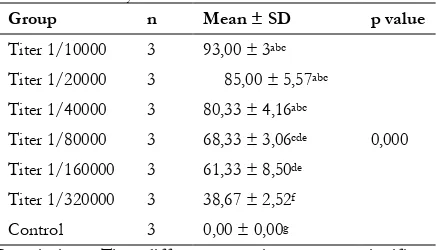
Brown color indicates a positive result, it means that there is a perfect bond of antigen and antibody, while the blue color indicates that the antigen-antibody bond is not perfect. Brown enterocytes cells were calculated to 100 entero-cytes and repeated for three times. Statistical analyses employing a One Way ANOVA and Tukey post hoc analysis were done in order to determine whether there are differences between the various dilution titers with antigen-antibody reaction. The results can be seen in Table 2. Results of One Way Anova analysis showed that there were significant differences between the various dilution titer with antigen-antibody reaction (p=0.000) (95% confidence level). The results of comparative analysis of each group through post hoc test (Tukey) can be seen in Table 2.
In Figure 4, it appeared that the lowest dilution of antigen (1/10000) showed a positive result (brown color) in almost all enterocytes cells, then by increasing dilution (decreased concentration) brown color that appeared decreased until blue colors fully appeared in control. Many antibodies
JTLS |J. Trop. Life. Science 20 Volume 4 | Number 1 |January | 2014
of hemagglutinin pili protein bound to the receptors appeared at the lowest dilution, so the brown color produced were also many. Appearance of color gradation changes from brown to blue along with the decreased of pili proteins that bind to these antibodies can be seen in Figure 4 (1/20000-1/320000 titer). These results demonstrate the hemagglutinin pili protein MW 12.8 kDa of K.pneumoniae really an adhesion protein that has the ability to bind enterocytes cells. These results are similar to previous studies that the pili pro-tein is an adhesion molecule via immuno-cytochemistry test [9,29]. Based on Table 2, there was a significant difference between the antigen-antibody reaction with various dilution titers of hemagglutinin pili protein MW 12.8 kDa of K.pneumoniae (p= 0.000 with a confidence
interval (α)=95%).
Table 2. The relationship between various dilution titers with antigen-antibody reaction using immunocyto chemistry test
Group n Mean ± SD p value
Titer 1/10000 3 93,00 ± 3abc
0,000 Titer 1/20000 3 85,00 ± 5,57abc
Titer 1/40000 3 80,33 ± 4,16abc
Titer 1/80000 3 68,33 ± 3,06cde
Titer 1/160000 3 61,33 ± 8,50de
Titer 1/320000 3 38,67 ± 2,52f
Control 3 0,00 ± 0,00g
Description: The different notation means significant difference (p<0.05) and when it contains the same notation it means no significant difference (p> 0.05).
This study was the first which characterized pili protein of K. pneumoniaetaken from bronchial aspirates of VAP cases. This identified pili protein can be used to develop a diagnostic kit and a vaccine against K. pneumoniae. It could help clinicians to treat their patients admitted to the Intensive Care Unit so it can reduce morbidity and mortality because of Ventilator Associated Pneumoniae caused by K. pneumoniae. Different specimen from the same bacteria could result in different pili protein to be identified, as proven by the research conducted by Sukarjati using hemagglutinin pili protein MW 32.23 kDa of
E.coli taken from semen specimen of infertile man, and another study by Sudana et al. using hemagglutinin pili protein MW 37 kDa of E. coli
taken from midstream urine of UTI patients [32,33]. Further researches are needed to examine hemagglutinin pili protein of K.pneumoniae taken from other specimen and other cases.
CONCLUSION
Based on the results and discussion, it can be concluded that antibodies of hemaglutinin pili protein with MW 12.8 kDa of K. pneumoniae can act as an adhesion molecule. Through adhesion inhibition and immunocytochemistry test, it has been found out that hemagglutinin pili protein with MW 12.8 kDa of K. pneumoniae antibody can inhibit the adhesion of K. pneumoniae to the enterocytes of mice. The need to do further re-search is to find the characters of molecular sub-units in pili adhesion protein of K. pneumoniae
found in other isolates and other cases apart from VAP.
ACKNOWLEDGMENTS
We send our deepest thank to Soeyati Poejiati, Ali Sabet, and Tarina Widaningrum for their good assistance in the study.
REFERENCES
1. Abbott SL (2007) Klebsiella, enterobacter, citrobacter, serratia, plesiomonas, and Other Enterobacteriaceae. In: Murray PR, Baron EJ, Jorgensen JH, Landry ML, Pfaller MA, eds. Manual of Clinical Microbiology 9th edition. ASM Press. Washington.
2. Podschun R, Ullmann U (1998) Klebsiella spp. as nosocomial pathogens: epidemiology, taxonomy, typing methods, and pathogenicity factors. Clinical Microbiology Reviews. 11: 589-603. 3. Jones RN (2010) Microbial etiologies of
hospital-acquired bacterial pneumonia and ventilator-associated bacterial pneumonia. Clinical Infectious Disease. 51: 81-87.
4. Meatherall BL, Gregson D, Ross T, Pitour JDD, Laupland KB (2009) Incidence, risk factors, and outcomes of Klebsiella pneumoniae bacteremia. The American journal of medicine. 122: 866-873. 5. Limbago BM, Rasheed JK, Anderson KF, Zhu
W, Kitchel B, Watz N, Munro S, Gans H, Banaei N, Kallen AJ (2011) IMP-producing carbapenem-resistant Klebsiella pneumoniae in the United States. Journal of Clinical Microbiology. 49: 4239-4245. 6. Brisse S, Fevre C, Passet V, Issenhuth-Jeanjean S,
Tournebize R, Diancourt L, Grimont P (2009) Virulent clones of Klebsiella pneumoniae: identification and evolutionary scenario based on genomic and phenotypic characterization. Plos One. 4: e4982.
7. Wilson BA, Salyers AA, Whitt DD, Winkler ME, eds (2010) Bacterial pathogenesis: a molecular approach. 3rd Edition. ASM Press. USA.
8. Sanarto S (2002) Protein adhesin Salmonella typhi sebagai faktor virulensi berpotensi imunogenik
JTLS | J. Trop. Life. Science 21 Volume 4 | Number 1 | January | 2014
terhadap produksi S-IgA protektif. PhD thesis. Airlangga University. Surabaya.
9. Sumarno, Yanuhar U, Winarsih S, Islam S, Santoso S (2012) Detection of molecule adhesion sub-unit pili 48 kDa Salmonella typhi by immunochemistry method using sera patients suffering from typhoid fever. J.Basic. Appl. Sci. Res. 2: 8527-8532.
10. Noorhamdani (2005) Protein fimbria 16 Kda bakteri Acinetobacter baumannii dari urin penderita infeksi saluran kemih berperan sebagai protein hemaglutinin dan adhesin. Jurnal Kedokteran Brawijaya. 21: 44-59.
11. Suswati E, Mufida DC (2010) Protein
haemaglutinin outer membran protein (OMP) 35 kDa sebagai protein adhesin Proteus mirabilis pada vesika urinaria kelinci. Jurnal Natur Indonesia. 12: 136-142.
12. Hidayati DYN (2010) Identifikasi molekul adhesi pili P.aeruginosa pada HUVECS culture. J. Exp. Life. Sci. 1: 7-14.
13. Sumarno, Susanto A, Ismanoe G, Wienarsih (2011) Combinations of protein sub-unit pili 37.8 KDA V. Cholerae with Cholera toxin sub-unit B V. Cholerae can protect come out of the solution in the intestinal mice. J. Pharm. Biomed. Sci. 1: 154-160.
14. Nelson DL, Cox MM, eds (2005) Lehninger; principles of biochemistry. 4th Edition. W.H. Freeman and Company. New York. 14.
15. Ling JM, Hui YW, French GL (1988) Evaluation of the Microbact-24E bacterial identification system. J Clin Pathol. 41: 910-914.
16. Sikarwar AS, Batra HV (2011) Identification of Klebsiella Pneumoniae by capsular polysaccharide polyclonal antibodies. International Journal of Chemical Engineering and Applications. 2: 130-134.
17. Ehara M, Ishibashi Y, Ichinose M, Iwanaga S, Shimotori TN (1970) Purification and partial characterization of pili of Vibrio cholerae O1. Vaccine. 5: 283-288.
18. Laemli UK (1970) Cleavage of structural protein during the assembly of the head of bacteriophage T4. Nature. 227: 680-685.
19. Li X, Johnson ED, Mobley HL (1999) Requirement of MrpH for mannosa resistent Proteus like fimbriae mediated hemaglutination by Proteus mirabilis. Infection and Immunity. 67: 2822-2833.
20. Goetsch L, Plotnicky-Gilquin AGH, Haeuw JF, Beck A, Bonnefoy JY, Corvaia N (2001) Targeting of nasal mucosa-associated antigen-presenting cells in vivo with an outer membrane protein a derived from Klebsiella pneumoniae. infection and immunity. 69: 6434-6444.
21. Digiandomenico A, Rao J, Harcher K, Zaidi TS, Gardner J, Neely AN, Pier GB, Goldberg, JB
(2007) Intranasal immunization with
heterologously expressed polysaccharide protects against multiple Pseudomonas aeruginosa infections. PNAS. 104: 4624-4629.
22. Horzempa J, Held TK, Cross AS, Fusrt D,
Qutyan M, Neely AN, Castric P (2008)
Immunization with a Pseudomonas aeruginosa 1244 pilin provides O-antigen-specific protection. Clinical and Vaccine Immunology. 15: 590–597.
23. Harlow E, Lane D (1988) Antibodies: a
laboratory manual. Cold Spring Harbor
Laboratory. New York. 386.
24. Nagayama KT, Oguchi M, Arita T, Honda (1995) Purification and characterization of a cell
associated haemagglutinin of Vibrio
parahaemolyticus. Infection and Immunity. 63: 1987–1992.
25. Key M (2009) Immunohistochemical Staining Methods. 5th Edition. Dako. California. 57. 26. Noorhamdani (2004) Aktivitas hemaglutinasi
bakteri Acinetobacter baumanni yang berasal dari specimen klinik dan lingkungan. Jurnal Kedokteran Brawijaya. 20: 105-109.
27. Mufida DC, Sumarno, Santoso S (2010) Identifikasi protein adhesi pili proteus mirabilis P355 dan protein reseptor pada vesika urinaria kelinci. J.Exp.Life Sci. 1: 1-6.
28. Sukrama IDM (2011) A novel of replacing
carcinoma colon-2 (CACO-2) cell with
enterocyte mice to determine bacteria adhesion activity in vitro. Journal of US-China Medical Science.8: 292-297.
29. Agustina W, Fitri LE, Raras TYM, Siswanto B, Sumarno (2012) Antibody protein hemagglutinin subunit pili with MW 49.8 kDa Shigella dysenteriae adhesion on mice enterocyte. IOSR Journal of Pharmacy. 2: 13-20.
30. Anam K (2012) Identifikasi protein Hemaglutinin sub unit pili 49.8 kDa dan anti hemaglutinin 7.9 kDa serta uji respon imun reaksi silang Shigella spp. Unpublished. Faculty of Medicine of Brawijaya University. Malang.
31. Di Martino P, Bertin Y, Girardeau JP, Livrelli V, Joly B, Darfeuille-Michaud A (1995) Molecular characterization and adhesive properties of CF29K, an adhesion of Klebsiella pneumoniae strains involved in nosocomial infections. Infection and Immunity. 63: 4336-4344.
32. Sukarjati, Soebadi DM, Hinting A (2013) Role of Escherichia coli pili adhesion molecule to inhibit Escherichia coliadhesion to human spermatozoa in vitro. Androl Gynecol: Curr Res. 1: 1-7.
33. Sudhana IW, Suwitra K, Sumarno (2009) Characteristics of pili hemaglutinin protein and it role in the pathogenesis of urinary tract infection with uropathogenic Escherichia coli. Indonesian Journal of Biomedical Science. 3: 1-7.