Empirical investigations into the tunica structuring point of the
shoot apex of
Pelargonium zonale
Jens Wegner *
Department of Plant Breeding,Faculty of Agriculture and Horticulture,Humboldt Uni6ersity of Berlin,Wendenschloßstr.254,
12557Berlin,Germany
Received 26 May 1999; received in revised form 24 August 1999; accepted 7 December 1999
Abstract
A tunica structuring point of 3.85 cell units was formally predicted for the shoot apex. This point is decisive for the genesis of the tunica corpus structure as such and for the number of tunica layers in an apex at any given moment. Here, a method to determine, in empirical investigations, the theoretical number of tunica layers in real apices is demonstrated. Furthermore, results of an empirical investigation into the postulated tunica structuring point are presented and causes for deviations of the observed values from the theoretical values are discussed. © 2000 Elsevier Science Ireland Ltd. All rights reserved.
Keywords:Apex; Number of tunica layers;Pelargonium zonale; Tunica corpus structure; Tunica structuring point
www.elsevier.com/locate/plantsci
radius of 3.85 cell units behaves like a tunica. Furthermore, a mathematical formula was de-duced to calculate the theoretical number of tunica layers in a shoot apex at any given moment [1]. Here, a method to determine in empirical investi-gations the theoretical number of tunica layers in real apices is demonstrated. Besides that, results of an empirical investigation into the postulated tu-nica structuring point are presented and causes for deviations of observed values from calculated val-ues are discussed.
2. Material and methods
For investigations Pelargonium zonale (L.) L’He´rit. ex Ait. ‘Kleiner Liebling’ was used. Pre-served slides of the shoot apices of these plants were made. To judge the layered structure, median longitudinal sections through the shoot apex are necessary which can be found in the series of microcuttings. The preparation process was car-ried out following Romeis [2] and modified by the following:
1. Introduction
Most of the higher plants are characterized by a multi-layered structure of their hemispherical shoot apex. This cell layer arrangement known as tunica corpus structure is based upon a specific way of cell division. While cells of the tunica divide in anticlinal direction (=at right angle to the surface) only, cells of the corpus divide anti-clinal as well as perianti-clinal (=parallel to the surface).
It could be shown that there is no primarily genetic fixation of this tunica corpus structure but apex-internal stress causes its development. This apex-internal stress is caused by a high accumula-tion of meristematic cells. For the shoot apex, a tunica structuring point of 3.85 cell units was predicted saying that every cell layer beyond the
* Present address: Laboratory of Plant Genetics, Faculty of Agri-culture, Tokyo University of Agriculture and Technology, 3-5-8 Saiwai-cho Fuchu-shi, Tokyo 183-8509, Japan. Tel./fax: + 81-42-3675625.
E-mail address:[email protected] (J. Wegner)
1. De-aerate and fixing: Carnoy’s solution (six parts 96% ethanol, three parts chloroform, one part acetic acid) for 2 h
2. Dehydration: 96% ethanol (1 day), propanol (1 day), dyeing the plasma in a 1:1 propanol:eosin mixture (2 h), xylol (2 days)
3. Embedding: 1:1 xylol:paraffin mixture (at 60°C in the incubator for 2 days), allow the xylol to evaporate out of the mixture (at 60°C for 3 days), embedding in paraffin
4. Slicing: 8mm thick at the microtome stretching
the sections with some drops of water (drying the preparations for 1 day)
5. Dyeing: xylol (10 min); propanol, two times 96% ethanol, 70% ethanol, water (always dip-ping shortly), dyeing in haemalum (1 min, rinse of excess dye with running water), dyeing in eosin (20 min, removing excess dye by short dipping in water)
6. Conservation: 70% ethanol, 96% ethanol, two times propanol, xylol (always dipping shortly), conservation in Canada balsam
3. Results
To decide the theoretical number of tunica lay-ers, it is necessary to determine the radius of the shoot apex. However, since apices are practically never absolutely equal to a complete hemisphere one cannot simply measure straight across the picture.
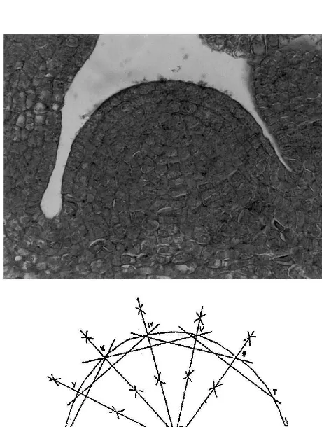
The deviation of the real apex section from a half circle results in more than one central point and more than one radius. Therefore, for every apex several ‘central points’ and ‘radiuses’ were determined (compare Fig. 1, below). First, the contours were drawn using photographs of apex longitudinal sections. Then, five strings were drawn in each apex outline. The strings were cut in the middle at right angles by a straight line (=the central perpendicular). In a half circle (or arc of a circle), all the central perpendiculars would cross at exactly one point (=the central point of the circle). In the investigated longitudinal sections, however, three to ten points of intersection were found. At the contours of the apex (that is analogous to the arc of the circle), seven points were set up. Five of them were the points of intersection of the central perpendicular and the apex contours. The other two points were the outermost intersections of the strings and the apex outline. By this procedure, the measuring points are equally distributed over the entire apex contours.
The Fig. 1 (above) shows the median longitudi-nal section through a shoot apex of P.zonale. The sectional view seems to be equivalent to a half circle. The geometrical construction to find the central point of the apex outline, however, reveals its deviations from the idealized model. In this, the distances of the ‘central points’ to each other can be regarded as a measurement of deviation from the model. The radiuses were determined by mea-suring the distances from each apex ‘central point’ to each measuring point of the apex outline.
To calculate the theoretical number of tunica layers the cell diameter must be measured. For calculations, the average cell diameter was deter-mined. Only entirely meristematic cells were
Table 1
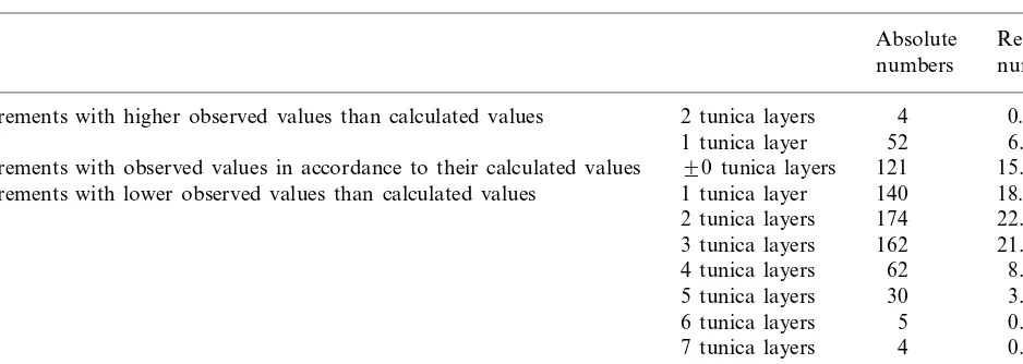
Quantitative distribution of the deviations of the theoretical tunica numbers from their observed values (764 pairs of values)
Absolute Relative numbers numbers
4
Measurements with higher observed values than calculated values 2 tunica layers 0.52
52 6.81
1 tunica layer
121
90 tunica layers 15.84 Measurements with observed values in accordance to their calculated values
Measurements with lower observed values than calculated values 1 tunica layer 140 18.32 174
2 tunica layers 22.77 162 21.20 3 tunica layers
62
4 tunica layers 8.12 5 tunica layers 30 3.93
5
6 tunica layers 0.65 7 tunica layers 4 0.52
4
8 tunica layers 0.52
cluded in these measurements; that means they had not started expanding yet. (The meristematic state of these cells was recognized through their very small size and through their very high plasma concentration resulting in a very intensive eosin dyeing during preparation.) The theoretical num-ber of tunica layers which exists when assuming the tunica structuring point of 3.85 cell units is calculated from the average cell diameter and the radius of the apex. The calculation of these theo-retical numbers followed the formula:
nT=
rA−3.85dCell
dCell
wherenT=number of tunica layers;rA=radius of the apex; dCell=cell diameter (measured in direc-tion of the radius).
A total of 764 calculated values were compared to their corresponding observed values.
In these investigations two to six tunica layers were observed and, by contrast, theoretical tunica numbers ranging from 2 to 12 were calculated. Table 1 shows the distribution of deviation of the theoretical values from the observed values. It is especially emphasized that the observed values refer to a specific radius and therefore incomplete tunica layers are included. Counting incomplete tunica layers is in contrast to Schmidt [3] and other authors who regarded only those layers as tunicas which were entirely free from periclinal divisions. It has been explained in a previous paper why their view must be objected [1].
The results show that generally fewer tunica layers were observed than theoretically predicted.
On average, 1.85 tunica layers were calculated more than observed.
4. Discussion
The calculations of the theoretical numbers of tunica layers are based on the assumption that the apex is equal to a hemisphere. Casting a quick glance over the preparations of P. zonaleone will find that the sections match a half circle quite well. Furthermore, in all apices a higher number of tunica layers can be found so that it is never just a yes/no decision when comparing real apices to the theoretical model. In that regard,P. zonaleis a quite suitable object for empirical investigations into the tunica structuring point.
The three to ten ‘central points’ found in the constructions for the apex central point reveal the deviations of real apices from their model. Greater divergences of the apex outline from a half circle resulted in points of intersection being in particu-lar far outside of the plot which therefore gave particularly small or large radiuses. Scattered points emerged in this way, however; they resulted more often in too high than in too low theoretical numbers of tunica layers.
arranged evenly enough — for the genesis of such very even cell rows, particularly high pressure from the corpus and strong tension inside the tunica, are necessary.
The major source of error in verifying the tunica structuring point is probably the beginning expan-sion of apical cells which can be seen in the longitudinal section by a lighter staining and larger diameter of the more central cells (Fig. 1, above). The differences in staining intensity reflect the reduced plasma density in growing cells. The presented photograph can be regarded as represen-tative of the sampling. For the calculations equal size and the same frequency of division of all apical cells were assumed. Even though these cells keep dividing it must be assumed that along with the beginning expansion and differentiation the direction of cell division follows a genetically given purpose.
The calculation of the theoretical number of tunica layers is affected by the cell size. For the determination of the average cell size only entirely meristematic (meaning the smallest) cells were con-sidered. Since tunica cells come under increasingly strong tangential tension along with their increas-ingly outward position they will appear thinner and so the average cell size is distorted down-wards. A too small cell size results in calculating too high a number of theoretical tunica numbers. By contrast, the too large cell diameters of ex-panding cells distort the theoretical tunica num-bers below the observed numnum-bers.
When calculating the theoretical numbers of tunica layers the three dimension of the apex are considered. When analyzing longitudinal sections, however, only two dimensions are looked at mean-ing that no volumes but areas are investigated. Outer factors which might have an influence on the apex shape, e.g. lateral pressure from attached leaves, cannot be seen in a longitudinal section if these factors lay before or behind the sectional view.
At this point, some investigations by Thielke are worth quoting. She examined apices of different species ofSaccharum[4 – 9] andErianthus[10]. Her investigations lead her to the conclusion that the development of a tunica is clone specific. For unlayered apices she assumed an abnormal genetic constitution [4]. In her investigations, she recog-nized that unlayered apices consist of only few but large cells. In subsequent investigations she
re-ported variations in the number of tunica layers at different developmental stages and she saw the connection of apex size and the development of a tunica layer [5,7 – 10]. However, in her reports she never established a connection between cell size and the apex radius. Apical cells were just de-scribed as ‘to be of large size’. Even though Thielke does not give any suitable measured values to recognize that the ratio of apex to apical cells is the cause for the development of a layered struc-ture in the shoot tip, her observations can be very well regarded as a lump verification of the exis-tence of the tunica structuring point as postulated earlier [1].
With this paper a method to determine the number of tunica layers in real apices is demon-strated with the purpose of clarifying the develop-mental nature of the tunica corpus structure of the shoot apex. Furthermore, results of an empirical verification of the tunica structuring point are presented and causes for deviations of observed values from theoretical values are discussed.
Acknowledgements
I thank F. Pohlheim of the Humboldt Univer-sity of Berlin and K. Klopfer of the UniverUniver-sity of Potsdam for discussions. I received financial sup-port from the state of Berlin and the FAZIT-Stiftung Frankfurt.
References
[1] J. Wegner, A theoretical approach to the genesis of cell layer arrangements in undifferentiated tissues, Plant Sci. 153 (2000) 177 – 183.
[2] B. Romeis, in: P. Bo¨ck (Ed.), Mikroskopische Technik, 17th ed., Urban and Schwarzenberg, Mu¨nchen, 1989. [3] A. Schmidt, Histologische Studien an phanerogamen
Vegetationspunkten, Bot. Arch. 8 (1924) 345 – 404. [4] Ch. Thielke, Der Sproßscheitel der GattungSaccharum,
Naturwissenschaften 46 (1959) 478 – 479.
[5] Ch. Thielke, Histologische Untersuchungen am Sproßscheitel von Saccharum spontaneum L. 1. Der Sproßscheitel vonSaccharum spontaneumL., Ber. Dtsch. Bot. Ges. 73 (1960) 147 – 154.
[6] Ch. Thielke, U8ber Vegetationskegelstrukturen in der Gattung Saccharum, Ber. Dtsch. Bot. Ges. 74 (1961) 67 – 68.
[8] Ch. Thielke, Histologische Untersuchungen am Sproßscheitel vonSaccharum. III. Der Sproßscheitel von Saccharum robustum Brandes und Jeswiet., Ber. Dtsch. Bot. Ges. 76 (1963) 265 – 275.
[9] Ch. Thielke, Histologische Untersuchungen am
Sproßscheitel vonSaccharum. Der Sproßscheitel von Sac-charum officinarumL., Planta 62 (1964) 332 – 349. [10] Ch. Thielke, Der Strukturwandel am vegetativen
Sproßscheitel von Erianthus Michx., Planta 59 (1963) 587 – 599.