Research Report
Radiological anatomy analysis of uncinate process, concha bullosa,
and deviated septum in chronic rhinosinusitis
Retno Sulistyo Wardani*, Arroyan Wardhana**, Endang Mangunkusumo*, Vally Wulani***, Brent Anthony Senior****
*Rhinology Division, Otorhinolaryngology Department, Faculty of Medicine, Universitas Indonesia – Dr. Cipto Mangunkusumo Hospital, Jakarta, Indonesia
**Ear Nose Throat Clinic, Koja Hospital, Jakarta, Indonesia
***Radiodiagnostic Department, Faculty of Medicine, Universitas Indonesia – Dr. Cipto Mangunkusumo Hospital, Jakarta, Indonesia.
****Otolaryngology/Head and Neck Surgery, University of North Carolina, USA, Adjunct Professor of Otorhinolaryngology, Universitas Indonesia – Jakarta, Indonesia
ABSTRACT
Background: Risk factors of chronic rhinosinusitis include epithelial disease related to disturbance of mucociliary transport system, anatomical variations obstructing the ostiomeatal complex and dysfunction of neural regulation. Purpose: To find the prevalence of deviated septum, concha bullosa and lateral deflection of the uncinate process in ipsilateral chronic rhinosinusitis. Methods: A retrospective study of computed tomographic (CT) scans of chronic rhinosinusitis patients who were not cured by 6 weeks of maximal medical treatment of rhinosinusitis. Pneumatization of the middle turbinate and deviation of the septum were evaluated on coronal slices, while lateral deflection of the uncinate process was also assessed by calculating the angle using Osirix software (Pixmeo, Geneva, Switzerland). Results: One hundred ninety-three CT scans comprising 386 nasal sides were analyzed. The prevalence of the combination of deviated septum, concha bullosa and lateral deflection of the uncinate process occurring concurrently with ipsilateral maxillary sinusitis was found to be 32,9% (p=0.01, OR=9.1, 95% CI=1.2-69.7). The prevalence of deviated septum with concha bullosa was found in 40.9% with ipsilateral maxillary sinusitis (p=0.03, OR=3.8, 95% CI=1.1-13.3). One single anatomical variation of lateral deflection of the uncinate process had a proportion of 71.5% in ipsilateral maxillary sinusitis (p=0.03, OR=2.7 95% CI=1.1-6.9). Conclusion: Deviated septum, concha bullosa and lateral deflection of the uncinate process were frequently present in the existence of ipsilateral chronic maxillary rhinosinusitis. It suggested greater association compared to one or a combination of two anatomical variation in the role of local factors contributing to sinonasal epithelial dysfunction.
Keywords: chronic rhinosinusitis, deviated septum, concha bullosa, lateral deflection of uncinate process
ABSTRAK
Latar belakang: Rinosinusitis kronis adalah penyakit inflamasi yang mengakibatkan gangguan
keseimbangan homeostasis fungsi hidung dan sinus paranasal. Patofisiologinya terjadi melalui kombinasi gangguan transpor sistim mukosiliar, variasi anatomi yang menyempitkan kompleks ostiomeatal serta gangguan regulasi persarafan sensorik, simpatis dan parasimpatis. Tujuan: Untuk mengidentifikasi
prevalensi septum deviasi, konka bulosa dan prosesus unsinatus defleksi lateral, serta hubungannya dengan rinosinusitis kronik yang terjadi pada sisi yang sama (ipsilateral). Metode: Penelitian retrospektif potong
lintang pada tomografi komputer (TK) pasien dengan rinosinusitis kronik yang tidak sembuh dengan terapi medikamentosa maksimal selama 6 minggu. Pneumatisasi konka media dan deviasi septum dievaluasi pada potongan koronal, sedangkan defleksi prosesus unsinatus ke lateral dinilai dan diukur sudutnya dengan menggunakan piranti lunak Osirix (Pixmeo, Geneva, Switzerland). Hasil: Analisis dilakukan
32,9% (p=0,01, OR=9,1, 95% CI=1,2-69,7). Prevalensi septum deviasi dengan konka bulosa ditemukan pada 40.9% sinusitis maksila ipsilateral (p=0,03, OR=3,8, 95% CI=1,1-13,3). Variasi anatomi tunggal yaitu prosesus unsinatus defleksi lateral memiliki proporsi 71,5% pada sinusitis maksila ipsilateral. (p=0,03, OR=2,7 95% CI=1,1-6,9). Kesimpulan: Septum deviasi, konka bulosa dan prosesus unsinatus
defleksi lateral sering ditemukan bersamaan dengan kejadian sinusitis maksila ipsilateral. Kombinasi tiga variasi anatomi yang menyempitkan kompleks ostiomeatal (KOM) lebih kuat daripada hanya satu variasi anatomi atau kombinasi 2 variasi anatomi, sebagai faktor lokal yang berkontribusi terhadap disfungsi epitel sinonasal serta gagalnya resolusi inflamasi kronik.
Kata kunci: Rinosinusitis kronis, septum deviasi, konka bulosa, prosesus unsinatus defleksi lateral
Corresponding author: Retno S. Wardani, MD, PhD. Rhinology Consultant, Otorhinolaryngology Department Head and Neck Surgery Department, Faculty of Medicine Universitas Indonesia, Dr. Cipto Mangunkusumo Hospital. Jl. Diponegoro 71, Jakarta 10430. Indonesia.
Email: retno.wardani@ui.ac.id
INTRODUCTION
Chronic rhinosinusitis (CRS) is a disease impairing quality of life that results in emotional dysfunction equal to diseases such as diabetes mellitus and heart failure. Rhinosinusitis is also highly prevalent, accounting for 2 out of 100 outpatient clinic visits, while also ranking 10th in disease occurrence in the 85% economically productive-age group.1,2
The definition of chronic rhinosinusitis is inflammation of the nose and paranasal sinuses lasting more than 12 weeks. Anatomical variation obstructing ostiomeatal complex (OMC), dysfunction of mucociliary transport system, bacterial or viral infection, allergic rhinitis and laryngopharyngeal reflux are all postulated as risk factors for dysfunction of sinonasal epithel leading to unresolved inflammation process as well as acquired and innate immune system dysfunctions. Previous studies of anatomical variation in the nose and sinuses have confirmed their presence in about 1/3 of those without rhinosinusitis symptoms, in contrast to other studies that have found their prevalence in the presence of rhinosinusitis to be 64.9 – 86%.1,2
The specific anatomical variations of deviated septum, concha bullosa and lateral deflection of uncinate process have all been
populations without rhinosinusitis, concha bullosae are found in 13.2-50% cases, while the percentage increases to 33.8-72.6% in population reporting rhinosinusitis symptoms. Deviated septum more than 3 mm from midline is also reported to have a correlation to rhinosinusitis.1-5
Stalman et al6 performed a retrospective study evaluating clinical symptoms of chronic rhinosinusitis and its relationship to ipsilateral concha bullosa and contralateral septal deviation in 1095 CT-scans. There were no statistical relationship between either unilateral concha bullosa or deviated septum with any sinus disease (on either side). There was, however, a strong relationship between the presence of a unilateral concha bullosa and its contralateral deviated septum. It was hypothetized that septal deviation is not due to direct result of pressure effect from concha bullosa because the air channel between concha bullosa and nasal septum was preserved.
presence of maxillary sinusitis, anterior ethmoid sinusitis, and frontal sinusitis; but the association was not significant between presence of a concha bullosa and anterior ethmoid sinusitis and no relationship was seen between severity and location of deviated septum with the incidence of maxillary, anterior ethmoid and frontal sinusitis.7
Variations of anatomical structure related to OMC obstruction may be identified through CT analysis and should be performed before any FESS intervention. CT plays a central role in the modern management of CRS due to its ability to delineate mucosal disease, to demonstrate a primary obstructive pathology and to image distal structures such as the posterior ethmoid sinus that cannot be viewed with direct nasoendoscopy.1-2
Additionally, CT may help to identify some uncommon anatomic variations that may increase the risk of surgical complications especially for a novice surgeon. Pre-operative CT analysis is an important tool to avoid high risk areas, providing good resolution differentiating soft tissue and bone structure. It is time efficient to perform with relatively low radiologic exposure.
At Dr. Cipto Mangunkusumo Hospital, the guideline for CT examination for chronic rhinosinusitis without any symptom or sign of orbital or intracranial complication is at least after 6 weeks of maximal medical treatment for the comorbidities or risk factors identified.
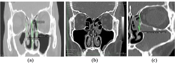
The open source software OsiriX (Pixmeo, Geneva, Switzerland) has been widely used to support CT analysis, providing additional understanding of paranasal sinus radiological anatomy. In this study, OsiriX has been used to measure the angle of septal deviation and the lateral deflection of the uncinate process in a much more rapid and accurate approach.
METHOD
A cross sectional retrospective study was performed to analyze the association between deviated septum, concha bullosa and lateral deflection of uncinate process separately and in combination with chronic rhinosinusitis. All archived CT scans of chronic rhinosinusitis cases from 2008 – 2011 in Radiodiagnostic Department and Rhinology Division of Otorhinolaryngology - HNS Department of Dr. Cipto Mangunkusumo Hospital were analyzed. Inclusion criteria for chronic rhinosinusitis samples included age more than 12 years with the coronal CT imaging of suitable quality, ie, with a minimum 3-5 mm slice thickness. Radiological description of any rhinosinusitis appearance was based on the existence of mucosal thickening, air fluid level, partial or total opacification. The exclusion criteria were unavailability of digital data, CT images with unidentified lateral nasal wall due to obscuring opacification, destruction of bone or cases with evidence of previous surgery. Each side of the CT scans was evaluated for deviated septum, concha bullosa and uncinate process deflection.
Evaluation and analysis of the CT scans was performed and the agreement of the evaluators was considered definitive. Chi square and bivariate analysis were then performed for anatomical variation risk factors for chronic rhinosinusitis, with significance considered with a P value less than 0.05 and 95% confidence interval more than one.
RESULTS
The research was carried out starting from October 2011 until August 2012 in Rhinology Division, Otorhinolaryngology Head Neck Surgery Department and the Radiology Department, Dr. Cipto Mangunkusumo Hospital. CT scans in the form of digital data in 212 compact discs (CD) were obtained from the Radiology Department. Out of 212 CDs, 193 CDs met the inclusion criteria, while 10% were excluded. CT analysis was performed using the OsiriX (Open-Source Software for Navigating in Multidimensional DICOM Images) programme.
The 193 CT scans consisted of 100 male subjects (51,8%) and 93 female subjects (48,2%). Youngest age of the subjects was 12 years old and the oldest was 79 years old with a median of 36 years old. For statistical analysis, calculation was made utilizing 193 subjects samples and 386 sides. Findings of rhinosinusitis including either mucosal thickening, or opacification, or air-fluid levels
were mostly found in the anterior ethmoid sinuses (378/386=97.9%), followed by the maxillary sinuses (337/386=87.3%) and the frontal sinuses (337/386=86.3%).
Unilateral, c-shaped deviated septums were found in 75.1% subjects (145/193) while bilateral, s-shaped or z-shaped deviated septums were found in 47 of 193 subjects (24.4%). No septal deviation was only found in 1 subject (0.05%).
A lamellar type of concha bullosa was most commonly found in 36.8% (142/386) sides, followed by extensive and bulbous type of 17.4% (67/386) sides and 6.9% (27/386) sides while a middle turbinate without pneumatisation was found in 38.9% (150/386) of sides.
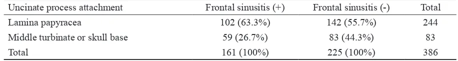
Lateral deflection of the uncinate process (UP) of less than 40o was more commonly found than the medial deflection of more than 400. The lateral deflection was found in 70.7% (243/386) sides, while medial deflection was found in 29.3% (113/386) of sides. Attachment of UP to lamina papyracea, skullbase and middle turbinate was found in 244/386 (63.2%), 102/386 (26.4%) and 40/386 (10.4%) of sides.
Cross-tabulation correlations were made to analyze single anatomical variation, combinations of two anatomical variations and all three variations toward ipsilateral maxillary, anterior ethmoid and frontal
(a) (b) (c)
1 and 2. Table 3 shows analysis of association between superior attachment of UP to lamina papyracea with ipsilateral frontal sinusitis.
Bivariate statistical analysis was used to evaluate the association of anatomical variations and the existence of ipsilateral rhinosinusitis. The data found significant consistency of anatomical variations towards ipsilateral maxillary sinusitis that would be specifically assessed and presented. Lateral deflection of UP occurring towards ipsilateral maxillary sinusitis was found in 71.5% sides (OR=2,7; 95% CI 1.1 – 6.9; P=0.03). Combination of concha bullosa and deviated septum towards ipsilateral maxillary sinusitis was found in 40.9% sides (OR=3.8; 95% CI 1.1 – 13.3; P=0.02). Occurence of lateral deflection of UP, deviated septum and concha bullosa towards maxillary sinusitis
ipsilaterally was found in 32.9% sides (OR=9.1; 95% CI 1.2 – 69.7; P= 0.01).
DISCUSSION
Certain anatomic variations of the lateral nasal wall are believed to be important risk factors because they can contribute to the blockage of the ostiomeatal complex, impacting drainage and ventilation, and thereby increasing the risk of sinus mucosal disease. However, the relative importance of specific anatomical variations is still a matter of discussion and previous reports have been variable and contradictory.1-2,9 For our mutual understanding, blockage of ostiomeatal complex due to certain anatomical variations should be viewed as one local factor impacting the same side Table 1. Single anatomical variations towards ipsilateral sinusitis extension
Single anatomical variations Maxillary sinusitis (n= 337)
Anterior ethmoid sinusitis (n = 378 units)
Frontal sinusitis (n = 333)
Deviated septum 215 (63.7%) 235 (62.1%) 209 (62.5%)
Concha bullosa 210 (62.3%) 234 (61.9%) 204 (60.0%)
Lateral deflection of UP 241 (71.5%)1 266 (70.3%) 235 (69.7%) 1OR=2,7; 95% CI 1.1 – 6.9; P= 0.03
Table 2. Two and three anatomical variations towards ipsilateral sinusitis extension
Anatomical variations Maxillary sinusitis (n= 337)
Anterior ethmoid sinusitis (n = 378)
Frontal sinusitis (n=333)
Deviated septum, concha bullosa 138 (40.9%)2 147 (38.9%) 128 (38.4%)
Deviated septum, lateral deflection of UP 160 (47.5%) 173 (45.8%) 156 (46.8%) Concha bullosa,
lateral deflection of UP 161 (47.5%) 175 (46.3%) 154 (46.2%)
All three 111 (32.9%)3 117 (30.9%) 105 (31.5%)
2OR=3.8; 95% CI 1.1 – 13.3; P=0.02 3OR=9.1; 95% CI 1.2 – 69.7; P= 0.01
Table 3. Association of uncinate process superior attachment with ipsilateral frontal sinusitis
Uncinate process attachment Frontal sinusitis (+) Frontal sinusitis (-) Total
Lamina papyracea 102 (63.3%) 142 (55.7%) 244
Middle turbinate or skull base 59 (26.7%) 83 (44.3%) 83
Total 161 (100%) 225 (100%) 386
of affected maxillary, anterior ethmoid and frontal sinusitis. This is not the one and only risk factor in complex orchestrated inflammatory process leading to sinonasal epithelial dysfunction and its homeostatic imbalance. Chronic rhinosinusitis is one example of disease as a result of homeostatic imbalance.
This study reviewed rhinosinusitis subjects resistant to appropriate medical treatment, with CT scan evidence of sinus inflammation. The most common findings were evidence of anterior ethmoid sinuses inflammation (97.9%), which were in accordance with Dua10 who reported 88% subjects with anterior ethmoid sinuses being the predominant affected sinuses. However when anatomical variations were analyzed concurrently with the sinusitis disease location (table 1 and 2), the ipsilateral maxillary sinus was the most commonly affected sinus with any single and combined anatomical variations of deviated septum, concha bullosa and lateral deflection of UP.
Interestingly, only 1 subject (0.05%) of rhinosinusitis had a straight septum, as the other 192 subjects (99.95%) had deviated septums either of unilateral c-shape, or bilateral of s-shape or z-shaped. Gadda9 observed 82 (58.5%) deviated septum in 140 CT scans of rhinosinusitis subjects who were failed to medical therapy. In general population, deviated nasal septum is present in 20-31%, and severe deviation has been noted as a contributing factor for sinusitis. In this study a deviated septum as a single risk factor of anatomical variation of CRS could not be proven, as it needed other coinciding factors to be related to sinusitis. Previously Gadda9 found a statistically significant correlation between unilateral deviated septum and ipsilateral maxillary sinusitis (P<0.01). One postulate for this pathologic condition was due to an increase of air turbulence in the nasal cavity causing physical wall stress.
shedding may occur and the damage to the mucosa may lead to inflammation in the OMC and subsequent ipsilateral maxillary sinusitis.11 Alternatively, a compensatory inferior turbinate hypertrophy opposite the side of septal deviation may play a role with deviated septum towards contralateral maxillary sinusitis due to narrowing of the nasal valve, resulting in an increase in the airflow turbulence, and later causing epithelial shedding.9,11,12
Harar et al13 postulated that a deviated septum caused an increase in the airflow around ostiomeatal complex (OMC) causing disturbance of mucociliary clearance, while the concha bullosa narrowed the semilunar hiatus, obstructing the infundibular drainage thereby resulting in ipsilateral maxillary sinusitis. In a group of 76 subjects with sinusitis and headache predominant symptoms, Hatipoglu et al14 found a concurrent deviated septum and concha bullosa in 44% of the cases. The overall incidence of concha bullosa was 31.52%, and was increased in patients with deviated septum (45.34%) while decreased in patients without deviated septum (18.95%). Stallman6 also reported concurrent deviated septum and concha bullosa in 79% of 998 CT scans but could not demonstrate a significant relationship to sinusitis. Sinusitis is believed to have a relationship with concha bullosa by causing a negative pressure in the paranasal sinus ventilation, disrupting the mucociliary clearance and hence triggering recurrent sinusitis.15 To date, our study is the first study of CT scans with prior clinically defined CRS showing an association of deviated septum with concha bullosa as independent variables in the development of ipsilateral maxillary sinusitis.
lateral deflection of the UP was observed in 21.4% of cases, and pneumatized UP in 2.8% of patients. In our study of patients with CT evidence of sinus inflammation, the median value of lateral UP deflection was 38.40 and to the medial was 41.80. The prevalence of lateral deflection of UP in ipsilateral maxillary sinusitis was 71.5%, and it was statistically significant with P=0.03, OR=2.7 95% CI=1.1-6.9. A previous study conducted in our institution by Riyadi7 reported that lateral deflection of UP yielded a two-fold increase in the incidence of maxillary sinusitis compared to medial deflection of UP. It is postulated that lateral deflection of UP disrupted the drainage of maxillary sinus ostium due to narrowing of infundibulum ethmoid, causing ipsilateral maxillary sinusitis.
In another study, Sousa et al16 in a retrospective study of 312 patient CT scans found 18 cases of ”silent sinus syndrome” involving the maxillary sinus, with anatomical variations found in 77.8% (n=14) and lateral deflection of the UP in 61.1% (n=11). The predominant lateral deflection of the UP in our Indonesian population study may indicate a racial predilection. Further study is also needed to explore this, since Gadda6 found statistically significant medial deflection of the UP associated with ipsilateral ethmoid sinusitis, while contrastingly in our study, lateral deflection of the UP poses an increased association of 2.7 times with ipsilateral maxillary sinusitis compared to medial deflection of the UP.
Han17 reported the prevalence of a terminal recess in 89.1% of Chinese subjects without frontal sinus disease symptoms. This study found that proportion of the predominant attachment of UP to lamina papyracea (102 sides) forming terminal recess associated with ipsilateral frontal sinusitis in 63.3% (102/161) cases. Terminal recess may play a role in the pathogenesis of frontal sinusitis due to lack of an anatomical barrier against ascending irritants, allergen,
and/or rhinogenic infection.1-2,18 Turgut18 found superior attachment of UP to lamina papyracea were 226 (63%) out of the 361 sides. The prevalence of frontal sinusitis was 41% in those with the frontal sinus outflow tract medially to the superior attachment of the UP having drainage to the middle meatus, and 23% in those with frontal sinus outflow tract laterally to the superior attachment of the UP and drained into the ethmoid infundibulum. A group with UP superior attachment to lamina papyracea had a statistically significant higher rate of frontal sinusitis. (χ2 = 12.11; P<0.001. Several other factors have been discussed regarding the pathophysiologic process of chronic frontal sinusitis. Kuhn classified a number of cells that can lead to obstruction of the frontal recess and cause frontal sinusitis. In addition to anatomical obstruction, mucosal obstruction of the frontal recess plays an important role in chronic frontal sinusitis. There are also different factors such as hypoxia, dehydration, infection, foreign bodies, environmental irritants, trauma, tumor, and allergens that can affect thefrontal sinus physiologic functions by disrupting the mucociliary clearance.1,2,8,18
In this research, the combination of deviated septum, concha bullosa and lateral deflection of UP was more highly related to ipsilateral maxillary sinusitis, compared with a combination of two anatomical variations or a single anatomical variation. Variations of anatomical structures obstructing ostiomeatal complex designating the role of local factor that may contribute to sinonasal epithelial dysfunction.
REFERENCE
1. Chan Y, Kuhn FA. An update on the classifications, diagnosis and treatment of rhinosinusitis. Curr opin Otolaryngol Head and Neck Surg. 2009;17:204-8.
2. F o k k e n s W J , L u n d V J , M u l l o l J . Epidemiology and predisposing factors. In: Fokkens WJ, Lund VJ, Mullol J, editors. European Position Paper on Rhinosinusitis and Nasal Polyps; 2012. p. 10-8.
3. Yasan H, Dogru H, Baykal B, Doner F, Tuz M. What is the relationship between chronic sinus disease and isolated nasal septal deviation? Otolaryngol Head and Neck Surg 2005;133:190-3.
4. Orlandi RR. A systematic analysis of septal deviation associated with rhinosinusitis. Laryngoscope 2010;120:1687-95.
5. Li L, Han D, Zhang L, Li Y, Zang H, Wang T et al. Aerodynamic investigation of the correlation between nasal septal deviation and cronic rinosinusitis. Laryngoscope;2012:1-5.
6. Stallman JS, Lobo JN, Som PM. The Incidence of concha bullosa and its relationship to nasal septal deviation and paranasal sinus disease. AJNR 2004; 25:1613-8.
7. Riyadi V. Anatomical Variations Obstructing O s t i o M e a t a l C o m p l e x i n C h r o n i c Rhinosinusitis. Otorhinolarygology Head Neck Surgery Residency Thesis. Faculty of Medicine Universitas Indonesia, 2009.
8. Bolger WE. Anatomy of the paranasal sinuses. In: Kennedy DW, Bolger WE, Zeinreich SJ, editors. Diseases of the sinuses: diagnosis and management. Canada : BC Decker. 2001. p. 1-11.
9. Gadda GL, Rosso S, Aversa S, Petrelli A, Ondolo C, Succo G. Parametric statistical correlations between paranasal sinus anatomic variations and chronic rhinosinusitis. Acta Otolaryngol Itali 2012;32:244-51.
10. Dua K, Chopra H, Khurana AS, Munjal M. Ct scan variations in chronic sinusitis. Ind J Radiol Imag 2005;15:3;315-20.
11. Caughey RJ, Jameson MJ, Gross CW, Han JK. Anatomic risk factors for sinus disease: fact or fiction. Am J Rhino 2005;19:334-9.
12. Berger G, Bernheim J, Ophir D. Epithelial shedding of the inferior turbinate in perennial allergic and nonallergic rhinitis. Arch Otolaryngol Head and neck Surg 2007; 133:78-82.
13. Harar RPS, Chadha NK, Rogers G. The role of septal deviation in adult chronic rhinosinusitis: a study of 500 patients. Rhinology 2004;42:126-130.
14. Hatipoglu HG, Cetin MA, Yuksel E. Concha bullosa types: their relationship with sinusitis, ostiomeatal and frontal recess disease. Diagn Intervent Radiol 2005;11:145-9.
15. Kayalioglu G, Oyar O, Govsa F. Nasal cavity and paranasal sinus bony variations: a computed tomographic study. Rhinology 2000; 38:108-113.
17. Han D, Zhang L, Ge W, Tao J, Xian J, Zhou B. Multiplanar computed tomographic analysis of the frontal recess region in Chinese subjccts without frontal sinus disease symptoms. ORL J Otorhinolaryngol Relat Spec.2008;70(2)104-12.
18. Turgut S, Ercan I, Sayin I, Basak M. The relationship between frontal sinusitis and localization of the frontal sinus outflow tract: a computer-assisted anatomical and clinica study. Arch Otolaryngol Head Neck Surg. 2005;131(6):518-22.