LUNG FIELD SEGMENTATION ON COMPUTED TOMOGRAPHY discussed by the researchers. Automatic lung nodule detection generally consists of two steps, namely lung nodule candidate detection and classification to determine nodule. Detection of nodule candidates begins with segmenting lung field. Current method is widely used for lung field segmentation of CT scan image is Thresholding and Active Contour, which relies on contrast of gray values between lung parenchyma and surrounding tissues. Drawback of both methods is that if nodule is large and located on borders of lung, then nodules will not be included in the image of lung (nodules contained will be lost). This means that segmentation of lung field was considered a failure, because image of nodule is object of attention will be lost.
Purpose of this research is lung field segmentation that contain abnormalities in CT scan image using registration methods. Registration is used namely elastic registration (non-rigid registration). We also perform lung fields segmentation using thresholding and Active Contour, as a comparison with method we proposed. Results of our study show that segmentation with registration approach has accuracy 95.5%, sensitivity 91.5%, and specificity of 96.6%. Segmentation with threshold has accuracy 92.2%, sensitivity 91.3%, and specificity 92.4%. While segmentation with Active Contour has accuracy 92.4%, sensitivity 87.5%, and specificity of 93.7%.
Keywords: active contour, non-rigid registration, lung field, thresholding.
INTRODUCTION
Lung nodules are one type of abnormality in lung organ can also be possibility of lung cancer. While lung cancer is all of malignancies disease in lung. Malignancy can be derived from lung itself (primary) or spread (metastasis) of tumors from other organs (Joseph, 2005). Computed Tomography (CT) is considered the most accurate modality available for early detection and diagnosis of pulmonary nodules and cancer.
and morphological processes. The next study is Dolejs, lung segmentation using thresholding and binary operations.
Drawbacks of both methods is that if nodule is large and located on the edge of boundary lungs, causing lung borders are not clear, so if done segmentation, nodule will not be included in lung image (part of lung that contained nodule will be lost). This means that segmentation of lung field was considered a failure, because nodules image that are focus of research will be lost. Purpose of our research is segmentation of lung field that contains abnormalities in CT scan image using registration. Registration method is used for lung segmentation is non-rigid registration methods. Non-rigid registration method consists of two activities, namely drawing lung image of defects (boundary edges damaged by large nodules), and the registration between the image that has been painted with lung image that has boundary edges abnormal.
Results of registration process is lungs image that have clear borders. Next process is the segmentation by mathematical morphology. To test accuracy of segmentation method with registration, then results will be compared with manual segmentation by groundtruth.
METHOD
In this study we have been implementing three segmentation methods, segmentation methods based on thresholding, active contour method, and elastic registration (non-rigid registration). The third method can be described as follows:
Segmentation with Elastic Registration
Steps of lung segmentation with an elastic registration can be shown in figure 1 below.
Figure 1 Steps of Lung Segmentation System Design
Registration
Calculation Number of Pixels Detected Pre-processing
Lung Segmentation
Comparing with Groundthruth
Pre-processing
Steps in pre-processing include: 1. Setting of Intensity Value
Intensity Adjustment works by performing a linear mapping of intensity values in histogram of beginning intensity values of a new histogram.
Figure 2 Intensity Adjustment Process Results
2. Histogram Equalization
Histogram equalization is a non-linear process that aims to give a light image corresponding to human visual analysis. Histogram equalization aims to change image, so resulting image has a histogram that is more evenly, which can make all level brightness a histogram can be used. Histogram equalization is a method to improve image quality by changing distribution of gray level images (Gonzales, 1987). It is intended that a more equitable distribution of gray levels compared to original image (Tinku, 2005).
Figure 3 Results of Histogram Equalization Registration
Registration method used is non-rigid registration with Sum of Squared Differences (SSD) method. Sum of Squared Differences (SSD) is one of simplest similarity matrix. Square of difference in image intensity and intensity remains respective moving images accumulated over all voxels of image remains. Image registration are assumed identical, except for Gaussian noise. SSD can be shown by the following formula:
(𝐼1 𝑖,𝑗 −
𝑖,𝑗 ∈𝑊 𝐼2(𝑥+𝑖,𝑦+𝑗))2 (1)
1. Input to registration process is a reference image (CT image of lung abnormalities present), and image registration is a CT image containing defects and edge boundaries are drawn manually by user using matlab command imfreehand. Registration and a reference image can be seen in Figure 3 below.
(a) (b)
Figure 4 Original CT image is used as reference image (a), Image Registration (Image has been Drawn Manual by User) (b)
2. Perform non-rigid registration process from input image above, which will produce a CT image of diseased who have clear borders.
(a) (b) (c) (d) Figure 4. Non-Rigid Registration Process
Reference Image (a), Registration Image (b), Registration Process (c), Image of Non Rigid Registration Result Using SSD (d)
Lung Segmentation
Steps in segmentation of lung field can be explained as follows:
1. Edge Detection
Edge detection is process of detecting significant changes in gray level of an image. Changes in level of intensity is measured by a gradient image. For example, an image f (x, y) is a function of two-dimensional, then gradient vector of x and y respectively, the first derivative with respect to x and y can be written in form of equation (Mark, 2008).
𝐺 = 𝐺𝑥𝐺𝑦 =
𝜕𝑓(𝑥,𝑦)
𝜕𝑥 𝜕𝑓(𝑥,𝑦)
𝜕𝑦
(2)
Gradient operator to calculate changes in gray level intensity and direction changes. Changing are calculated by difference in value of pixel's neighbors. In two-dimensional image, gradient approximated using equation.
𝐺 = 𝐺𝑥𝐺𝑦 = 𝑓 𝑥𝑓 𝑥+1,𝑦 −(𝑓 𝑥,𝑦
,𝑦+1 −𝑓(𝑥,𝑦) (3)
In the above equation can be written in matrix form
And
𝐺𝑦= −11 (5)
While magnitude of gradient can be calculated using multiple equation models, one of equations are:
𝐺 𝑓(𝑥,𝑦) = 𝐺𝑥2+𝐺
𝑦2 (6)
Edge detection is used in this study are: edge detection with convolution prewit method. Prewit measure used is3x3with horizontal elements of middle to Gx is equal to 0 and vertical elements in the middle for Gy is also equal to 0, as
Thresholding is a process of separation of pixels based on degree of gray. Gray pixels that have degrees less than specified limit will be given a value of 0, while gray pixels that have a greater degree than limit value will be converted into one.
3. Dilation
Dilatation function is adding pixels on each edge of a binary object that is an area that has a value of 1. Where dilatation of adding 8 pixels which are interconnected to surrounding objects. And dilation is a process of incorporation of background points (0) become part of object (1). Using of dilation is to place pivot point S at point A. Give the number 1 for all points (x, y) are exposed to or affected by the structure of S at that position. Dilation equation shown in equation 9 below:
D(A,S)=A+S (9)
4. Filling Image Areas
To fill the image area used an algorithm based on morphological reconstruction. Image area is an area of dark pixels surrounded by lighter pixels. If location of the image is determined, the next operation is to fill location using the 4-connected background neighbors for image input 2-D and 6-connected background neighbors for 3-D input. The output of this process is the area surrounded by a light-colored pixels will have value one.
5. Erosion
Erosion function is to remove 8 pixels of binary object associated with edges of object. Erosion is removal of object point (1) as part of background (0). Proccese of erosion is put pivot point S at point A . If any part of S which are beyond pivot point A, then removed or used as background. Erosion are shown by equation10 below.
E(A,S)=AxS (10)
6. Multiplication
Image of multiplication performed to obtain final image of lung. Final lung is defined as a lung image that separated from surrounding tissue. Multiplication of image can be done by multiplying each pixel with a certain value. Can be modeled mathematically using the equation11 below.
∀𝑁𝑓 𝑥,𝑦 ;𝑖𝑓 𝑁𝑓 𝑥,𝑦 > 255 𝑡ℎ𝑒𝑛𝑁𝑓 𝑥,𝑦 = 255 (12)
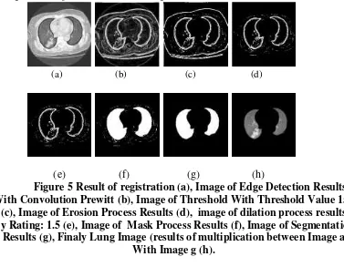
With Th 1, F (x, y) is the original image, Nf (x, y) is Result of multiplication between original image intensity with value of Th. In our study process of multiplication is multiplication of lung mask image segmentation results with lung CT images was done histogram equalization process. Results of lung field segmentation process can be seen in figure 6 below:
(a) (b) (c) (d)
(e) (f) (g) (h)
Figure 5 Result of registration (a), Image of Edge Detection Results With Convolution Prewitt (b), Image of Threshold With Threshold Value 155
(c), Image of Erosion Process Results (d), image of dilation process results By Rating: 1.5 (e), Image of Mask Process Results (f), Image of Segmentation
Results (g), Finaly Lung Image (results of multiplication between Image a With Image g (h).
Calculate Pixels That Are Detected
Image from segmentation result converted into a binary image and calculated number of pixels that are detected by displaying histogram.
Comparing with Groundthruth
Lung field segmentation process by groundtruth done by marking borders of lung on original CT image using Paint program. Image of marking of Groundtruth converted into a binary image and calculated number of pixels that are detected by displaying histogram. In this study groundtruth is a radiologist from Bethesda Hospital Yogyakarta.
Calculate of Accuracy, Sensitivity and Specificity
were identified as lung pixels. While FN is not detectable lung pixels. Then to test performance of proposed segmentation method by calculating accuracy, sensitivity and specificity. Accuracy indicates performance of proposed method. Sensitivity indicates lung pixels are detected correctly. Equation of accuracy, sensitivity and specificity shown in equation 7, 8, 9 below:
𝐴𝑐𝑐𝑢𝑟𝑎𝑐𝑦= 𝑇𝑃+𝑇𝑁
𝑇𝑃+𝐹𝑃+𝐹𝑁+𝑇𝑁 (7)
𝑆𝑒𝑛𝑠𝑖𝑡𝑖𝑣𝑖𝑡𝑦 = 𝑇𝑃
𝑇𝑃+𝐹𝑁 (8)
𝑆𝑝𝑒𝑐𝑖𝑓𝑖𝑐𝑖𝑡𝑦 =𝑇𝑁+𝐹𝑃𝑇𝑁 (9)
Segmentation Using Thresholding
Steps of system design for lung segmentation with thresholding similar to steps in segmentation of lung image using registration method. That distinguish between is, in the process of segmentation by thresholding there is no registration process.
Segmentation Using Active Contour
Steps of segmentation with active contour can be described as follows:
a. Perform early initialization of active contour
𝐶 = 𝑥,𝑦 :𝜑 𝑥,𝑦 = 0 ∀(𝑥,𝑦) ∈Ω (10)
b. Calculating value of u and v, which represents interior and exterior region
𝑢 = 𝐼 𝑥Ω ,𝑦 𝐻(𝜑(𝑥,𝑦))𝑑𝑥𝑑𝑦
𝐻Ω (𝜑(𝑥,𝑦)))𝑑𝑥𝑑𝑦 (11) c. Calculate energy of contour function
FF=(I-v)2-λ2(I-u)2 (12) d. Calculate flow
𝜕𝜑
𝜕𝑡 =
𝐹𝐹
𝐹𝐹 +𝜇.𝐶𝑢𝑟𝑣𝑎𝑡𝑢𝑟𝑒(𝜑) (13) e. An update to its contour
𝜑𝑡+1=𝜑𝑡+∆𝑡.𝑓𝑙𝑜𝑤 (14)
f. Process of step a, b, c, d to convergent conditions
EXPERIMENT AND RESULTS
(a) (b)
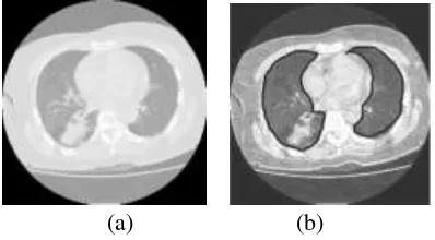
Figure 6 Examples of CT images Lung With Axial slices
Image Normal Ct Scan Lung (a), Abormally Lung CT Scan Image (b)
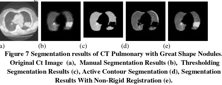
The first experiment is segmentation of lung CT image slice number 19 with big abnormality (beyond lung). Results of this experiment shows that thresholding method, image of nodules (disease) is not in image of lung. This indicates a failure of segmentation with the method. More results as shown in Figure 9 following:
(a) (b) (c) (d) (e)
Figure 7 Segmentation results of CT Pulmonary with Great Shape Nodules. Original Ct Image (a), Manual Segmentation Results (b), Thresholding Segmentation Results (c), Active Contour Segmentation (d), Segmentation
Results With Non-Rigid Registration (e).
Results of three segmentation methods are used can be seen that segmentation with registration have a higher accuracy compared with thresholding and active contour method. The results of the calculation accuracy of the methods used are listed in Table 2.
Table 1 Calculation Results of Accuracy With Large Abnormalities Forms Segmentation
Methods
Accuracy
%
Sensitivity
%
Specificity
%
Threshold 94.2 95.1 93.9
Active Contour
92.45 92.49 92.44
Registration 95.4 90.8 97
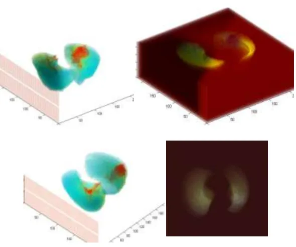
Figure 8 Results of 3-D Visualization
The next experiment is segmentation of one slice normal lung CT image. Results of this experiment shows that for normal lung, thresholding method has a higher accuracy rate than registration method. More results as shown in Figure 11 below:
(a) (b) (c) (d) (e)
Figure 9 Segmentation Results of the Normal Lung Original Ct image (a), Manual Segmentation (b), Thresholding Segmentation (c), Active Contour Segmentation (d), Segmentation Results
With Non-Rigid Registration (e).
Table 2 Accuracy of the Normal Lung Segmentation
Segmentation Methods
Accuracy
%
Sensitivity
%
Specificity
%
Threshold 93.3 88.3 94.5
Active Contour
94.9 88 96.9
3-D reconstructions of normal lung can be shown in the following figure.
Figure 10 Visualization of 3-D Image of Normal Lung
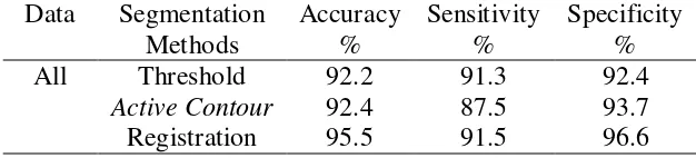
Experimental results using 20 slice CT scans of patients can be seen that registration method has higher accuracy than other two methods, namely the threshold and the active contour. This shows registration segmentation method is suitable to segments lung with containing abnormalities. Result of comparison of three methods used are listed in Table 4.
Table 3 Segmentation results Using Three Methods With All Data Data Segmentation
Methods
Accuracy %
Sensitivity %
Specificity %
All Threshold 92.2 91.3 92.4
Active Contour 92.4 87.5 93.7
Registration 95.5 91.5 96.6
CONCLUSION
Conclusion from above experiments is that segmentation by thresholding and active contour methods fail to segment pulmonary CT images containing disease (abnormalities). This is due in pulmonary with large nodules size have lung borders are not clear (low contrast value). Segmentation by registration can be identified region of lung CT with large abnormalities form. Segmentation by thresholding and active contour can run well on lung CT scan image that have small diseases shape or in lung that had no disease (normal lung).
DISCUSSION
REFERENCES
[1] D. Cascio, S. C. Cheran, A. Chincarini, G. De Nunzio, Automated Detection of Lung Nodules in Low-dos Computed Tomography, International Journal of Computer assisted Radiology and Surgery, Vol 2, Supplement 1, June 2007.
[2] Gonzales, R. C., Woods, R. E. And Eddins, S., Digital Image Processing Using Matlab, 1st Edn, Printice Hall, 2003.
[3] Hohne, K. H., M. Bomans, et al. (1990). "Rendering Tomographic Volume Data: Adequacy of Methods for Different Modalities and Organs." 3-D Imaging in Medicine F60: 197-215.
[4] Heidi C Roberts, Anna Walsham, Errol Colak, Hany Kashani, Chris Mongiardi, Demetris Patsios, The Utility of Computer-aided-detection for the Assessment of Pulmonary Arterial Filling detects at CT Angiography, University Healt Network, Toronto, ON, Canada, International Journal of Computer assisted Radiology and Surgery, Vol 2, Supplement 1, June 2007.
[5] Ilaria Gori, A Multi-Scale Approach to Lung Nodule detection in Computed Tomography, Instituto Nasionale di Fisica Nucleare, Sezione di Pisa Italy, International Journal of Computer assisted Radiology and Surgery, Vol 2, Supplement 1, June 2007.
[6] Jusuf A, Harryanto A, Syahruddin E, Endardjo S, Mudjiantoro S, Sutandio N. Type of lung cancer non-small cell carcinoma. National guidelines for diagnosis and management in Indonesia, 2005. PDPI dan POI, Jakarta, 2005.
[7] L. Borosky, M. C. Lee, L. Zhao, L. Agnihotri, C. A. Powell, Computer-aidec Diagnosis for Lung cancer Using a Classifier Ensemble, College of Physicians and Surgeons, New York, NY, USA, International Journal of Computer assisted Radiology and Surgery, Vol 2, Supplement 1, June 2007.
[8] Martin Dolej s, Jan Kybic, The Lung TIME Annotated Lung Nodule Dataset and Nodule Detection Framework, Faculty of Electrical Engineering, Czech Technical University in Prague, Czech Republic.
[9] Mark S. Nixon A and Alberto S. Aguado, (2008), “Feature Extraction And
Image Processing”, Second Edition, AcademicPress is an imprint of Elsevier.
[10] M. Syamsa Ardisasmita, Image Segmentation and Reconstruction Organ In Three Dimensions Using Mathematical Morphology and triangulated, Center for Computing and Information Technology Development BATAN.
[12] Takeshi Hara, Xiangrong Zhou, Shoji Okura, Hirosi Fujita, Takuji Kiryu, Hiroaki hoshi, Nodule Detection Methods Using autocorrelation Feature on 3D chest CT Scans, Departement of Intelligent.
[13] T.Kobota, A. Jerebko, M. Salganicoff, A. Krishnan, A Segmentation Algorithm With Competition-diffusion and Distance Transform For Automated Estimated of Pulmonary Nodule Diameter, Susquehanna University, Mathematical Sciences, Selingrove PA USA, International Journal of Computer assisted Radiology and Surgery, Vol 2, Supplement 1, June 2007.