Cytotoxicity of Physic Nut (Jatropha curcas
L.,
Euphorbiaceae)
Latex by Agar-Overlay
Fazwishni Siregar
Dentistry Study Programme, Faculty of Medicine, YARSI University, Jakarta
KEYWORDS Agar Overlay Technique; Cytotoxicity Test; Fibroblast L929; Jatropha Curcas Latex; Toxicology
ABSTRACT Jatropha curcas L. latex has been used as traditional medicine to cure various
infections among others are to cure toothache, apthae, as a mouth rinse to treat bleeding gums, as a hemostatic, and a wound dressing. To be used as herbal medicine, scientific investigation including toxicological studies must be conducted. The aim of these studies was to evaluate the cytotoxicity of J. curcas latex by agar overlay technique as part of toxicological study. Fibroblasts L929 were cultured in Petri dishes to confluence, and a layer of agar was then added. After staining the cells with neutral red, diluted latex (0.4-15% w/v) imbibed in filter paper were applied to the agar surface and incubated for 24 hours. Evaluation was based on the zone of decoloration and lysis indices. Result showed that there was zone of decoloration from 2-5 mm in diameter that was all in the index 2 (less than 5 mm) from 5 index decoloration classification. Lyses cells were not observed in the decoloration zone, which could be interpreted as a coagulative necrosis. It may be concluded that J. curcas latex has a moderate cytotoxic activity and necrotic coagulation in an agar- overlay technique using fibroblast L929.
INTRODUCTION
Jatropha curcas, Euphorbiaceae, is a small tree found in tropical areas. Phytochemical screening in J. curcas
latex identified the main chemical compounds which were sterols, flavone aglycones in the ether extract, tannins, reducing compounds, sterol glycosides in the ethyl extract, and tannins,
reducing compounds, poliose, saponins in the aqueous extract (Siregar et al, 2001).
Corespondence:
Using an alcohol and acetone precipitation, a proteolytic enzyme, curcain, can be obtained from the latex of J. curcas L. Curcain is a phytotoxin which is a toxic protein molecule (Nath and Dutta, 1991; Nath and Dutta, 1989). The Jatropha curcas latex is applied topically to wasp and bee stings. In tropical Africa and Southeast Asia the latex is used as a hemostatic and a wound dressing and is said to be efficacious in treating scabies, eczema, and ringworm. Furthermore, it is used as a mouth rinse to treat bleeding gums and to soothe a baby’s inflamed tongue. In Indonesia and the Philippines, a little latex on absorbent cotton is used to cure toothache (Perry and Metzger, 1980: Heyne, 1987: Suwondo, 1993). The latex is used as anti-inflammatory by massaging the latex on the traumatic area (de Feo, 1989). Observation and information from healers, village heads and elderly people in India revealed that latex was used in 8 ailments for some herbal medicinal formulations (Goswani et al, 2013). It is also used as wound healing (Prasadet al, 2012).
Eventhough the latex has been used traditionally as herbal medicine, scientific investigation including toxicological studies was very limited. Toxicological studies of a chemical or substance are very important to know its safety level that should be done for those that will be used for medicine, food additive, and cosmetics. Cytotoxicity test by MTT (3-[4,5-
dimethylthiazol-2-yl]-2,5-diphenyltetrazolium bromide) assay revealed that the latex is cytotoxic to human gingival fibroblast cells (Siregar and Akbar, 2000). Ames test showed no mutagenicity activity (Siregar and Kristiani, 2007).
Study on cytotoxic assay revealed that J. curcas latex extract was toxic to epithelial and fibroblast cell on dose dependent (Dewi Fatma et al, 2012).The objective of this study was to assess the cytotoxicity of the latex in cell cultures using the agar-overlay technique as a part of the toxicological studies (International Standard Organization, 1997; Messer and Feigel, 1985; Oh and Kim, 2005; Park et al, 2013; Dos Santos et al, 2008; Tunc et al. 2009; Velasco-Ortega et al, 2010).
MATERIALS AND METHODS
Latex was collected by incising the young bark of J. curcas trees at The Institute for Research and Development of Spices and Medical Plants, Bogor, Indonesia*. After freeze-drying for 50 hours, it was stored at -20 oC. The dry
weight per fresh volume obtained was around 15%. This freeze-dried latex was used as a test sample, and not the fresh latex, throughout the experiments. The materials used were phosphate buffered saline, neutral red, RPMI medium, fetal bovine serum (FBS), penicillin-streptomycin, fungizone. Fibroblast L929 cell line was kindly donated by G. Moekti from Balitvet, Bogor, Indonesia.
Fibroblasts L929 were cultured with RPMI medium supplemented with FBS and added with 100.000 IU/L penicillin, 100 mg/L streptomycin, and 2.5 mg/L fungizone on Petri dishes, at 37 oC and 5% CO2 on air. Medium was
changed two times a week or if the pH was lowered signed by color changing in medium. After confluence, the medium was aspirated leaving a monolayer of cells at the bottom of the dishes. Then agar medium (45-48 oC)
(30 minutes). Cells were stained with 5 ml freshly prepared neutral red vital stain for 15 minutes. Excess staining solution was removed by aspiration.
Various dilutions of J. curcas
latex in saline (0, 4, 9, 18, 37, 75, and 150 mg/L or from 0.4% to 15%) were imbibed (10 μl) into a 5 mm Whatman filter paper No. 1 and applied to the agar surface. The Whatman filter paper without sample and paper with saline were used as paper and solvent controls. Three-fold replication was employed throughout. Cells were then incubated for 24 hours. Evaluation was based on zone and lysis indices.
Zone index was determined by the size of the decolorized zone while the lysis index was determined by the extent of lysis within the zone. Decoloration means the cells died. The zone index is: 0 = no detectable zone around or under the sample; 1 = zone limited to area under the sample; 2 = zone not greater than 5 mm in extension from sample; 3 = zone more than 5 mm but less than 10 mm in extension from sample; 4 = zone greater than 10 mm in extension from sample but not involving entire plate; 5 = zone involving the entire plate. The lysis index is: 0 = no observable lysis; 1 = up to 20% of zone lysis; 2 = 20-40% of zone
lysis; 3 = 40-60% of zone lysis; 4 = 60-80% of zone lysis; 5 = more than 60-80% lyses within the zone (International Standard Organization, 1997; Messer and Feigel, 1985; Oh and Kim, 2005; Park et al, 2013; Dos Santos et al, 2008; Tunc et al. 2009; Velasco-Ortega et al, 2010).
RESULTS
Zone of decoloration was not observed around and under the paper and solvent control. Parallel with increasing concentration from 0.4% to 15% latex, decoloration zones were enlarged from 2 mm to 4-5 mm in diameter (Table 1). However, all the increased zone of decoloration was extended not greater than 5 mm from samples. Thus they were all in zone 2.
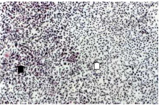
Lysed cells were not observed in the decoloration zone around and under control and samples. Under light microscope, the shape of fibroblast cells was seen although their processes appeared smaller than those observed in the color zone (Figure 1). Zone of decoloration were not observed around the control (0% concentration), while 2-5 mm extensions of zone decoloration were observed that were included in index zone 2.
Table 1. The extention of zone decoloration from samples at cytotoxicity test with agar-overlay technique.
J. curcasconcentration on saline (%)
Width of zone of decoloration (mm)
0 0.4 0.9 1.6 3.75
7.5 15
Figure 1. Cytotoxicity test agar-overlay technique forJ. curcaslatex. Black arrow is normal zone of fibroblast L929 cells. White arrow is fibroblast L929 in decoloration zone around the disc imbibed with 3.7%
latex, no lysis was observed. Magnification 100X.
DISCUSSION
Among its traditional uses,
Jatropha curcas latex was used to cure toothache, apthae, to stop cutaneous bleeding, and wound healing. From personal communication it was known that the fresh latex was imbibed to a small ball of absorbent cotton and put directly into the cavity of dental caries. While to aphtae and bleeding on skin, the fresh latex was put directly to the wound. There was a similarity in the usage that fresh latex was applied directly to the open tissue surface.
To consider its possible use in dentistry, many experiments should be completed and the safety level should first be determined by toxicological study. This is necessary because J. curcas has variable content and toxic substances in the plant. Cytotoxicity is a part of toxicity study that should be established to determine the toxic effect of a substance toward cells by cell culture method. There are several methods for cytotoxicity study, and
disadvantage. Using more than one method to test a substance will be complementary. By the method of MTT assay, it was revealed thatJ. curcaslatex is cytotoxic (Siregar and Akbar, 2000). However, the morphology of cells could not be seen. It was known, by this method, that the latex precipitated medium component that supplemented with FBS. The consequence was only very low concentration, and not high concentration, of latex that could be evaluated. By agar overlay technique, high concentration of latex (15%) could be imbibed into the paper that was then put on the agar surface. Thus the latex did not influence the growth medium directly and high concentration of fresh latex (15%) could be evaluated. Moreover, the morphology of cells could be observed under light microscope by this method.
Latex was then lyophilized for 50 hours and stored at -20 oC to reduced
chemical reaction. The lyophilized latex was dissolved in saline, and used as sample.
In the cytotoxicity test with an agar-overlay technique, there was a widening zone of decoloration parallel with the increasing concentration of latex. Thus there was a dose-respons relation. However, all zones that formed from all concentration (0.4 – 15%) tested were not higher than classification 2 out of 5 zones decoloration. Zone 2 is zone not greater than 5 mm in extension from sample, while zone 5 is decoloration involving the entire plate. This could be interpreted that latex is moderately cytotoxic. Decoloration means the cells died. However, there were no lysed cells. It could then be concluded that the effect was a coagulative necrosis. This effect was probably because of the tannins content of latex.
Phytochemical screening revealed that J. curcas latex contains tannins.1,3 Tannins precipitate albumin,
starch, gelatin, most of alkaloid, metallic salt, and is used as astringent. In veterinary, it is used as astringent, hemostatic, and in solution for burning. Tannins on the surface of an opened tissue have the effect to coagulate protein forming a protective shield, and tissue regeneration was formed underneath (Windholz, 1979; Tyler, 1988). The coagulative effect of latex is also used as hemostatic on bleeding (Nath and Dutta, 1991; Kone-Kamba, 1987). Tannins coagulate the cell wall proteins and have bactericidal activity in high concentrations (Siregar and Kristiani, 2007). relieve pulpalgia in dental caries where it might succeed by devitalizing the dental pulp and no more pulpal pain.
Acknowledgment
This research was funded by URGE, 051/ HTPP-III/URGE/1997. Thanks to The Research Institute of Herbal Medicine and Spices, Indonesian Research Center for Veterinary Science (Balai Besar Penelitian Veteriner), Indonesia, and Prof. Dr. drg. S. M. Soerono Akbar, Sp.KG for helping to realize this research.
*The plant has been identified as Jatropha curcas L (Euphorbiaceae) by Herbarium Bogoriense, Bogor, Indonesia.
REFERENCES
de Feo V 1989. Uso di planti ad azione antiinflammatoria nell’Alto Ucayall, Peru Orientale. Fitoterapia. 62: 481-494. Dewi Fatma S, Widurini, Farida R, Irmaleny 2012. In vitro cytotoxicity of Jatropha curcas in epithelial and fibroblast cells. J Nat Prod. 5: 214-221. Dos Santos RL, Pithon MM, de Oliveira
MV, da Silva Mendas G, Romanos MTV, de Oliveira Ruellas AC 2008. Cytotoxicity of intraoral orthodontic elastics. Braz J Oral Sci. 7: 1520-1525. Goswani NK, Saharia D, Kar A 2013.
Traditional uses of Jatropha curcas Linnaeus (euphorbiaceae) as medicine by differnet communities in Northeast India. Pleione. 7: 66-72.
Translated by Litbang Kehutanan. p 1180-1182.
International Standard organization 1997. Biological Evaluation of Dental Materials. Technical Report No 7405. Kone-Bamba D, Pelissier Y, Ozoukou ZF
and Kouao D 1987. Haemostatic activity of fifteen medicinal plants of the traditional medicine of the Ivory Coast. Plant Med Phytother. 21: 122-130.
Messer HH, Feigal RJ 1985. A comparison of the antibacterial and cytotoxic effects of paraphenol. J Dent Res. 64: 818-821.
Nath LK, Dutta SK 1991. Extraction and purification of curcain, a protease from the latex of Jatropha curcas Linn. Indian J Pharmacol. 43: 111-114.
Nath LK, Dutta SK 1989. A kinetic study on curcain, a protease from the latex of Jatropha curcas Linn. Indian J Pharmaceut Sci. 51: 43-47.
Oh KT, Kim KN 2005. Ion release and cytotoxicity of stainless steel wires. Europ J Orthod. 27(6): 533-540.
Park YJ, Song YH, An JH, Song HJ, Anusavice K 2013. Cytocompatibility of pure metals and experimental binary titanium alloys for implant materials. J Dent. 41: 1251-1258.
Perry LM, Metzger J 1980. Medicinal Plants of East and Southeast Asia. Cambridge: MIT Press. p 246-247. Prasad, DMR, Izam A, Khan MdMR 2012.
Jatropha curcas: plant of medicinal benefits. J Med Plant Res. 6: 2691-2699. Siregar F, Soerono Akbar SM, Chairul 2001.
Phytochemical screening and
hemolytic activity of Jatropha curcas L. (Euphorbiaceae) latex. Proceeding International Seminar on Natural Product Chemistry and Utilization of Natural Resources. Depok.
Siregar F, Akbar SMS 2000. Cytotoxicity of Jatropha curcas (Euphorbiaceae) latex on fibroblast by MTT assays. Med J Indones. 9: 253-257.
Siregar F, Kristiani I 2007. Mutagenicity activity of Jatropha curcas L (Euphorbiaceae) latex. Berkala Ilmu Kedokteran. 39: 23-26.
Suwondo S 1993. Improving the use of the Indonesian traditional herbal medicine for the prevention of caries and gingivitis on the basis of antibacterial activity test against streptococcus mutans and clinical trial on gingivitis. Dissertation, Faculty of Dentistry, University of Padjadjaran, Bandung. Tunc ES, Oxer L, Sari S, Cetiner S 2009.
Cytotoxic effects of halogen and light emitting diode cured compomers on human pulp fibroblast. Int J Peadiatric Dent. 19: 55-60.
Tyler VE, Brady LR, Robbers JE 1988. Pharmacognosy. 9thed., Lea & Febiger, Philadelphia. p 67-68, 77-78, 97-98. Velasco-Ortega E, Jos A, Camean AM,
Pato-Mourelo J, Segura-Egea JJ 2010. In vitro evaluation of cytotoxicity and genotoxicity of a commercial titanium alloy for dental implantology. Mutat. Res.: Genet. Toxicol. Environ. Mutagen.
doi:10.1016/j.mrgentox.2010.06.013 Windholz ML 1976. The Merck Index.