T h e n e w e n g l a n d j o u r n a l o f m e d i c i n e
n engl j med 355;16 www.nejm.org october 19, 2006 1699
Drug Therapy
Management of Sepsis
James A. Russell, M.D.
From the University of British Columbia, Critical Care Medicine, St. Paul’s Hospital, Vancouver, BC, Canada. Address reprint requests to Dr. Russell at the University of British Columbia, Critical Care Medicine, St. Paul’s Hospital, 1081 Burrard St., Vancouver, BC V6Z 1Y6, Canada, or at [email protected].
N Engl J Med 2006;355:1699-713.
Copyright © 2006 Massachusetts Medical Society.
A
better understanding of the inflammatory, procoagulant, andimmunosuppressive aspects of sepsis has contributed to rational therapeu-tic plans from which several important themes emerge.1 First, rapid diagno-sis (within the first 6 hours) and expeditious treatment are critical, since early, goal-directed therapy can be very effective.2 Second, multiple approaches are necessary in the treatment of sepsis.1 Third, it is important to select patients for each given therapy with great care, because the efficacy of treatment — as well as the likeli-hood and type of adverse results — will vary, depending on the patient.
T H E S P E C T R U M O F S E P S I S
Nomenclature is important when it helps us understand the pathophysiology of a disease. This is true for sepsis, since nomenclature has informed the design of ran-domized, controlled trials and, ultimately, the prognosis of sepsis. Sepsis is defined as suspected or proven infection plus a systemic inflammatory response syndrome (e.g., fever, tachycardia, tachypnea, and leukocytosis).3 Severe sepsis is defined as sepsis with organ dysfunction (hypotension, hypoxemia, oliguria, metabolic acidosis, throm-bocytopenia, or obtundation). Septic shock is defined as severe sepsis with hypoten-sion, despite adequate fluid resuscitation. Septic shock and multiorgan dysfunction are the most common causes of death in patients with sepsis.4 The mortality rates associated with severe sepsis and septic shock are 25 to 30%5 and 40 to 70%,6 respec-tively.
There are approximately 750,000 cases of sepsis a year in the United States,7 and the frequency is increasing, given an aging population with increasing numbers of patients infected with treatment-resistant organisms, patients with compromised im-mune systems, and patients who undergo prolonged, high-risk surgery.7
P A T H O P H Y S I O L O G Y
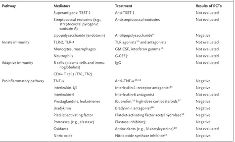
Sepsis is the culmination of complex interactions between the infecting microorgan-ism and the host immune, inflammatory, and coagulation responses.8 The rationale for the use of therapeutic targets in sepsis has arisen from concepts of pathogenesis (Table 1).
Both the host responses and the characteristics of the infecting organism influ-ence the outcome of sepsis. Sepsis with organ dysfunction occurs primarily when host responses to infection are inadequate. In addition, sepsis often progresses when the host cannot contain the primary infection, a problem most often related to charac-teristics of the microorganism, such as a high burden of infection and the presence of superantigens and other virulence factors, resistance to opsonization and phago-cytosis, and antibiotic resistance.
review article
INNATE IMMUNITY AND INFLAMMATION
IN EARLY SEPSIS
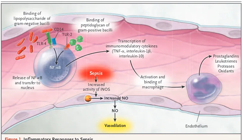
Host defenses can be categorized according to in-nate and adaptive immune system responses. The innate immune system responds rapidly by means of pattern-recognition receptors (e.g., toll-like re-ceptors [TLRs]) that interact with highly conserved molecules present in microorganisms10 (Fig. 1). For example, TLR-2 recognizes a peptidoglycan of gram-positive bacteria, whereas TLR-4 recognizes a lipopolysaccharide of gram-negative bacteria (Fig. 1). Binding of TLRs to epitopes on microor-ganisms stimulates intracellular signaling, increas-ing transcription of proinflammatory molecules such as tumor necrosis factor α (TNF-α) and in-terleukin-1β, as well as antiinflammatory cytokines such as interleukin-10.32 Proinflammatory cyto-kines up-regulate adhesion molecules in neutro-phils and endothelial cells. Although activated neu-trophils kill microorganisms, they also injure endothelium by releasing mediators that increase vascular permeability, leading to the flow of pro-tein-rich edema fluid into lung and other tissues. In addition, activated endothelial cells release ni-tric oxide, a potent vasodilator that acts as a key mediator of septic shock.
SPECIFICITY AND AMPLIFICATION OF THE IMMUNE
RESPONSE BY ADAPTIVE IMMUNITY
Microorganisms stimulate specific humoral and cell-mediated adaptive immune responses that amplify innate immunity. B cells release immuno-globulins that bind to microorganisms, facilitating their delivery by antigen-presenting cells to natural killer cells and neutrophils that can kill the micro-organisms.
T-cell subgroups are modified in sepsis. Helper (CD4+) T cells can be categorized as type 1 helper (Th1) or type 2 helper (Th2) cells. Th1 cells gen-erally secrete proinf lammatory cytokines such as TNF-α and interleukin-1β, and Th2 cells se-crete antiinf lammatory cytokines such as in-terleukin-4 and interleukin-10, depending on the infecting organism, the burden of infection, and other factors.33
DISTURBANCE OF PROCOAGULANT–
ANTICOAGULANT BALANCE
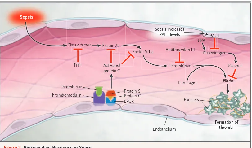
Another important aspect of sepsis is the alteration of the procoagulant–anticoagulant balance, with an increase in procoagulant factors and a decrease in anticoagulant factors (Fig. 2). Lipopolysaccha-ride stimulates endothelial cells to up-regulate
tis-Table 1. Pathways and Mediators of Sepsis, Potential Treatments, and Results of Randomized, Controlled Trials (RCTs).*
Pathway Mediators Treatment Results of RCTs
Superantigens: TSST-1 Anti-TSST-1 Not evaluated
Streptococcal exotoxins (e.g., streptococcal pyrogenic exotoxin A)
Antistreptococcal exotoxins Not evaluated
Lipopolysaccharide (endotoxin) Antilipopolysaccharide9 Negative
Innate immunity TLR-2, TLR-4 TLR agonists10 and antagonists Not evaluated
Monocytes, macrophages GM-CSF, interferon gamma11 Not evaluated
Neutrophils G-CSF† Not evaluated
Adaptive immunity B cells (plasma cells and
immu-noglobulins) IgG Not evaluated
CD4+ T cells (Th1, Th2)
Proinflammatory pathway TNF-α Anti–TNF-α13,14 Negative
Interleukin-1β Interleukin-1–receptor antagonist15 Negative
Interleukin-6 Interleukin-6 antagonist Not evaluated Prostaglandins, leukotrienes Ibuprofen,16 high-dose corticosteroids17 Negative
Bradykinin Bradykinin antagonist18 Negative
Platelet-activating factor Platelet-activating factor acetyl hydrolase19 Negative
Proteases (e.g., elastase) Elastase inhibitor‡ Negative Oxidants Antioxidants (e.g., N-acetylcysteine)20 Not evaluated
drug ther a py
n engl j med 355;16 www.nejm.org october 19, 2006 1701
sue factor, activating coagulation. Fibrinogen is then converted to fibrin, leading to the formation of microvascular thrombi and further amplifying injury.
Anticoagulant factors (e.g., protein C, protein S, antithrombin III, and tissue factor–pathway in-hibitor) modulate coagulation. Thrombin-α binds to thrombomodulin to activate protein C by bind-ing to endothelial protein C receptor.34 Activated protein C inactivates factors Va35 and VIIIa36 and inhibits the synthesis of plasminogen-activator in-hibitor 1.37 Activated protein C decreases apopto-sis,38 adhesion of leukocytes,39 and cytokine pro-duction.40
Sepsis lowers levels of protein C, protein S, antithrombin III, and tissue factor–pathway in-hibitor.41 Lipopolysaccharide and TNF-α decrease the synthesis of thrombomodulin and endothelial protein C receptor, impairing the activation of protein C,42 and increase the synthesis of
plas-minogen-activator inhibitor 1, thus impairing fi-brinolysis.
Key to an understanding of sepsis is the recog-nition that the proinflammatory and procoagulant responses can be amplified by secondary ischemia (shock) and hypoxia (lung injury) through the re-lease of tissue factor and plasminogen-activator inhibitor 1.43
IMMUNOSUPPRESSION AND APOPTOSIS
IN LATE SEPSIS
Host immunosuppression has long been consid-ered a factor in late death in patients with sepsis,44 since the sequelae of anergy, lymphopenia,45 hypo-thermia, and nosocomial infection all appear to be involved.46 When stimulated with lipopolysac-charide ex vivo, monocytes from patients with sep-sis express lower amounts of proinflammatory cy-tokines than do monocytes from healthy subjects, possibly indicating relative immunosuppression.47 Table 1. (Continued.)
Pathway Mediators Treatment Results of RCTs
Procoagulant pathway Decreased protein C Activated protein C5 Positive
Decreased protein S Protein S22 Not evaluated
Decreased antithrombin III Antithrombin III23 Negative
Decreased tissue factor–
pathway inhibitor Tissue factor–pathway inhibitor
24 Negative
Increased tissue factor Tissue factor antagonist25 Not evaluated
Increased
plasminogen-activator inhibitor 1 Tissue plasminogen activator Not evaluated Antiinflammatory Interleukin-10 Interleukin-10§ Not evaluated
TNF-α receptors TNF-α receptors13 Negative
Hypoxia Hypoxia-inducing factor 1α, vascular endothelial growth factor
Early, goal-directed therapy2
Supernormal oxygen delivery Erythropoietin26
Positive Negative Not evaluated Immunosuppression or
apoptosis
Lymphocyte apoptosis Anticaspases27 Not evaluated
Apoptosis of intestinal epithelial cells
Anticaspases27 Not evaluated
Endocrine Adrenal insufficiency Corticosteroids28 Mixed results¶
Vasopressin deficiency Vasopressin29 Not evaluated
Hyperglycemia Intensive insulin therapy30,31 Not evaluated∥
* TSST denotes staphylococcal toxic shock syndrome toxin 1, GM-CSF granulocyte–macrophage colony-stimulating factor, G-CSF granulocyte colony-stimulating factor, Th1 type 1 helper T cells, and Th2 type 2 helper T cells. Organism features means components of bacteria that are toxic to the host and that are potential therapeutic targets in sepsis.
† G-CSF is effective in patients with sepsis who have profound neutropenia.12
‡ Elastase inhibitor was ineffective in a phase 2 trial involving patients with acute lung injury. § Interleukin-10 was ineffective in a phase 2 trial involving patients with acute lung injury.
¶ Corticosteroids had no effect on overall 28-day mortality but decreased mortality in a subgroup of patients with no response to corticotropin (see text for details). Additional trials of corticosteroids in patients with septic shock are in progress.
∥ Intensive insulin therapy decreased the mortality rate among critically ill surgical patients but has not yet been evaluated in patients with sepsis.
Multiorgan dysfunction in sepsis may be caused, in part, by a shift to an antiinflammatory pheno-type and by apoptosis of key immune, epithelial, and endothelial cells. In sepsis, activated helper T cells evolve from a Th1 phenotype, producing proinflammatory cytokines, to a Th2 phenotype, producing antiinflammatory cytokines.48 In ad-dition, apoptosis of circulating and tissue lympho-cytes (B cells and CD4+ T cells) contributes to immunosuppression.49 Apoptosis is initiated by proinflammatory cytokines, activated B and T cells, and circulating glucocorticoid levels, all of which are increased in sepsis.50 Increased levels of TNF-α
and lipopolysaccharide during sepsis may also induce apoptosis of lung and intestinal epithe-lial cells.51
SEPSIS AND WIDESPREAD ORGAN DYSFUNCTION
The altered signaling pathways in sepsis ultimate-ly lead to tissue injury and multiorgan dysfunction. For example, cardiovascular dysfunction is charac-terized by circulatory shock and the redistribution of blood flow, with decreased vascular resistance, hypovolemia, and decreased myocardial contrac-tility associated with increased levels of nitric ox-ide,52 TNF-α,53 interleukin-6,54 and other media-Binding of
lipopolysaccharide of
gram-negative bacilli peptidoglycan of Binding of gram-positive bacilli
Release of NF-κB and transfer to
nucleus
Transcription of immunomodulatory cytokines
(TNF-α, interleukin-1β, interleukin-10)
Increased activity of iNOS
Increased NO
Endothelium TLR-2
TLR-4 CD14
Vasodilation
Sepsis
Activation and binding of macrophage
Prostaglandins Leukotrienes
Proteases Oxidants
NF-κB
NO
Figure 1. Inflammatory Responses to Sepsis.
drug ther a py
n engl j med 355;16 www.nejm.org october 19, 2006 1703
tors. Respiratory dysfunction is characterized by increased microvascular permeability, leading to acute lung injury. Renal dysfunction in sepsis, as discussed recently by Schrier and Wang,55 may be profound, contributing to morbidity and mortality.
T R E A T M E N T A C C O R D I N G T O T H E
e a r l y a n d l a t e r S T A G E S O F S E P S I S
Consensus guidelines for the management of sep-sis have recently been published.56 The following therapeutic plan, informed by such guidelines, considers emergency care for the early stage of sep-sis (0 to 6 hours) and treatment for patients in later stages who require critical care.
Early, Goal-directed Therapy
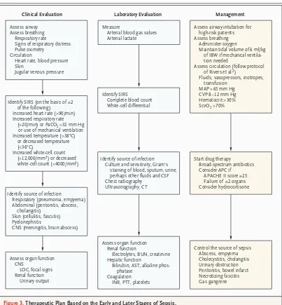
The cornerstone of emergency management of sep-sis is early, goal-directed therapy,2 plus lung-pro-tective ventilation,1 broad-spectrum antibiotics,57,58 and possibly activated protein C5 (Fig. 3 and Ta-ble 2). Rivers and colleagues2 conducted a random-ized, controlled trial in which patients with severe sepsis and septic shock received early, goal-direct-ed, protocol-guided therapy during the first 6 hours after enrollment or the usual therapy. In the group receiving early, goal-directed therapy, central ve-nous oxygen saturation was monitored continu-ously with the use of a central venous catheter. The level of central venous oxygen saturation served to trigger further interventions recommended in the
Tissue factor
Activated protein C
Endothelium Thrombin-α
Thrombin-α
Fibrin t-PA
Fibrinogen
Protein S Protein C EPCR Thrombomodulin
Platelets Factor Va
Factor VIIIa
Sepsis
TFPI
Plasminogen Antithrombin III
Plasmin PAI-1
Formation of thrombi Sepsis increases
PAI-1 levels
Figure 2. Procoagulant Response in Sepsis.
Sepsis initiates coagulation by activating endothelium to increase the expression of tissue factor. Activation of the coagulation cascade, and especially factors Va and VIIIa, leads to the formation of thrombin-α, which converts fi-brinogen to fibrin. Fibrin binds to platelets, which in turn adhere to endothelial cells, forming microvascular throm-bi. Microvascular thrombi amplify injury through the release of mediators and by microvascular obstruction, which causes distal ischemia and tissue hypoxia. Normally, natural anticoagulants (protein C and protein S), antithrombin III, and tissue factor–pathway inhibitor (TFPI) dampen coagulation, enhance fibrinolysis, and remove microthrombi. Thrombin-α binds to thrombomodulin on endothelial cells, which dramatically increases activation of protein C to activated protein C. Protein C forms a complex with its cofactor protein S. Activated protein C proteolytically inacti-vates factors Va and VIIIa and decreases the synthesis of plasminogen-activator inhibitor 1 (PAI-1). In contrast, sep-sis increases the synthesep-sis of PAI-1. Sepsep-sis also decreases the levels of protein C, protein S, antithrombin III, and TFPI. Lipopolysaccharide and tumor necrosis factor α (TNF-α) decrease the synthesis of thrombomodulin and en-dothelial protein C receptor (EPCR), thus decreasing the activation of protein C. Sepsis further disrupts the protein C pathway because sepsis also decreases the expression of EPCR, which amplifies the deleterious effects of the sep-sis-induced decrease in levels of protein C. Lipopolysaccharide and TNF-α also increase PAI-1 levels so that fibrino-lysis is inhibited. The clinical consequences of the changes in coagulation caused by sepsis are increased levels of markers of disseminated intravascular coagulation and widespread organ dysfunction. t-PA denotes tissue plasmin-ogen activator.
Identify SIRS
Complete blood count White-cell differential
Identify source of infection Culture and sensitivity, Gram’s
staining of blood, sputum, urine; perhaps other fluids and CSF Chest radiography
Ultrasonography, CT
Assess organ function Renal function
Electrolytes, BUN, creatinine Hepatic function
Bilirubin, AST, alkaline phos-phatase
Coagulation INR, PTT, platelets Identify SIRS (on the basis of ≥2
of the following)
Increased heart rate (>90/min) Increased respiratory rate
(>20/min) or PaCO2 <32 mm Hg or use of mechanical ventilation Increased temperature (>38°C)
or decreased temperature (<36°C)
Increased white-cell count (>12,000/mm3) or decreased
white-cell count (<4000/mm3)
Identify source of infection
Respiratory (pneumonia, empyema) Abdominal (peritonitis, abscess,
cholangitis) Skin (cellulitis, fasciitis) Pyelonephritis
CNS (meningitis, brain abscess)
Assess organ function CNS
LOC, focal signs Renal function
Urinary output
Start drug therapy
Broad-spectrum antibiotics Consider APC if
APACHE II score ≥25 Failure of ≥2 organs Consider hydrocortisone
Control the source of sepsis Abscess, empyema Cholecystitis, cholangitis Urinary obstruction Peritonitis, bowel infarct Necrotizing fasciitis Gas gangrene Measure
Arterial blood gas values Arterial lactate Assess airway
Assess breathing Respiratory rate
Signs of respiratory distress Pulse oximetry
Circulation
Heart rate, blood pressure Skin
Jugular venous pressure
Assess airway intubation for high-risk patients Assess breathing
Administer oxygen
Maintain tidal volume of 6 ml/kg of IBW if mechanical ventila-tion needed
Assess circulation (follow protocol of Rivers et al.2)
Fluids, vasopressors, inotropes, transfusion
MAP >65 mm Hg CVP 8–12 mm Hg Hematocrit >30% ScvO2 >70%
Clinical Evaluation Laboratory Evaluation Management
Figure 3. Therapeutic Plan Based on the Early and Later Stages of Sepsis.
In the author’s approach, emergency management should focus on simultaneous evaluation and resuscitation. Ear-ly diagnosis is critical because of the efficacy of earEar-ly, goal-directed therapy in the first 6 hours.2 Critical care
man-agement requires frequent, thorough reassessment and supportive measures for organ dysfunction. Assessment focuses on refinement of the antibiotic regimen, control of the source of sepsis, and evaluation for resolution of the signs of the systemic inflammatory response syndrome (SIRS). Supportive measures for organ dysfunction include ongoing cardiovascular support, continued use of lung-protective mechanical ventilation with a tidal volume of 6 ml per kilogram of ideal body weight (IBW),1 and activated protein C (APC) in appropriate patients for 96 hours. The
use of vasopressin, intensive insulin, and corticosteroids is controversial. Critical care management of sepsis also requires attention to new problems such as immunosuppression, nosocomial infection, and persistent ARDS. PaCO2 denotes partial pressure of arterial carbon dioxide, CNS central nervous system, LOC level of consciousness,
dru
g
t
h
e
r
a
p
y
n engl j med
355;16
www.nejm.org october
19, 2006
1705
Table 2. Results of Positive Randomized, Controlled Trials.*
Group Study
No. of
Patients Intervention Group Control Group Mortality Rate† NNT‡
Level of Evidence
Intervention Group
Control Group
%
Patients with acute lung in-jury and ARDS§
ARDS Clinical Trials
Network1 861 Low tidal volume (6 ml/kg of ideal body weight) High tidal volume (12 ml/kg of ideal body weight) 31 40 11 I Patients with severe sepsis
and septic shock
Rivers et al.2 263 Early, goal-directed therapy Usual therapy 33 49 6 I
Patients with severe sepsis
and septic shock Bernard et al.
5 1690 Activated protein C Placebo 25 31 16 I
Patients with severe sepsis and septic shock, at in-creased risk for death¶
Bernard et al.5 817 Activated protein C Placebo 31 44 7.7 I
Patients in septic shock Annane et al.28 299 Hydrocortisone +
fludrocorti-sone Placebo 55 61 NA I–II∥
Patients in septic shock** Annane et al.28 229 Hydrocortisone +
fludrocorti-sone Placebo 53 63 10 I–II∥
Critically ill surgical patients Van den Berghe et al.31 1548 Intensive insulin (to maintain glucose level of 4.4–6.1 mmol/liter)
Usual insulin (to maintain glucose level of 10–11.1 mmol/liter)
4.6 8 29 I
Patients in medical ICU†† Van den Berghe et al.30 1200 Intensive insulin (to maintain glucose level of 4.4–6.1 mmol/liter)
Usual insulin (to maintain glucose level of 10–11.1 mmol/liter)
37 40 NA I
* The inclusion criteria were as follows: for the ARDS Clinical Trials Network,1 a ratio of the partial pressure of arterial oxygen to the forced inspiratory volume in 1 second of less than 300, pulmonary infiltrates, mechanical ventilation, no congestive heart failure; for Rivers et al.,2 sepsis plus either increased lactate levels (severe sepsis) or hypotension (septic shock); for Bernard et al.,5 severe sepsis; for Annane et al.,28 vasopressor-dependent septic shock, mechanical ventilation, oliguria, and increased lactate levels. One study by Van den Berghe et al.31 involved patients in the surgical intensive care unit (ICU), 62% of whom had undergone cardiac surgery. The other study by Van den Berghe et al.30 involved patients in the medical ICU. † The 28-day mortality rate is shown for all groups except those studied by Van den Berghe, for which the intensive care unit (ICU)31 or in-hospital30 mortality rate is shown.
‡ Values are the number needed to treat (NNT) to save one life. § Many of the patients had sepsis.
¶ An increased risk of death was defined by an Acute Physiology and Chronic Health Evaluation (APACHE) II score of at least 25.
∥ The level of evidence is I for the overall trial, but only II for the subgroup of patients with no response to the corticotropin stimulation test. ** The patients had no response to a corticotropin stimulation test with 250 μg of corticotropin.
†† This trial is included in the table because its results contrast with those of a similar positive trial involving patients in the surgical ICU.31
protocol. Crystalloids were administered to main-tain central venous pressure at 8 to 12 mm Hg. Vasopressors were added if the mean arterial pres-sure was less than 65 mm Hg; if central venous oxygen saturation was less than 70%, erythrocytes were transfused to maintain a hematocrit of more than 30%. Dobutamine was added if the central venous pressure, mean arterial pressure, and he-matocrit were optimized yet venous oxygen satu-ration remained below 70%. Early, goal-directed therapy in that study decreased mortality at 28 and 60 days as well as the duration of hospitalization. Patients in the early, goal-directed therapy group received more fluids, transfusions, and dobutamine in the first 6 hours, whereas control subjects ceived more fluids and more control subjects re-ceived vasopressors, transfusion, and mechani-cal ventilation for a period of 7 to 72 hours. The mechanisms of the benefit of early, goal-directed therapy are unknown but may include reversal of tissue hypoxia and a decrease in inflammation and coagulation defects.59
VENTILATION
Once early, goal-directed therapy has been initiat-ed, lung-protective ventilation should be consid-ered. Acute lung injury often complicates sepsis, and lung-protective ventilation — meaning the use of relatively low tidal volumes — is thus another important aspect of management. Furthermore, lung-protective ventilation decreases mortality1 and is beneficial in septic acute lung injury.60 Ex-cessive tidal volume and repeated opening and closing of alveoli during mechanical ventilation cause lung injury. Lung-protective mechanical ven-tilation, with the use of a tidal volume of 6 ml per kilogram of ideal body weight (or as low as 4 ml per kilogram if the plateau pressure exceeds 30 cm H2O) as compared with 12 ml per kilogram of ideal body weight (calculated in men as 50 + 0.91 [height in centimeters – 152.4] and in women as 45.5 + 0.91 [height in centimeters – 152.4]) has been shown to decrease the mortality rate (from 40 to 31%), to lessen organ dysfunction, and to lower levels of cytokines.61 Positive end-expiratory pres-sure (PEEP) decreases oxygen requirements; how-ever, there is no significant difference in mortal-ity between patients treated with the usual PEEP regimen of the Acute Respiratory Distress Syn-drome (ARDS) Clinical Trials Network1 and those treated with higher PEEP levels.62
Patients receiving ventilation require appropri-ate but not excessive sedation, given the risks of prolonged ventilation and nosocomial pneumo-nia.63 Titrating sedation64 and interrupting seda-tion daily until patients are awake63 decrease the risks associated with sedation. Neuromuscular blocking agents should be avoided to reduce the risk of prolonged neuromuscular dysfunction.65
BROAD-SPECTRUM ANTIBIOTICS
Because the site of infection and responsible micro-organisms are usually not known initially in a pa-tient with sepsis, cultures should be obtained and intravenous broad-spectrum antibiotics adminis-tered expeditiously while the host immune status is ascertained. The rising prevalence of fungi, gram-positive bacteria, highly resistant gram-neg-ative bacilli, methicillin-resistant Staphylococcus aureus, vancomycin-resistant enterococcus, and pen-icillin-resistant pneumococcus,66 as well as local patterns of antibiotic susceptibility, should be con-sidered in the choice of antibiotics. Observation-al studies indicate that outcomes of sepsis67 and septic shock57 are worse if the causative microor-ganisms are not sensitive to the initial antibiotic regimen.
ACTIVATED PROTEIN C
drug ther a py
n engl j med 355;16 www.nejm.org october 19, 2006 1707
of infection or the infecting microorganism, pos-sibly because all bacteria and fungi decrease pro-tein C levels.71
Recent trauma or surgery (within 12 hours), active hemorrhage, concurrent therapeutic antico-agulation, thrombocytopenia (defined as a plate-let count of less than 30,000 per cubic millime-ter), and recent stroke were exclusion criteria for safety reasons in the Recombinant Human Acti-vated Protein C Worldwide Evaluation in Severe Sepsis (PROWESS) trial of activated protein C.5 In that trial, there was a trend toward a higher rate of serious bleeding (defined as bleeding requiring the transfusion of 3 U of packed red cells over a period of 2 days or intracranial hemorrhage) among patients receiving activated protein C than among patients in the placebo group (3.5% vs. 2%, P = 0.06), especially during infusion of the activated protein C (2.4% vs. 1%).5 Intracranial hemorrhage occurred in two patients who received activated protein C and in one who received placebo.5 In the Extended Evaluation of Recombinant Human Ac-tivated Protein C United States (ENHANCE U.S.) trial, intracranial hemorrhage occurred in 0.6% of patients given activated protein C.72 Meningitis and severe thrombocytopenia may be risk factors for intracranial hemorrhage.69
When the data are examined together, activated protein C would appear to be cost-effective for patients with severe sepsis and a high risk of death, with the cost per quality-adjusted year of life gained ranging from $24,48473 to $27,400,74 which is similar to the costs of therapies such as organ transplantation75 and drug-eluting stents.76
The mechanism of action by which activated protein C improves the clinical outcome is un-known. Activated protein C was shown to increase protein C and decrease markers of thrombin gen-eration (e.g., d-dimer, a marker of disseminated intravascular coagulation) in one study.77 Although activated protein C prevents hypotension, it has little effect on coagulation in a human intrave-nous endotoxin model of sepsis,78 suggesting that modulation of coagulation may not be the primary mechanism underlying the cardiovascular bene-fit. Other anticoagulant therapies have included antithrombin III23 and tissue factor–pathway in-hibitor,24 yet only activated protein C was effective, perhaps because of its complex antiinflammato-ry,79 antiapoptotic, and anticoagulant37 actions.
treatment of ANEMIA IN SEPSIS
Anemia is common in sepsis80 in part because me-diators of sepsis (TNF-α and interleukin-1β) de-crease the expression of the erythropoietin gene and protein.81 Although treatment with recombi-nant human erythropoietin decreases transfusion requirements,26 its use in randomized, controlled trials failed to increase survival. Erythropoietin takes days to weeks to induce red-cell production and thus may not be effective.
Two trials used different transfusion strategies in different stages of sepsis.2,80 Rivers et al.2 used a hematocrit of 30% as a threshold for transfu-sion in early sepsis as part of a 6-hour protocol. Transfusion was associated with an improved out-come. Hebert et al. compared hemoglobin values of 70 and 100 g per liter as a threshold for trans-fusion later in the course of critical care.80 Patients were expected to stay in the intensive care unit (ICU) for more than 3 days, and two transfusion strategies were compared during their entire ICU stay. There was no significant difference in mor-tality between patients who received transfusion on the basis of higher hemoglobin levels (100 to 120 g per liter) and those who did so on the basis of lower levels (70 to 90 g per liter).80
Transfusion is worthwhile if needed during the emergency stage of sepsis; Rivers et al. observed a marked decrease in mortality when transfusion was provided early.2 Hebert et al. suggest main-taining hemoglobin levels at 70 to 90 g per liter after the first 6 hours to decrease transfusion re-quirements.80 (Because the protocol of Rivers et al. did not extend beyond 6 hours, it is not known whether a higher transfusion threshold would be useful after 6 hours.)
CORTICOSTEROIDS in PATIENTS WHO REQUIRE
CRITICAL CARE
Although corticosteroids have been considered for the management of sepsis for decades, random-ized, controlled trials suggest that an early, short course (48 hours) of high-dose corticosteroids does not improve survival in severe sepsis.82,83 Because adrenal insufficiency is being reconsidered as part of septic shock, there has been renewed interest in therapy with corticosteroids, with a focus on timing, dose, and duration. Several controversies over their use persist, however. First, the concept of adrenal insufficiency in sepsis is controversial.
Second, only two (of five)83 small randomized, controlled trials84 have shown that corticosteroid therapy (low-dose hydrocortisone) decreases the need for vasopressor support in patients with sep-sis. Third, only one adequately powered trial reported a survival benefit of such treatment in patients who had no response to a corticotropin-stimulation test.28
Annane and colleagues28 evaluated oliguric pa-tients with vasopressor-dependent septic shock who required ventilation. Patients underwent a 250-μg corticotrophin-stimulation test28 and were classified as having adrenal insufficiency (no re-sponse) when the serum total cortisol level rose by less than 10 μg per deciliter.85 Patients were then randomly assigned to receive placebo or hy-drocortisone plus fluhy-drocortisone for 7 days. Cor-ticosteroids significantly improved survival both in the overall cohort and in the prospectively de-fined subgroup of patients who had no response to corticotropin; however, over a 28-day period, the difference in mortality was not significant (P = 0.09). Patients without a response to cortico-tropin who received corticosteroids had signifi-cantly lower mortality than patients who received placebo. Subgroup analyses provide inadequate evidence for a change in therapy, however, given the many examples of therapies that were purport-edly successful according to subgroup analysis but were subsequently shown not to be useful in ade-quately powered, randomized, controlled trials.86
Observational studies87 offer no data that indi-cate how patients respond to corticosteroids and thus provide limited guidance as compared with randomized, controlled trials. Marik and Zaloga87 reported that 95% of patients in septic shock had serum cortisol levels under 25 μg per deciliter; another group85 have stated that during septic shock, cortisol levels of less than 15 μg per deci-liter should be used as an indicator of relative ad-renal insufficiency.
A recent study of serum free cortisol has added further complexity to the diagnosis of adrenal in-sufficiency in the critically ill.88 Serum total cor-tisol reflects both corcor-tisol bound to protein (corti-sol-binding globulin and albumin) and free cortisol (the physiologically active form). Patients with sep-sis who have low serum albumin levels may have low serum total cortisol levels (falsely suggesting adrenal insufficiency), despite normal or even in-creased serum free cortisol levels (indicating truly normal cortisol levels) ― a relevant point because
hypoalbuminemia is common in sepsis. Indeed, Hamrahian and colleagues88 reported that criti-cally ill patients with hypoalbuminemia had cor-ticotropin-stimulated serum total cortisol levels that were subnormal but corticotropin-stimulat-ed serum free cortisol levels that were higher than normal. When survivors were reassessed 6 to 10 weeks after hospital discharge, their corticotro-pin-stimulated serum free cortisol levels had de-clined to the normal range. Therefore, random and corticotropin-stimulated serum total cortisol levels must be interpreted cautiously in patients with sepsis and hypoalbuminemia. Annane and colleagues28 measured serum total cortisol to identify patients who would have a response to corticotropin. Further studies of corticotropin-induced changes in serum free cortisol levels dur-ing septic shock are needed.
Corticosteroids have also been considered for the treatment of persistent ARDS.89 Although mortality was lower among patients treated with methylprednisolone than among those given pla-cebo in one small trial,89 patients in the placebo group crossed over to the methylprednisolone group. A randomized, placebo-controlled trial of methylprednisolone for persistent ARDS, conduct-ed by the ARDS Network, showconduct-ed no difference between groups in 60-day mortality.90
Corticosteroids can have important adverse effects in patients with sepsis, including neuro-myopathy and hyperglycemia, as well as decreased numbers of lymphocytes, immunosuppression, and loss of intestinal epithelial cells through apo-pto sis. The immunosuppression that accompanies corticosteroid use in sepsis may lead to nosoco-mial infection and impaired wound healing.
Thus, the use of corticosteroids, as well as the diagnosis of adrenal insufficiency, in patients with sepsis is complex. Randomized, controlled trials indicate that early use of short-course, high-dose corticosteroids does not improve survival in se-vere sepsis.
E VA L U A T I O N A N D C O N T R O L
O F T H E S O U R C E O F S E P S I S
resis-drug ther a py
n engl j med 355;16 www.nejm.org october 19, 2006 1709
tant organisms. A thorough search for the source of sepsis may require imaging (e.g., ultrasonogra-phy or computed tomograultrasonogra-phy) and drainage (e.g., thoracentesis).
VASOPRESSIN
Vasopressin deficiency29 and down-regulation of vasopressin receptors92 are common in septic shock. Vasopressin dilates renal,93 pulmonary, ce-rebral, and coronary94 arteries. Intravenous infu-sion of low-dose vasopressin (0.03 to 0.04 U per minute) has been reported to increase blood pres-sure, urinary output, and creatinine clearance, per-mitting a dramatic decrease in vasopressor ther-apy.29,95 However, vasopressin therapy may cause intestinal ischemia,96 decreased cardiac output,95 skin necrosis, and even cardiac arrest, especially at doses greater than 0.04 U per minute.95 Virtually all studies of vasopressin in patients with sepsis have been small and have involved acute infusion (an infusion provided in 1 to a few hours as com-pared with 1 or more days). Inhibition of nitric oxide synthase with NG-methyl-L-arginine hydro-chloride also decreased vasopressor use but sig-nificantly increased mortality from septic shock,21 suggesting that apparent short-term improvement in surrogate markers such as hemodynamics can be associated with an increased risk of death.
HYPERGLYCEMIA AND INTENSIVE INSULIN THERAPY
Hyperglycemia and insulin resistance are virtually universal in sepsis. Hyperglycemia is potentially harmful because it acts as a procoagulant,97 in-duces apoptosis,98 impairs neutrophil function, in-creases the risk of infection, impairs wound heal-ing, and is associated with an increased risk of death. Conversely, insulin can control hyperglyce-mia and improve lipid levels99; insulin has antiin-flammatory,100 anticoagulant, and antiapoptotic101 actions.
The appropriate target glucose range and in-sulin dose in patients with sepsis are unknown, because no randomized, controlled trial has been conducted to specifically study patients with sep-sis. The results of a randomized, controlled trial of insulin in surgical patients suggested that in-tensive insulin therapy might be of benefit in sep-sis. Van den Berghe and colleagues31 randomly assigned critically ill surgical patients to receive insulin infusion to maintain blood glucose levels at 4.4 to 6.1 mmol per liter (intensive insulin dose) or 10.0 to 11.1 mmol per liter (conventional
in-sulin dose). The study involved intubated surgi-cal patients (primarily those undergoing cardiac surgery), not patients with sepsis. Intensive insu-lin therapy decreased the rate of death in the ICU, especially among patients who remained in the ICU for at least 5 days. Intensive insulin therapy also significantly decreased the prevalence of pro-longed ventilatory support, renal-replacement ther-apy, peripheral neuromuscular dysfunction, and bacteremia. A recent trial by the same group in medical ICU patients showed no significant dif-ference in mortality with the use of intensive or conventional insulin therapy; intensive insulin therapy decreased the rate of death among patients who remained in the ICU for 3 or more days30 but increased the rate of death among patients whose stay lasted fewer than 3 days.
The mechanisms by which intensive insulin therapy benefits surgical patients are not known, but they could include the induction of euglyce-mia, the benefits related to increased insulin levels, or both.101,102 Intensive insulin therapy is antiinflammatory100 and protects endothelial101 and mitochondrial103 function.
Although intensive insulin therapy appears to be beneficial in surgical patients, the lack of ef-ficacy in medical patients, combined with the risks involved for patients who have a short stay in the ICU, indicates clinical equipoise and the need for a randomized, controlled trial in patients with sepsis.30,31
RENAL DYSFUNCTION AND DIALYSIS
Acute renal failure is associated with increased morbidity, mortality, and resource use in patients with sepsis.55 Continuous renal-replacement ther-apy decreases the incidence of adverse biomarkers, but there is little evidence that it changes out-comes.104 Low-dose dopamine (2 to 4 μg per ki-logram per minute) neither decreases the need for renal support nor improves survival and, conse-quently, is not recommended.105 Lactic acidosis is a common complication of septic shock; howev-er, sodium bicarbonate improves neither hemo-dynamics nor the response to vasopressor medi-cations.106
S U P P O R T A N D G E N E R A L C A R E
The goal of cardiovascular support should be ad-equate perfusion, though whether it is beneficial to try to maintain central venous oxygen saturation
above 70%2 after the first 6 hours is unknown. Re-spiratory support requires continued application of a tidal volume of 6 ml per kilogram and a well-defined weaning protocol (e.g., that of the ARDS Clinical Trials Network1,62,90). Because sepsis in-creases the risk of deep venous thrombosis, pro-phylactic heparin — which can be added to acti-vated protein C — is recommended for patients who do not have active bleeding or coagulopathy.107
Enteral nutrition is important because it is gen-erally safer and more effective than total paren-teral nutrition.108 However, total parenteral nutri-tion may be required in patients who have had abdominal sepsis, surgery, or trauma. For patients with sepsis who are receiving mechanical venti-lation, stress ulcer prophylaxis with the use of histamine H2–receptor antagonists may decrease the risk of gastrointestinal hemorrhage.109 Pro-ton-pump inhibitors may be effective but have not been fully evaluated for stress ulcer prophylaxis.
Use of sedation, neuromuscular-blocking agents, and corticosteroids should be minimized because they can exacerbate the septic encephalopathy, polyneuropathy, and myopathy of sepsis. The use of immune support benefits specific subgroups of patients with sepsis (e.g., patients with neutrope-nia benefit from treatment with granulocyte col-ony-stimulating factor).12 The risk of nosocomial infection in patients with sepsis may be decreased by using narrow-spectrum antibiotics, weaning patients from ventilation, avoiding immunosup-pression, and removing catheters.
INEFFECTIVE THERAPIES
Several types of therapy have proven ineffective. Antilipopolysaccharide therapy was ineffective,9 perhaps because it was applied late (after the li-popolysaccharide peak in sepsis) or because the antibodies used lacked the ability to neutralize li-popolysaccharide. Numerous therapies that block proinflammatory cytokines have failed, perhaps because the approach was narrowly focused, path-ways are redundant, or cytokines are critical to
host defense and their blockade is excessively im-munosuppressive.15 Ibuprofen,16 platelet-activat-ing factor acetylhydrolase,19 bradykinin antago-nists,18 and other therapies110 have not improved survival among patients with sepsis.
POTENTIAL NEW THERAPIES
Superantigens and mannose are bacterial products that may be potential therapeutic targets (Table 1). Inhibition of tissue factor, a proximal target, might mitigate excessive procoagulant activity. Strategies to boost immunity could improve the outcome of sepsis when applied early in sepsis if measures of immune competence indicate impaired immuni-ty or when applied late in sepsis. Interferon gam-ma improved gam-macrophage function and increased survival in one study of sepsis.11 Inhibition of apo-ptosis (e.g., with anticaspases) improved survival in an animal model of sepsis.27 Lipid emulsion (which binds and neutralizes lipopolysaccharide) is being evaluated in a phase 3 trial; lipids may modulate innate immunity by inhibiting lipopoly-saccharide.
S U M M A R Y
Optimal management of sepsis requires early, goal-directed therapy; lung-protective ventilation; an-tibiotics; and possibly activated protein C.56 The use of corticosteroids, vasopressin, and intensive insulin therapy requires further study. Later in the course of sepsis, appropriate management neces-sitates organ support and prevention of nosoco-mial infection. Studies focused on novel targets, mechanisms of action, and combination therapy may improve current treatment.
Supported by the University of British Columbia.
No potential conflict of interest relevant to this article was reported.
I am indebted to my colleagues in the ICU and the Division of Critical Care Medicine (especially Dr. Keith Walley) of St. Paul’s Hospital for their care of patients, education, and assistance in my critical care research; to Dr. Barry Kassen for his review of the manuscript; and to the late Diane Minshall for her assistance with Figures 1 and 2.
References
The Acute Respiratory Distress Syn-drome Network. Ventilation with lower tidal volumes as compared with tradition-al tidtradition-al volumes for acute lung injury and the acute respiratory distress syndrome. N Engl J Med 2000;342:1301-8.
Rivers E, Nguyen B, Havstad S, et al. Early goal-directed therapy in the treatment 1.
2.
of severe sepsis and septic shock. N Engl J Med 2001;345:1368-77.
Bone RC, Balk RA, Cerra FB, et al. Def-initions for sepsis and organ failure and guidelines for the use of innovative thera-pies in sepsis. Chest 1992;101:1644-55.
Russell JA, Singer J, Bernard GR, et al. Changing pattern of organ dysfunction in 3.
4.
early human sepsis is related to mortality. Crit Care Med 2000;28:3405-11.
Bernard GR, Vincent JL, Laterre PF, et al. Efficacy and safety of recombinant hu-man activated protein C for severe sepsis. N Engl J Med 2001;344:699-709.
Annane D, Aegerter P, Jars-Guincestre MC, Guidet B. Current epidemiology of 5.
drug ther a py
n engl j med 355;16 www.nejm.org october 19, 2006 1711
septic shock: the CUB-Rea Network. Am J Respir Crit Care Med 2003;168:165-72.
Martin GS, Mannino DM, Eaton S, Moss M. The epidemiology of sepsis in the United States from 1979 through 2000. N Engl J Med 2003;348:1546-54.
Hotchkiss RS, Karl IE. The pathophys-iology and treatment of sepsis. N Engl J Med 2003;348:138-50.
Ziegler EJ, Fisher CJ Jr, Sprung CL, et al. Treatment of gram-negative bactere-mia and septic shock with HA-1A human monoclonal antibody against endotoxin: a randomized, double-blind, placebo-con-trolled trial. N Engl J Med 1991;324:429-36.
Modlin RL, Brightbill HD, Godowski PJ. The toll of innate immunity on micro-bial pathogens. N Engl J Med 1999;340: 1834-5.
Docke WD, Randow F, Syrbe U, et al. Monocyte deactivation in septic patients: restoration by IFN-gamma treatment. Nat Med 1997;3:678-81.
Lyman GH. Guidelines of the Na-tional Comprehensive Cancer Network on the use of myeloid growth factors with cancer chemotherapy: a review of the evidence. J Natl Compr Canc Netw 2005;3:557-71.
Abraham E, Laterre PF, Garbino J, et al. Lenercept (p55 tumor necrosis factor receptor fusion protein) in severe sepsis and early septic shock: a randomized, double-blind, placebo-controlled, multi-center phase III trial with 1,342 patients. Crit Care Med 2001;29:503-10.
Abraham E, Wunderink R, Silverman H, et al. Efficacy and safety of monoclo-nal antibody to human tumor necrosis factor alpha in patients with sepsis syn-drome: a randomized, controlled, double-blind, multicenter clinical trial. JAMA 1995; 273:934-41.
Fisher CJ Jr, Dhainaut JF, Opal SM, et al. Recombinant human interleukin 1 re-ceptor antagonist in the treatment of pa-tients with sepsis syndrome: results from a randomized, double-blind, placebo-con-trolled trial. JAMA 1994;271:1836-43.
Bernard GR, Wheeler AP, Russell JA, et al. The effects of ibuprofen on the phys-iology and survival of patients with sep-sis. N Engl J Med 1997;336:912-8.
Bone RC, Fisher CJ Jr, Clemmer TP, Slotman GJ, Metz CA, Balk RA. A con-trolled clinical trial of high-dose methyl-prednisolone in the treatment of severe sepsis and septic shock. N Engl J Med 1987;317:653-8.
Fein AM, Bernard GR, Criner GJ, et al. Treatment of severe systemic inflamma-tory response syndrome and sepsis with a novel bradykinin antagonist, deltibant (CP-0127): results of a randomized, dou-ble-blind, placebo-controlled trial. JAMA 1997;277:482-7.
Opal S, Laterre PF, Abraham E, et al. Recombinant human platelet-activating 7.
factor acetylhydrolase for treatment of se-vere sepsis: results of a phase III, multi-center, randomized, double-blind, place-bo-controlled, clinical trial. Crit Care Med 2004;32:332-41.
Bernard GR, Wheeler AP, Arons MM, et al. A trial of antioxidants N-acetylcyste-ine and procysteN-acetylcyste-ine in ARDS. Chest 1997; 112:164-72.
Lopez A, Lorente JA, Steingrub J, et al. Multiple-center, randomized, placebo-con-trolled, double-blind study of the nitric oxide synthase inhibitor 546C88: effect on survival in patients with septic shock. Crit Care Med 2004;32:21-30.
Fourrier F, Chopin C, Goudemand J, et al. Septic shock, multiple organ failure, and disseminated intravascular coagula-tion: compared patterns of antithrombin III, protein C, and protein S deficiencies. Chest 1992;101:816-23.
Warren BL, Eid A, Singer P, et al. High-dose antithrombin III in severe sep-sis: a randomized controlled trial. JAMA 2001;286:1869-78. [Erratum, JAMA 2002; 287:192.]
Abraham E, Reinhart K, Opal S, et al. Efficacy and safety of tifacogin (recombi-nant tissue factor pathway inhibitor) in severe sepsis: a randomized controlled trial. JAMA 2003;290:238-47.
Carraway MS, Welty-Wolf KE, Miller DL, et al. Blockade of tissue factor: treat-ment for organ injury in established sep-sis. Am J Respir Crit Care Med 2003;167: 1200-9.
Corwin HL, Gettinger A, Rodriguez RM, et al. Efficacy of recombinant human erythropoietin in the critically ill patient: a randomized, double-blind, placebo-con-trolled trial. Crit Care Med 1999;27:2346-50.
Hotchkiss RS, Chang KC, Swanson PE, et al. Caspase inhibitors improve sur-vival in sepsis: a critical role of the lym-phocyte. Nat Immunol 2000;1:496-501.
Annane D, Sebille V, Charpentier C, et al. Effect of treatment with low doses of hydrocortisone and f ludrocortisone on mortality in patients with septic shock. JAMA 2002;288:862-71.
Patel BM, Chittock DR, Russell JA, Walley KR. Beneficial effects of short-term vasopressin infusion during severe septic shock. Anesthesiology 2002;96:576-82.
Van den Berghe G, Wilmer A, Her-mans G, et al. Intensive insulin therapy in the medical ICU. N Engl J Med 2006;354: 449-61.
Van den Berghe G, Wouters P, Week-ers F, et al. Intensive insulin therapy in critically ill patients. N Engl J Med 2001; 345:1359-67.
Brown MA, Jones WK. NF-kappaB ac-tion in sepsis: the innate immune system and the heart. Front Biosci 2004;9:1201-17.
Abbas AK, Murphy KM, Sher A. Func-20.
tional diversity of helper T lymphocytes. Nature 1996;383:787-93.
Esmon CT. Structure and functions of the endothelial cell protein C receptor. Crit Care Med 2004;32:Suppl 5:S298-S301.
Walker FJ, Sexton PW, Esmon CT. The inhibition of blood coagulation by acti-vated protein C through the selective in-activation of activated factor V. Biochim Biophys Acta 1979;571:333-42.
Fulcher CA, Gardiner JE, Griffin JH, Zimmerman TS. Proteolytic inactivation of human factor VIII procoagulant pro-tein by activated human propro-tein C and its analogy with factor V. Blood 1984;63:486-9.
van Hinsbergh VW, Bertina RM, van Wijngaarden A, van Tilburg NH, Emeis JJ, Haverkate F. Activated protein C decreas-es plasminogen activator-inhibitor activi-ty in endothelial cell-conditioned medium. Blood 1985;65:444-51.
Joyce DE, Gelbert L, Ciaccia A, De-Hoff B, Grinnell BW. Gene expression profile of antithrombotic protein C defines new mechanisms modulating inflamma-tion and apoptosis. J Biol Chem 2001;276: 11199-203.
Grinnell BW, Hermann RB, Yan SB. Human protein C inhibits selectin-medi-ated cell adhesion: role of unique fucosyl-ated oligosaccharide. Glycobiology 1994;4: 221-5.
Murakami K, Okajima K, Uchiba M, et al. Activated protein C prevents LPS-induced pulmonary vascular injury by in-hibiting cytokine production. Am J Physi-ol 1997;272:L197-L202.
Creasey AA, Reinhart K. Tissue factor pathway inhibitor activity in severe sep-sis. Crit Care Med 2001;29:Suppl 7:S126-S129.
Liaw PC, Esmon CT, Kahnamoui K, et al. Patients with severe sepsis vary mark-edly in their ability to generate activated protein C. Blood 2004;104:3958-64.
Lawson CA, Yan SD, Yan SF, et al. Monocytes and tissue factor promote thrombosis in a murine model of oxygen deprivation. J Clin Invest 1997;99:1729-38.
Meakins JL, Pietsch JB, Bubenick O, et al. Delayed hypersensitivity: indicator of acquired failure of host defenses in sepsis and trauma. Ann Surg 1977;186:241-50.
Hotchkiss RS, Tinsley KW, Swanson PE, et al. Sepsis-induced apoptosis causes progressive profound depletion of B and CD4+ T lymphocytes in humans. J Immu-nol 2001;166:6952-63.
Oberholzer A, Oberholzer C, Moldaw-er LL. Sepsis syndromes: undMoldaw-erstanding the role of innate and acquired immunity. Shock 2001;16:83-96.
Ertel W, Kremer JP, Kenney J, et al. Downregulation of proinflammatory cy-tokine release in whole blood from septic patients. Blood 1995;85:1341-7.
Gogos CA, Drosou E, Bassaris HP, Skoutelis A. Pro- versus anti-inflamma-34.
tory cytokine profile in patients with se-vere sepsis: a marker for prognosis and future therapeutic options. J Infect Dis 2000;181:176-80.
Hotchkiss RS, Swanson PE, Freeman BD, et al. Apoptotic cell death in patients with sepsis, shock, and multiple organ dysfunction. Crit Care Med 1999;27:1230-51.
Ayala A, Herdon CD, Lehman DL, De-Maso CM, Ayala CA, Chaudry IH. The in-duction of accelerated thymic programmed cell death during polymicrobial sepsis: control by corticosteroids but not tumor necrosis factor. Shock 1995;3:259-67.
Crouser ED, Julian MW, Weinstein DM, Fahy RJ, Bauer JA. Endotoxin-induced ile-al mucosile-al injury and nitric oxide dys-regulation are temporally dissociated. Am J Respir Crit Care Med 2000;161:1705-12.
Herbertson MJ, Werner HA, Walley KR. Nitric oxide synthase inhibition par-tially prevents decreased LV contractility during endotoxemia. Am J Physiol 1996;270: H1979-H1984.
Herbertson MJ, Werner HA, Goddard CM, et al. Anti-tumor necrosis factor-alpha prevents decreased ventricular contractil-ity in endotoxemic pigs. Am J Respir Crit Care Med 1995;152:480-8.
Cain BS, Meldrum DR, Dinarello CA, et al. Tumor necrosis factor-alpha and in-terleukin-1beta synergistically depress hu-man myocardial function. Crit Care Med 1999;27:1309-18.
Schrier RW, Wang W. Acute renal fail-ure and sepsis. N Engl J Med 2004;351:159-69.
Dellinger RP, Carlet JM, Masur H, et al. Surviving Sepsis Campaign guidelines for management of severe sepsis and sep-tic shock. Crit Care Med 2004;32:858-73. [Errata, Crit Care Med 2004;32:1448, 2169-70.]
Ibrahim EH, Sherman G, Ward S, Fra-ser VJ, Kollef MH. The influence of inad-equate antimicrobial treatment of blood-stream infections on patient outcomes in the ICU setting. Chest 2000;118:146-55.
Leibovici L, Shraga I, Drucker M, Ko-nigsberger H, Samra Z, Pitlik SD. The benefit of appropriate empirical antibiotic treatment in patients with bloodstream infection. J Intern Med 1998;244:379-86.
Kietzmann T, Roth U, Jungermann K. Induction of the plasminogen activator inhibitor-1 gene expression by mild hy-poxia via a hyhy-poxia response element bind-ing the hypoxia-inducible factor-1 in rat hepatocytes. Blood 1999;94:4177-85.
Eisner MD, Thompson T, Hudson LD, et al. Efficacy of low tidal volume ventila-tion in patients with different clinical risk factors for acute lung injury and the acute respiratory distress syndrome. Am J Respir Crit Care Med 2001;164:231-6.
Ranieri VM, Suter PM, Tortorella C, et 49.
al. Effect of mechanical ventilation on in-flammatory mediators in patients with acute respiratory distress syndrome: a ran-domized controlled trial. JAMA 1999;282: 54-61.
Brower RG, Lanken PN, MacIntyre N, et al. Higher versus lower positive end-expiratory pressures in patients with the acute respiratory distress syndrome. N Engl J Med 2004;351:327-36.
Kress JP, Pohlman AS, O’Connor MF, Hall JB. Daily interruption of sedative in-fusions in critically ill patients undergo-ing mechanical ventilation. N Engl J Med 2000;342:1471-7.
Ely EW, Truman B, Shintani A, et al. Monitoring sedation status over time in ICU patients: reliability and validity of the Richmond Agitation-Sedation Scale (RASS). JAMA 2003;289:2983-91.
Segredo V, Caldwell JE, Matthay MA, Sharma ML, Gruenke LD, Miller RD. Per-sistent paralysis in critically ill patients after long-term administration of vecuroni-um. N Engl J Med 1992;327:524-8.
National Nosocomial Infections Sur-veillance (NNIS) system report, data sum-mary from January 1992–April 2000, is-sued June 2000. Am J Infect Control 2000; 28:429-48.
Harbarth S, Garbino J, Pugin J, Ro-mand JA, Lew D, Pittet D. Inappropriate initial antimicrobial therapy and its effect on survival in a clinical trial of immuno-modulating therapy for severe sepsis. Am J Med 2003;115:529-35.
Vincent JL, Angus DC, Artigas A, et al. Effects of drotrecogin alfa (activated) on organ dysfunction in the PROWESS trial. Crit Care Med 2003;31:834-40.
Ely EW, Laterre PF, Angus DC, et al. Drotrecogin alfa (activated) administra-tion across clinically important subgroups of patients with severe sepsis. Crit Care Med 2003;31:12-9.
Abraham E, Laterre PF, Garg R, et al. Drotrecogin alfa (activated) for adults with severe sepsis and a low risk of death. N Engl J Med 2005;353:1332-41.
Opal SM, Garber GE, LaRosa SP, et al. Systemic host responses in severe sepsis analyzed by causative microorganism and treatment effects of drotrecogin alfa (ac-tivated). Clin Infect Dis 2003;37:50-8.
Bernard GR, Margolis BD, Shanies HM, et al. Extended Evaluation of Recom-binant Human Activated Protein C United States Trial (ENHANCE US): a single-arm, phase 3B, multicenter study of drotreco-gin alfa (activated) in severe sepsis. Chest 2004;125:2206-16.
Manns BJ, Lee H, Doig CJ, Johnson D, Donaldson C. An economic evaluation of activated protein C treatment for severe sepsis. N Engl J Med 2002;347:993-1000.
Angus DC, Linde-Zwirble WT, Cler-mont G, et al. Cost-effectiveness of drotrecogin alfa (activated) in the treat-62.
ment of severe sepsis. Crit Care Med 2003; 31:1-11.
Whiting JF, Kiberd B, Kalo Z, Keown P, Roels L, Kjerulf M. Cost-effectiveness of organ donation: evaluating investment into donor action and other donor initia-tives. Am J Transplant 2004;4:569-73.
Weintraub WS. Economics of siroli-mus-eluting stents: drug-eluting stents have really arrived. Circulation 2004;110: 472-4.
Dhainaut JF, Yan SB, Margolis BD, et al. Drotrecogin alfa (activated) (recombi-nant human activated protein C) reduces host coagulopathy response in patients with severe sepsis. Thromb Haemost 2003; 90:642-53.
Derhaschnig U, Reiter R, Knobl P, Baumgartner M, Keen P, Jilma B. Recom-binant human activated protein C (rhAPC; drotrecogin alfa [activated]) has minimal effect on markers of coagulation, fibrino-lysis, and inflammation in acute human endotoxemia. Blood 2003;102:2093-8.
Riewald M, Petrovan RJ, Donner A, Ruf W. Activated protein C signals through the thrombin receptor PAR1 in endothelial cells. J Endotoxin Res 2003;9:317-21.
Hebert PC, Wells G, Blajchman MA, et al. A multicenter, randomized, controlled clinical trial of transfusion requirements in critical care. N Engl J Med 1999;340:409-17. [Erratum, N Engl J Med 1999;340: 1056.]
Jelkmann W. Proinflammatory cyto-kines lowering erythropoietin produc-tion. J Interferon Cytokine Res 1998;18: 555-9.
Sprung CL, Caralis PV, Marcial EH, et al. The effects of high-dose corticoste-roids in patients with septic shock: a pro-spective, controlled study. N Engl J Med 1984;311:1137-43.
Annane D, Bellissant E, Bollaert PE, Briegel J, Keh D, Kupfer Y. Corticosteroids for severe sepsis and septic shock: a sys-tematic review and meta-analysis. BMJ 2004;329:480.
Briegel J, Forst H, Haller M, et al. Stress doses of hydrocortisone reverse hy-perdynamic septic shock: a prospective, randomized, double-blind, single-center study. Crit Care Med 1999;27:723-32.
Cooper MS, Stewart PM. Corticoste-roid insufficiency in acutely ill patients. N Engl J Med 2003;348:727-34.
Assmann SF, Pocock SJ, Enos LE, Kas-ten LE. Subgroup analysis and other (mis)uses of baseline data in clinical tri-als. Lancet 2000;355:1064-9.
Marik PE, Zaloga GP. Adrenal insuf-ficiency during septic shock. Crit Care Med 2003;31:141-5.
Hamrahian AH, Oseni TS, Arafah BM. Measurements of serum free cortisol in critically ill patients. N Engl J Med 2004; 350:1629-38.
drug ther a py
n engl j med 355;16 www.nejm.org october 19, 2006 1713
al. Effect of prolonged methylprednisolone therapy in unresolving acute respiratory distress syndrome: a randomized con-trolled trial. JAMA 1998;280:159-65.
Steinberg KP, Hudson LD, Goodman RB, et al. Efficacy and safety of cortico-steroids for persistent acute respiratory distress syndrome. N Engl J Med 2006;354: 1671-84.
Fisher CJ Jr, Agosti JM, Opal SM, et al. Treatment of septic shock with the tumor necrosis factor receptor:Fc fusion protein. N Engl J Med 1996;334:1697-702.
Grinevich V, Knepper MA, Verbalis J, Reyes I, Aguilera G. Acute endotoxemia in rats induces down-regulation of V2 vaso-pressin receptors and aquaporin-2 content in the kidney medulla. Kidney Int 2004;65: 54-62.
Tamaki T, Kiyomoto K, He H, et al. Vasodilation induced by vasopressin V2 receptor stimulation in afferent arterioles. Kidney Int 1996;49:722-9.
Okamura T, Ayajiki K, Fujioka H, Toda N. Mechanisms underlying arginine vaso-pressin-induced relaxation in monkey iso-lated coronary arteries. J Hypertens 1999; 17:673-8.
Holmes CL, Walley KR, Chittock DR, Lehman T, Russell JA. The effects of vaso-pressin on hemodynamics and renal function in severe septic shock: a case se-ries. Intensive Care Med 2001;27:1416-21. van Haren FM, Rozendaal FW, van der Hoeven JG. The effect of vasopressin on gastric perfusion in catecholamine-depen-dent patients in septic shock. Chest 2003; 124:2256-60.
Carr ME. Diabetes mellitus: a hyper-coagulable state. J Diabetes Complica-tions 2001;15:44-54.
Ortiz A, Ziyadeh FN, Neilson EG. Ex-pression of apoptosis-regulatory genes in renal proximal tubular epithelial cells exposed to high ambient glucose and in diabetic kidneys. J Investig Med 1997; 45:50-6.
Mesotten D, Swinnen JV, Vanderhoy-donc F, Wouters PJ, Van den Berghe G. Contribution of circulating lipids to the improved outcome of critical illness by gly-cemic control with intensive insulin thera-py. J Clin Endocrinol Metab 2004;89:219-26.
Dandona P, Aljada A, Mohanty P, et al. Insulin inhibits intranuclear nuclear factor kappaB and stimulates IkappaB in mononuclear cells in obese subjects: evi-dence for an anti-inflammatory effect? J Clin Endocrinol Metab 2001;86:3257-65.
Langouche L, Vanhorebeek I, Vlas-selaers D, et al. Intensive insulin therapy protects the endothelium of critically ill patients. J Clin Invest 2005;115:2277-86.
Vanhorebeek I, Langouche L, Van den Berghe G. Glycemic and nonglycemic effects of insulin: how do they contribute to a better outcome of critical illness? Curr Opin Crit Care 2005;11:304-11.
Vanhorebeek I, De Vos R, Mesotten D, Wouters PJ, De Wolf-Peeters C, Van den Berghe G. Protection of hepatocyte mito-chondrial ultrastructure and function by strict blood glucose control with insulin in critically ill patients. Lancet 2005;365: 53-9. A phase II randomized, controlled trial of continuous hemofiltration in sepsis. Crit Care Med 2002;30:100-6.
Bellomo R, Chapman M, Finfer S, Hickling K, Myburgh J. Low-dose dopa-mine in patients with early renal dysfunc-tion: a placebo-controlled randomised tri-al. Lancet 2000;356:2139-43.
Cooper DJ, Walley KR, Wiggs BR, Russell JA. Bicarbonate does not improve hemodynamics in critically ill patients who have lactic acidosis: a prospective, controlled clinical study. Ann Intern Med 1990;112:492-8.
Samama MM, Cohen AT, Darmon JY, et al. A comparison of enoxaparin with placebo for the prevention of venous throm-boembolism in acutely ill medical patients. N Engl J Med 1999;341:793-800.
Marik PE, Zaloga GP. Early enteral nutrition in acutely ill patients: a system-atic review. Crit Care Med 2001;29:2264-70. [Erratum, Crit Care Med 2001;29: 2387-8.]
Cook D, Guyatt G, Marshall J, et al. A comparison of sucralfate and ranitidine for the prevention of upper gastrointesti-nal bleeding in patients requiring mechan-ical ventilation. N Engl J Med 1998;338: 791-7.
Eichacker PQ, Parent C, Kalil A, et al. Risk and the efficacy of antiinflamma-tory agents: retrospective and confirma-tory studies of sepsis. Am J Respir Crit Care Med 2002;166:1197-205.
Copyright © 2006 Massachusetts Medical Society. 104.
FULLTEXTOFALLJOURNALARTICLESONTHEWORLDWIDEWEB
Access to the complete text of the Journal on the Internet is free to all subscribers. To use this Web site, subscribers should go to the Journal’s home page (www.nejm.org) and register by entering their names and subscriber numbers as they appear on their mailing labels. After this one-time registration, subscribers can use their passwords to log on for electronic access to the entire Journal from any computer that is connected to the Internet. Features include a library of all issues since January 1993 and abstracts since January 1975, a full-text search capacity, and a personal archive for saving articles and search results of interest. All articles can be printed in a format that is virtually identical to that of the typeset pages. Beginning six months after publication, the full text of all Original Articles and Special Articles is available free to nonsubscribers who have completed a brief registration.


