Photoinduced transformation of 14-F-bacteriorhodopsin
gelatin films based on both wild type and D96N mutant
A.B. Druzhko
a,*, V.Yu. Shakhbazian
a, R. Alvarez
b, A.R. de Lera
b,
H.H. Weetall
caInstitute of Theoretical and Experimental Biophysics Russian Academy of Sciences,Pushchino,Moscow region142290Russia bDepartment of Organic Chemistry,Uni6ersity de Vigo,Vigo,Spain
cNational Institute of Standards and Technology,Maryland,USA
Received 11 September 2000; received in revised form 9 November 2000; accepted 12 November 2000
Abstract
Spectral and kinetic transformations were studied in gelatin films made with 14-F wild type (WT) bacteri-orhodopsin (BR) and 14-F D96N mutant BR. Unlike the recent study of water suspensions of the same pigments, where a red shifted species at 660 nm was shown to form under the light in 14-F WT only, there are no drastic differences in photoinduced behavior between gelatin films based on 14-F WT and 14-F D96N. It is not observed any photoinduced formation of red shifted species at 660 nm for both types of films as it is observed for corresponding pigments in water suspension. The observed results are explained in a terms of relationship between the rates of two photoinduced processes that occur in suspensions and films of corresponding pigments. Kinetic characteristics of the photoinduced processes for the films with chemical additives suggest that there are no advantages in using 14-F D96N films when compared to films based on 14-F WT. © 2001 Elsevier Science Ireland Ltd. All rights reserved.
Keywords:Biomaterials; Bacteriorhodopsin derivatives; Fluorine analogs; Gelatin films; Optical spectroscopy
www.elsevier.com/locate/biosystems
1. Introduction
Over the last few decades the example of bacte-riorhodopsin (BR) has demonstrated that biologi-cal systems potentially can be used to solve material requirements in technical devices (Oester-helt et al., 1991; Birge, 1995). Indeed,
pho-tochromic, electrochromic and non-linear-optical properties of BR allow the potential utilization of this naturally found molecular device (Druzhko et al., 1995; Hampp and Silber, 1996; Kolodner et al., 1997; Hampp, 2000).
BR is a unique light-energy-transducing
molecule, that is the photocycling protein in the
purple membrane of the bacteriumHalobacterium
salinarium (Ebrey, 1992). It has a remarkable feature of forming a hexagonal crystalline array of BR molecule trimers. Seven transmembrane
a-helices are arranged in a circular manner with
* Corresponding author. Tel.: +7-095-9239668; fax: + 7-0967-790553.
E-mail address: [email protected] (A.B. Druzhko).
chromophore inside the pore (Grigorieff et al., 1996). In native non-modified BR chromophore is retinal bound to the protein via the protonated
Schiff base at the a-helixG (Henderson et al.,
1990). Under the light BR molecules undergo a photocycle with a number of intermediates (J, K, L, M, N and O). The intermediate M is a key intermediate, where the release and uptake of the proton occur. The proton acceptor D85 is proto-nated in the L-to-M reaction, in which the Schiff base becomes deprotonated, whereas the donor D96 is deprotonated in the M-to-N transition, in which the Schiff base is reprotonated. For the wild-type BR protein in aqueous solutions at
room temperature, it takes around 50 ms for the
M-state to accumulate following the initial pho-ton absorption event (the B-to-M photoreaction, where B is initial state of BR) (Birge, 1990). After the formation of the M-state further thermal re-laxation occurs and the original B-state is regener-ated on a timescale of 10 ms (Birge, 1990), with associated spectral recovery. This time (10 ms) is considered to be too short for cache-memory applications. Dehydration of BR in air to yield either polymer- or non-polymer-based films in-creases, by several orders of magnitude, the life-time of the M-state (Korenstein and Hess, 1977; Vsevolodov et al., 1989). Moreover, speaking about applications, it is preferential to have BR in the form of thin films, as it is easier to manipulate with such type of sample. Chemical modification of polymer-based BR films increases the lifetime of the M-state (information storage time) to min-utes (Dyukova and Vsevolodov, 1996).
Some applications require significantly greater information storage time than minutes. We have attempted to solve this problem by substituting artificially synthesized analogs for the natural chromophore in native BR (Druzhko and Zhar-mukhamedov, 1985). This provides a range of
novel retinal complexes, that possess
pho-tochromic characteristics different from those in native BR. Polymeric films based on 4-keto BR (BR with 4-keto retinal as the chromophore) have a decay time of M-state intermediate not less than 30 min (Druzhko et al., 1995).
Combination of the two approaches — specific replacement of the chromophore in some mutants
and then comparison of photoinduced character-istics of 4-keto WT BR and 4-keto D96N BR demonstrates an increase in the contribution of the most long-lived component of the M-state decay for the films based on 4-keto D96N as compared to those for 4-keto WT (Druzhko and Weetall, 1997). It is this increase that extends the operating range of this film and therefore, it may offer an advantage as a recording media, when compared to films based on the 4-keto WT.
There are advantages and disadvantages in us-ing of 4-keto BR. The absorption maximum of
4-keto BR at 508 nm (Druzhko and
Chamorovsky, 1995) is blue shifted as compared to that in native BR. This is a major disadvantage of this analog as a photochromic material because of the smaller photochromic shift — only about 90 nm between ground state and M-state (for comparison about 150 nm for native BR). A greater photochromic shift would result in a bet-ter image contrast. In addition the greabet-ter
pho-tochromic shift would enable one to use
inexpensive semi-conductive lasers for recording of information, which begin to irradiate more than 600 nm range. This indicates that an absorp-tion maximum at a wavelength longer than 600 nm would be wanted and thus red shifted pig-ments would be more desirable.
There have been several efforts to prepare un-usually red-shifted pigments (Singh et al., 1996; Gat et al., 1997; Hoischen et al., 1997). We have recently demonstrated that 14-F retinal when in-corporated into apomembranes WT and D96N, produces pigments with drastically different pho-toinduced behavior (Druzhko et al., 1998).
Red-shifted pigment (lmax5680 nm) has been
previously observed as a minor component of the major 587-nm pigment in 14-F BR made with white membrane JW2N (Tierno et al., 1990). A similar red shifted pigment is formed under yellow
light (l\500 nm) only in the 14-F analogs
slower and more complicated photoinduced be-havior. Results of differential absorbance spectra, kinetic measurements together with pH-dependen-cies of light-on and light-off kinetics suggest exis-tence of two processes initiated in both 14-F WT and D96N by yellow light. For 14-F WT they occur simultaneously with the pH-dependent equilibrium between them. For 14-F D96N the photocycle with ground state at 588 nm is fol-lowed by the formation of the red species at 660 nm, which begins only after the light is turned off (Druzhko et al., 1998). Because of these unique properties of these analogs in suspension, it is of interest to determine how the peculiarities of pho-toinduced behavior of these two 14-F pigments — WT BR and D96N BR — manifest themselves for gelatin films, based on these pigments.
In the present work we have studied photoin-duced transformation of 14-F BR gelatin films based on both wild type and D96N mutant to learn whether either of these materials are prefer-able as a media for recording and processing of optical information.
2. Experimental details
Apomembranes (AM) from WT and D96N mu-tant were prepared in the course of reaction of photoinduced hydroxylaminolysis: aqueous sus-pensions of WT and D96N BR were bleached by
illumination with wavelength l\500 nm in the
presence of 0.5 M NH2OH, pH 8.2 – 8.4, at 7°C.
The further purification of AM was performed as
previously described (Druzhko and Weetall,
1997). The activity of AM was tested by
reconsti-tution with all-trans retinal (Sigma, St. Louis,
MO). The 13-cis-14-F retinal and all-trans-14-F retinal were prepared as previously reported (Francesch et al., 1997). Retinal analogues
dis-solved iniso-propanol (TLC Grade) were used for
reconstitution. The reconstitution procedure was carried out under dim red light at 25°C and described in detail in (Druzhko et al., 1998). Photochromic polymer (gelatin) films were pre-pared using a casting procedure, where the photo-sensitive mixture of BR derivatives, gelatin binder and chemical additives was introduced between
two glass supports separated by 1080mm spacers.
The process of film preparation has been
previ-ously described in detail (Dyukova and
Vsevolodov, 1996). The thickness of the dried
samples were approximately 70 mm. Chemical
ad-ditives diaminopropan (DAP) and guanidine hy-drochloride (GuHCl) were of reagent grade
(Sigma, St. Louis, MO). All UV/VIS absorption
spectral data and photoinduced absorption
changes were measured with diode-array UV/VIS
spectrophotometer (HP 8452A). The maximum power density of the excitation light (Kodak 300 W, 120 V bulb Ektagraphic projector, equipped
with a yellow long-pass filter (l\500 nm) was
approximately 30 mW cm−2.
3. Results and discussion
The absorption spectra of gelatin films made with 14-F WT and 14-F D96N BR are similar to the data observed for water suspensions of the same pigments (Druzhko et al., 1998). As indi-cated earlier, the absorption maxima of spectra for the water suspensions were red shifted relative to these pigments with natural chromophores (Druzhko et al., 1998). The same red shift has been observed for 14-F WT and D96N embedded into gelatin films. Both spectra have quite wide absorption maxima at 584 – 586 nm. Similar to the spectra for suspensions there are no sufficient differences between the initial spectra of different isomers 14-F pigments, WT as well as D96N. Most likely, there is an equilibrium between the
13-cis 14-F and all-trans 14-F chromophore
iso-mers after 1 h of reconstitution similar to Tierno et al.’s observation about distribution of isomers of 14-F retinal chromophore during reconstitution with white membranes under conditions similar to ours (Tierno et al., 1990). Actually it is more appropriate that our resulted pigments would not be further called as ‘all-trans’ and ‘13-cis’.
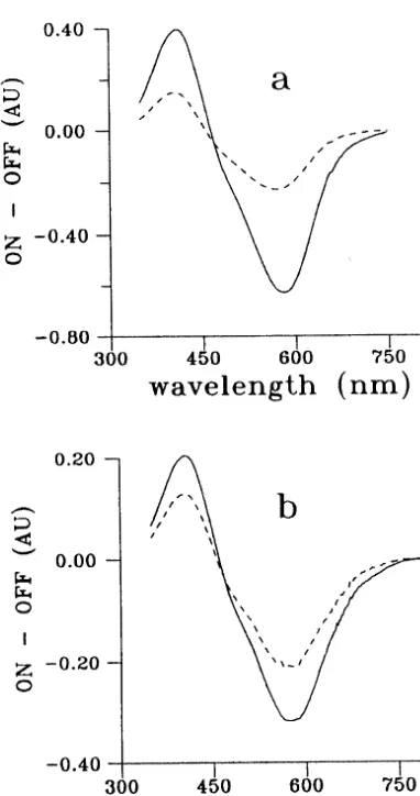
of 14-F WT and 14-F D96N measured in wide pH range (5 – 9), show a rise in absorbance at 660 nm only for 14-F WT BR water suspension and no rise in this spectral range for 14-F D96N water suspension (Druzhko et al., 1998). The differential absorbance spectra ‘light minus dark’ measured for gelatin films (Fig. 1) demonstrate no rise in 600-nm range for both WT and D96N of 14-F derivatives of BR. Thus, unlike the suspensions, the corresponding gelatin films demonstrate no drastic differences in photoinduced transforma-tions between 14-F WT and 14-F D96N.
Comparison of kinetic curves confirms this ob-servation. We have recently presented the kinetic curves of the photoinduced absorbance changes in 14-F WT and 14-F D96N water suspensions mon-itored at 412, 588 and 660 nm (Druzhko et al., 1998). In the case of the 14-F WT water suspen-sion, when the light is turned on, the 412 nm absorbance rises, the 588 nm absorbance
disap-pears and 660 nm absorbance again rises
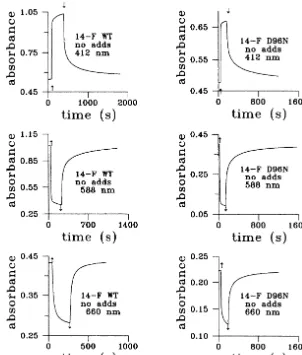
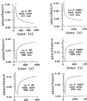
(Druzhko et al., 1998). In contrast, for 14-F D96N water suspension, when the light is turned on, the 660 nm absorbance disappears, and when the light is turned off, the 660 nm absorbance reforms and then decays very slowly (Druzhko et al., 1998). The kinetic curves of the photoinduced absorbance changes in 14-F WT and 14-F D96N gelatin films monitored at 412, 588 and 660 nm are presented in Fig. 2. As indicated in the Fig. 2, there are no drastic differences in photoinduced absorbance changes between 14-F WT and 14-F D96N gelatin films, nothing of the kind to those for suspensions. Unlike the rise of absorbance at 660 nm for suspensions — light-on for WT and light-off for D96N (Druzhko et al., 1998) — there is no rise at all in case of both types of films. Furthermore we have observed disappearance of absorbance at 660 nm. Chemical additives for film preparations (GuHCl and DAP) were used as in ref. Dyukova and Vsevolodov (1996). Kinetics for the films with chemical additives are presented in Fig. 3. This figure shows no principal differences in kinetics for gelatin films with chemical addi-tives compared with those films with no chemical additives. So, the main result of this study of films is that there is no formation of a red shifted species at 660 nm for both types of gelatin films as opposed to the observations of a red shift in the same pigments in water suspension. In addition, there are no fundamental differences in the pho-toinduced behavior between WT and D96N as observed with the water suspensions.
An explanation for these differences between the gelatin films and the water suspensions may be as follows. There was evidence of the existence of two processes initiated in both 14-F WT and D96N by yellow light (Druzhko et al., 1998). The one process is a photoinduced transformation of
Fig. 2. Photoinduced (l\500 nm) absorbance changes vs. time of 14-F WT and 14-F D96N gelatin films (no chemical additives) monitored at 412, 588 and 660 nm. denotes light is turned on,¡denotes light is turned off.
the initial ground state at 588 nm, and the an-other one is a formation and decay of red-shifted species at 660 nm. The relationship between the rates of these two processes might be the possible reason of the observed differences in photoin-duced behavior between 14-F WT and 14-F D96N in water suspension. In the case of fluori-nated analog based on D96N the photocycle with the ground state at 588 nm could not be as fast as that for WT. In the case of WT we couldn’t not observe the M-intermediate accumulation because of high rate of photocycle and it made possible to observe the red species formation and decay. Ac-tually the photocycle at 588 nm was partially
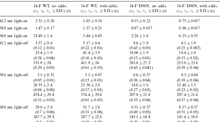
The time constants and corresponding relative amplitudes of exponential fittings of the light-on and light-off processes in 14-F WT and 14-F D96N gelatin films are presented in Table 1. The data characterize the numerical differences that the chemical additives contribute to the photoin-duced behavior of these two types of gelatin films. The effect of chemical additives is especially in evidence for light-off processes in 14-F WT films and is almost absent for 14-F D96N films. Earlier chemical additives as classified as di- and tri-amines were used for increasing the life time of the M-state of BR (Dyukova and Vsevolodov,
1996). Chemically modified films based on WT and D96N with native chromophore had a 9- to 12-fold increase in the lifetime of the M-state as compared with non-modified films. As for pig-ments with modified chromophores, chemically enhanced BR films based on 4-keto BR WT have been shown to exhibit the same tendency — 2- to 3-fold increase in the lifetime of the M-state (Druzhko and Weetall, 1997). Moreover, such a combination of chemical additives as GuHCl and DAP caused an increase in the contribution of the most long-lived component of the M-state decay for the films based on 4-keto D96N as compared
Table 1
Time constants of the one-, two- or three-exponential fits of the kinetic curves monitored at 412, 588 and 660 nm under the yellow light (l\500 nm) and after light exposurea
14-F D96N, no adds, 14-F WT, with adds,
14-F WT, no adds, 14-F D96N, with adds,
(t1,t2,t3 9S.D.) (s)
588 nm light-on 1.4790.17 0.9890.017
5.4490.45 2.2491.8
aThe light-on kinetic changes of absorbance were analyzed using one-exponential model. This function fits the experimental
curves with sufficiently small residuals 1–2%. For the second portion of the photocycle, when the protein donates a proton to the periplasm and the Schiff base takes the proton to the cytoplasm, more than one exponential fit is required (Varo and Lanyi, 1990). The sum of two or three exponentials fits the light-off kinetics with residuals about 1–3% of the measured value. The S.D. is from three measurements. The relative amplitudes Aiare given in brackets under the corresponding time constantsti. The experiments
were performed at room temperature (2291°C) and room relative humidity of 4594%.
with the observations for 4-keto WT, and it may offer an advantage as a recording media when compared to films based on the 4-keto WT (Druzhko and Weetall, 1997). In case of 14-F D96N this combination of chemical additives does not affect the lifetime of the M-state in a similar fashion (Table 1), thus it means the lack of possible advantages of 14-F D96N films when compared to films based on 14-F WT.
References
Birge, R.R., 1990. Photophysics and molecular electronic ap-plications of the rhodopsins. Ann. Rev. Phys. Chem. 41, 683 – 733.
Birge, R.R., 1995. Protein-based computers, Sci. Am. 90 – 95.
Druzhko, A., Chamorovsky, S., 1995. The cycle of pho-tochromic reactions of a bacteriorhodopsin analogs with 4-keto retinal. BioSystems 35, 133 – 136.
Druzhko, A., Weetall, H., 1997. Photoinduced transforma-tions of wild type and D96N-mutant 4-keto-bacteri-orhodopsin gelatin films. Thin Solid Films 293, 281 – 284. Druzhko, A., Zharmukhamedov, S., 1985. Biochrom’ films
based on some analogs of bacteriorhodopsin. In: Ivanitzky, G.R. (Ed.), Photosensitive Biological Complexes and Reg-istration of Optical Information. Pushchino, Moscow Re-gion Russia, pp. 129 – 136 in Russian.
Druzhko, A., Chamorovsky, S., Lukashev, E., Kononenko, A., Vsevolodov, N., 1995. 4-Keto-bacteriorhodopsin films as a promising photochromic and electrochromic material. BioSystems 35, 129 – 132.
Dyukova, T., Vsevolodov, N., 1996. U.S. Patent 5 518 858 ‘Photochromic compositions and materials containing bacteriorhodopsin’.
Ebrey, T., 1992. Light energy transduction in bacteri-orhodopsin. In: Jackson, M.B. (Ed.), Thermodynamics of Membrane Receptors and Channels. CRS Press, Boca Raton, FL, pp. 353 – 387.
Francesch, A., Alvarez, R., Lopes, S., de Lera, A., 1997. Synthesis of retinals fluorinated at odd-numbered side-chain positions and the corresponding fluorobacteri-orhodopsins. J. Org. Chem. 62, 310 – 319.
Gat, Y., Friedman, N., Sheves, M., Ottolenghi, M., 1997. Interaction between Asp-85 and the proton-releasing group in bacteriorhodopsin. A study of an O-like photocycle intermediate. Biochemistry 36, 4135 – 4148.
Grigorieff, N., Ceska, T., Downing, K., Baldwin, J., Hender-son, R., 1996. Electron-crystallographic refinement of the structure of bacteriorhodopsin. J. Mol. Biol 259, 393 – 421. Hampp, N., Silber, A., 1996. Functional dyes from nature: potentials for technical applications. Pure Appl. Chem. 68, 1361 – 1366.
Hampp, N., 2000. Bacteriorhodopsin as a photochromic reti-nal protein for optical memories. Chem. Rev. 100, 1755 – 1776.
Henderson, R., Baldwin, J.M., Ceska, T.A., Zemlin, F., Beck-mann, E., Downing, K., 1990. Model for the structure of bacteriorhodopsin based on high-resolution electron cryo-microscopy. J. Mol. Biol. 213, 899 – 929.
Hoischen, D., Steinmler, S., Grtner, W., Buss, V., Martin, H., 1997. Merocyanines as extremely bathochromically
ab-sorbing chromophores in the halobacterial membrane protein bacteriorhodopsin. Angew. Chem. Int. Ed. Engl. 36, 1630 – 1633.
Kolodner, P., Lukashev, E., Ching, Y., Druzhko, A., 1997. Electric-field and photochemical effects in D86N mutant bacteriorhodopsin substituted with 4-keto retinal. Thin Solid Films 302, 231 – 234.
Korenstein, R., Hess, B., 1977. Hydration effects oncis–trans isomerization of bacteriorhodopsin. Nature 270, 184 – 186. Oesterhelt, D., Bruchle, C., Hampp, N., 1991. A biological material for information processing. Quarterly Rev. Bio-phys. 24, 425 – 478.
Singh, A.K., Das, J., Majumdar, N., 1996. Novel bacteri-orhodopsin analogues based on azo chromophores. J. Am. Chem. Soc. 118, 6185 – 6191.
Tierno, M., Mead, D., Asato, E., Liu, R.S.H., Sekiya, N., Yoshihara, K., Chang, C., Nakanishi, K., Govindjee, R., Ebrey, T., 1990. 14-Fluorobacteriorhodopsin and other 14-substituted analogues. An extra unusually red-shifted pigment formed during dark adaptation. Biochemistry 29, 5948 – 5953.
Varo, G., Lanyi, J., 1990. Pathways of the rise and decay of the M photointermediate of bacteriorhodopsin. Biochem-istry 29, 2241 – 2250.
Vsevolodov, N., Druzhko, A., Dyukova, T., 1989. Actual possibilities of bacteriorhodopsin application in optoelec-tronics. In: Hong, F.T. (Ed.), Molecular Electronics — Biosensors and Biocomputers. Plenum, New York, pp. 381 – 384.