Two Phenolic Compounds from Chloroform Fraction of Syzygium Polycephalum MIQ. Stem Bark (Myrtaceae)
Tukiran1,*, Andika Pramudya Wardhana1, Nurul Hidajati1, Kuniyoshi Shimizu2
1Department of Chemistry, Faculty of Mathematics and Natural Sciences, Universitas Negeri Surabaya, Surabaya, Indonesia
2Department of Forests and Forest Products Sciences, Faculty of Agriculture, Kyushu University, Fukuoka, Japan
*email: [email protected]
Received October 12, 2017; Accepted April 24, 2018; Available online May 31, 2018
ABSTRACT
Syzygium polycephalum (Kupa) is a plant of the Myrtaceae family which is one of the endemic plants in Indonesia, commonly called as Gowok. The chemical components of the plant have not been reported so far. This study is intended to know the molecular structures of isolated compounds of chloroform fraction from S. polycephalum stem bark. The steam bark of the plant is dried, powdered and macerated with methanol to yield methanolic extract. The methanolic extract was then conducted to fractionation using hexane and chloroform to obtain hexane and chloroform fractions. The chloroform fraction was further subjected to separation using column chromatography to obtain pure isolates and followed by measuring of their spectroscopic evidences. The isolation of chloroform fraction had led to the findings of two pure isolates. Their structures of isolates were elucidated by extensive spectroscopic methods and by comparison with the literature data to gain two phenolic compounds that are gallic acid and 3,4,3’-tri-O-methylellagic acid.
Keywords: 3,4,3’-tri-O-methylellagic acid, gallic acid, Myrtaceae, phenolic compound, S. polycephalum.
INTRODUCTION
Myrtaceae family has about 130 genera and approximately 3800-5800 species of predominantly tropical and subtropical distribution. The Myrtaceae family is known to possess leaves with high concentrations of terpenes and considerable qualitative and quantitative variation in the types of terpenes, according to taxonomic identity and population and individual levels. These variations have pharmacological potential and many industrial applications (Barbosa, Cleber, Róbson, Renata, & Antônio, 2013).
One important genera of this family is Syzygium, which is one of the larger genera with around 500 species. They are usually trees and shrubs distributed in the tropics of the world from Africa to the West Pacific with major concentration in Malaysia. The genus is popular for the spice plant, i.e. Syzygium aromaticum (L.) Merr. & Perry which is native to Maluku Islands in Indonesia (Mohanan et al., 2015).
Syzygium can be found from sea level on swamp forests, lowland and montane forests to subalpine forests. Their habits are also vary, from canopy-emergent trees to canopy trees, understorey trees, treelets and shrubs. Indonesian Botanic Gardens (Bogor, Cibodas,
Purwodadi and Bali) have collected 40 species of Syzygium from all over Indonesia. Purwodadi Botanic Garden has collection of 15 species of Syzygium, 5 of which were from East Java. This number is still quite low compared to the total species of Syzygium in East Java (Ariyanti, Rony, Lia, & Deden, 2012).
The species of syzygium genus is well known for its medicinal properties. S. jambolana (S. cumini), popularly known as Jamun, has been the main ingredient of various medications of the traditional Indian system of medicine. Preclinical studies have shown that the various extracts of S. jambolana possess a range of pharmacological actions, such as antibacterial, antifungal, antiviral, anti-ulcerogenic, cardioprotective, anti-allergic, hepatoprotective and anti-diarrheal effects, thereby supporting its myriad traditional uses (Baliga, Harshith, Bantwal, Rajesh, & Princy, 2011). Studies in the past one decade have shown that Jamun possess antineoplastic, radioprotective and chemopreventive effects all of which are useful in the prevention and treatment of cancer (Preddy, 2014).
medicinal plant, and various parts of the tree have been used in traditional medicine, for instance as an antibiotic. S. aqueum leaf extracts have a significant composition of phenolic compounds, protective activity against free radicals as well as low pro-oxidant capability (Palanisamy et al., 2011).
The Syzygium species having appreciable medicinal properties have drawn the attention of the researchers in recent times included S. polycephalum. S. polycephalum, locally known as gowok or kupa or kepa, is an indigenous tree growth in Indonesia. It has synonyms: Eugenia polycephala Miq., Jambosa cauliflora DC., Jambosa polycephala (Miq.) Miq. and S. cauliflorum (DC.) Bennet. Gowok is indigenous to West and Central Malesia. It is common in Java and Kalimantan in Indonesia. It has been reported the presence of several compounds found in the plant that are ursolic acid, oleanolic acid, squalene, and -sitosterol from S. polycephalum leaves (Ragasa et al., 2014). It could be considered that all of these compounds are non phenolic compounds.
On the other hand, it was reported that the wood extracts (ethyl acetate extract) of S. polychephalum potentially contain anti-fungal compound (i.e. 3-O-glucosyl-3’,4’,5 -trihydroxyflavonol) to inhibit the growth of S. commune Fr. and Pleurotus sp fungi (Jemi, Syafii, Ferbianto, & Hanafi, 2010). Using the literature searching, there are no reports regarding the phenolic compounds of the stem bark of S. polycephalum. Therefore, it was of great interest to carry out a proper scientific investigation of the stem bark extract of this plant. The present study however, reports for the first time the isolation and structural elucidation of gallic acid (1) and 3,4,3’-tri-O -methylellagic acid (2) from the chloroform fraction of the stem bark of S. polycephalum.
EXPERIMENTAL SECTION
Materials
Chemicals and plant materials
The solvents used in this study are hexane, chloroform, ethyl acetate, and methanol that were of pro-analytical Grade (Grade AR) and silica gel obtained from E. Merck (Germany). The stem bark of S. polycephalum (c.a. 27 kg) was collected from a local area in Ngawi, East Java, Indonesia in December 2014. The identification of the plant
was performed by staff of Herbarium-LIPI, Purwodadi, East Java, Indonesia. A voucher sample is kept in the Herbarium of LIPI with Identification No. 0117/IPH.06/HM/I/2015, January 5, 2015.
Equipment and instruments
The equipment used to do extraction and fractionation (isolation) are filter paper, Buchner funnel, Hirsch funnel, Erlenmeyer flask, pippet, spatula, measuring glass, vials, containers, separating funnel, and vacuum rotary evaporator type BUCHI Rotavapor R-215. The equipment used to measure melting point of isolate is Fisher Scientific. Whereas, chromatographic techniques used to isolate phenolic compounds from chloroform fraction included Vacuum Liquid Chromatography (VLC) (using silica gel 60, 0.040-0.063 mm), Gravitational Column Chromatography (GCC) (silica gel 60, 0,063–0,200 mm and 0,200– 0,500 mm or 70–230 mesh ASTM), TLC analyses were carried out on silica gel 60 F254 chromaplates with the developing solvent systems. Checking the homogeneity of the compounds were made by TLC on Kieselgel gel 60 F254 pre-coated sheets (E.Merck) and the spots were detected by exposure to UV-lamp at 254 nm or 366 nm.
A number of instruments needed to identify and characterize an isolate included spectrophotometer FTIR-8400S SHIMADZU, spectrophotometer UV-1800 SHIMADZU. The
1H NMR spectra were recorded with a Bruker
DRX-600 NMR Spectrometer (600 MHz, CD3OD) instrument and the 13C NMR spectra
were obtained with the same instrument at 150 MHz in CD3OD. Chemical shifts are given in δ
(ppm) values relative to those of the solvent signal [CD3OD (δH 3.30; δC 49.0)] on the
tetramethylsilane (Sigma) scale. Procedures
Preparation of methanolic extract and its fractionation of S. polycephalum stem bark
again using chloroform and the last extract was subjected to investigate the chemical constituents.
Isolation and characterization of pure isolates The stem bark of S. polycephalum was macerated in MeOH at room temperature for 24 h and then filtered. The filtrate was concentrated under vacuum to give 349 g of crude residue. The crude (349 g) was suspended in methanol and defatted with hexane to gain hexane extract (22.38 g). This process was also repeatedly carried out by using chloroform to yield chloroform extract (5.66 g). The chloroform extract was then subjected to column chromatography (silica gel, n-hexane, n-hexane-CHCl3 and MeOH, in
order of increasing polarity) yielding 55 fractions that can be grouped to be 5 fractions [A (1-4), B (5-6), C (7-37), D (38-51), and E (52-55)]. The fraction A (1-4) was allowed to evaporate at room temperature and yielded a pure isolate as colorless needle crystal (10 mg) with mp. 256-257 oC. The crystal was
characterized by UV-Vis and FTIR and by comparison with literature data and determined its structure to be gallic acid (1). Then, fraction B (5-6) seemed that the fraction gave simple chromatogram profile and allowed to dry at fume hood yielded a pure enough isolate as off-white amorphous powder (10.3 mg) with mp. 267-269 oC. The isolate was then
characterized by UV-Vis, FTIR, LCMS and NMR spectroscopies and by comparison with literature data and determined its structure to be 3,4,3’-tri-O-methylellagic acid (2).
RESULTS AND DISCUSSION
The MeOH extract of S. polycephalum stem barks was partitioned successively with hexane and CHCl3. Successive column
chromatography of the CHCl3 extract over
silica gel using the various chromatographic techniques yielded compounds 1 and 2.
Gallic acid (1): a colorless needle crystal; MP. 256-257 C; UV-Vis (MeOH,
max): 216 and 271 nm; IR (KBr, max): = 3497,3368, 3292, 3065, 3005, 1709, 1620, 1541, 1443, 1246, 1026, and 702 cm−1; Authentic
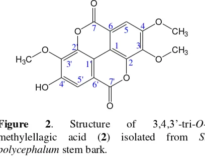
sample (gallic acid), as shown in Figure 1. An ellagic acid derivative, 3,4,3’ -tri-O-methylellagic acid (2): a white amorphous powder; MP. 267-269 oC; UV (MeOH,
max);
247 and 371 nm; IR (KBr,
max): = 3441, 2957,2918, 2851, 1753, 1728, 1611, 1578, 1493,
1361, 1298, 1115, 1092, 988 and 914 cm−1; 1H
NMR (600 MHz, DMSO-d6, ppm): δ (ppm) = 7.61 (1H, s, H-5) and 7.52 (1H, s, H-5’), 3.31 (1H, br, OH), 4.06, 4.04, and 3.99 (3H, s, -OCH3); 13C NMR (150 MHz, DMSO-d6): δ
(ppm) = 158.52 (C, C-7), 158.32 (C, C-7’), 153.71 (C, C-4), 153.06 (C, C-4’), 112.49 (C, C-5), 107.44 (C, C-5’), 141.48 (C, C-3), 140.28 (C, C-3’), 140.95 (C, C-2), 140.77 (C, C-2’), 113.44 (C, C-6), 111.84 (C, C-6’), 111.76 (C, C-1), 110.89 (C, C-1’); LC-ESI-MS (m/z 345.39 [M+H+] for C
17H12O8, as shown in
Figure 2.
Compound 1 was obtained as a colorless needle crystals (10 mg), m.p. 256-257 oC. The
UV-Vis (MeOH, max) spectrum of compound
1 showed maximum absorption at 216 and 271 nm indicating a phenolic compound
HO HO
HO
O
OH
Figure 1. Structure of gallic acid (1) isolated from S. polycephalum stem bark.
O O
O O O
O
O
HO
CH3 H3C
CH3
7
3'
5' 7'
1 3
5
1'
6 4
2
6' 4'
2'
Figure 2. Structure of 3,4,3’-tri-O -methylellagic acid (2) isolated from S. polycephalum stem bark.
The IR (KBr, νmax) spectrum exhibited the following absorption frequencies: 3497, 3368, 3292, 3065, 3005, 1709, 1620, 1541, 1443, 1246, 1026, and 702 cm−1. The IR spectrum of
it showed absorption bands broadly at 3497, 3368, 3292, 3065, and 3005 cm−1 indicating
hydroxyl group and at 1709 cm−1 indicating
carbonyl group. The absorption bands at 1620, 1541, and 1443 cm−1 indicated benzene ring
namely meta-position toward carbonyl group. The last absorption band at 702 cm−1 showed
substituted benzene. By comparing the IR data of compound 1 with that of authentic sample (gallic acid) and those reported in literature data (Tukiran, Mahmudah, Hidayati, & Shimizu, 2016), it was confirmed as gallic acid (1).
Compound 2 was obtained as an off-white amorphous powder (10.3 mg), m.p. 267-269 oC and its molecular formula C
17H12O8
was determined by the LC-ESI-MS (m/z 345.39 [M+H+]). The UV-Vis (MeOH,
max)
spectrum of compound 2 showed maximum absorption at 247 and 371 nm indicating phenolic compound with conjugated benzene
ring of carbonyl group. The IR (KBr, νmax) spectrum exhibited the following absorption frequencies: 3441, 2957, 2918, 2851, 1753, 1728, 1611, 1578, 1493, 1361, 1298, 1115, 1092, 988 and 914 cm−1. The IR spectrum of it
showed sharp absorption bands at 3441 cm−1
indicating hydroxyl group, at 2957, 2918, and 2851 cm−1 representing C-H stretching, and at
1753 and 1728 cm−1 revealing the presence of
two carbonyl groups. The characteristic absorption bands at 1611, 1578, and 1493 cm−1
indicated benzene ring system. The presence of methyl group is shown specifically at 1361 cm−1. For a while, absorption bands at 1298,
1115 and 1092 cm−1 indicated –O-aryl and –
O-CH3, respectively. The last absorption band at
988 and 914 cm−1 showed substituted benzene.
The 1H-NMR spectrum (600 MHz,
DMSO-d6, ppm) of compound 2 revealed the presence of six significant proton signals that could be explained as follows. Two signals located at δH 7.61 (1H, s) and 7.52 (1H, s) indicated two aromatic protons due to the ellagic acid skeleton. The spectrum of the compound also displayed one signal at δH 3.31 (1H, br) suggesting the presence of an aromatic hydroxyl group (aryl –OH). In addition, three signals located at δH 4.06, 4.04, and 3.99 (3H, s) showed three methoxyl groups.
The 13C-NMR spectrum (150 MHz,
DMSO-d6, ppm) of compound 2 displayed seventeen carbon signals that could be described as follows. The spectrum showed 17 signals, of which 14 signals were assigned to the ellagic acid portion and the rest signals were three methoxyl groups. Two carbon signals located at δC 158.52 and 158.32 confirming clearly for two carbonyl groups
[C-7(7’)] were attributed to ellagic acid lactone carbonyl signals, two carbon signals at δC
153.71 and 153.06 confirmed as benzene ring attached by methoxyl and hydroxyl groups
[C-4(4’)], and two carbon signals at δC 112.49 and 107.44 indicated benzene ring attached by hydrogen [C-5(5’)]. Then, two carbon signals
located at δC 141.48 and 140.28 with high intensity indicated as benzene ring attached by methoxyl groups [C-3(3’)]. For a while, two
carbon signals on the position of δC 140.95 and 140.77 represented benzene ring attached by the respect lactone groups [C-2(2’)] and δC 113.44 and 111.84 revealed benzene ring attached by carboxyl groups [C-6(6’)]. Finally, two carbon signals located at δC 111.76 and 110.89 revealed benzene ring attached by other phenyl group and vice versa [C-1(1’)] and assigned as ellagic acid skeleton.
The assignments of all protonated carbons of compound 2 were accomplished by interpretation of the HSQC NMR spectrum
indicated five connections between: δ 7.52 (H -5) and 107.44 (C-5), δ 7.61 (H-5’) and 112.49 (H-5’), δ 4.06 and 60.92 (3’-OCH3), δ 4.04 and 61.28 (3-OCH3), and δ 3.99 and 56.70 (4 -OCH3). The comparison of 1H- and 13C-NMR
spectral data of compound 2 and that were identical with those reported in literature data (Hiranrat, 2010; Gao, Hu, & Li, 2012) might be justified that the compound is 3,4,3’-tri-O -methylellagic acid.
There are no previous reports regarding the investigation of chemical components of S. polycephalum stem bark especially for phenolic compounds. In this study, two phenolic compounds were now successfully isolated from chloroform fraction of the plant that are gallic acid (1) and 3,4,3’-tri-O -methylellagic acid (2). Both compounds were found from the plant for the first time.
2015). These organic acids had also been found from 14 edible Myrtaceae fruits: Eugenia aggregata, E. brasiliensis, E. luschnathiana, E. reinwardtiana, Myrciaria cauliflora, M. dubia, M. vexator, Syzygium cumini, S. curranii, S. jambos, S. javanicum, S. malaccense, S. samarangense, and S. samarangense var. Taiwan pink (Shubhi, et al., 2013). But, in this study ellagic acid has not been found from the plant.
Meanwhile, 3,4,3’-tri-O-methylellagic acid (2) that is also phenolic compound is an ellagic acid derivative. In chemically, the structure of this compound possess ellagic skeleton as shown in Figure 3. Indeed, 3,4,3’ -tri-O-methylellagic acid (2) is ellagic acid with the hydroxyl (–OH) groups at position 3, C-4 and C-3’ are replaced by methoxyl (-OCH3)
groups. With methoxylated ellagic acid, this compound becomes more non-polar and insoluble in chloroform. So, 3,4,3’-tri-O -methylellagic acid (2) can be formed and derived from the chloroform extract.
The diversity and complexity of ellagic acid derivatives is actually determined on the type and amount of functional groups which can replace the hydroxyl groups either by methoxyl groups and the glycosyl groups mostly at position C-3, C-4, C-3’ and C-4’. From here,
for instance 3,3’-di-O-methylellagic acid (Reynertson, 2007), ellagic acid 4-O-α-L-2” -acetylrhamnopyranoside, 3-O-methylellagic
acid 3’-O-α-L-rhamnopyranoside, 3-O
-methylellagic acid 3’-O--D-glucopyranoside (Simoes-Pires et al., 2009) has been isolated from S. cumini, beside ellagic acid its self (Simoes-Pires et al., 2009; De Bona et al., 2016).
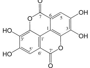
O O
OH OH
HO HO
O O
7 6 5
1 3
7' 6' 5'
3' 1'
Figure 3. Skeletal formula of ellagic acid The other examples of ellagic acid derivatives are 3-O-ellagic acid-4’-O-α -rhamnopyranoside, ellagic acid rhamnopyranoside, 3-O-methylellagic
acid-4’-O-α-2”-O-acetylrhamnopyranoside, and 3-O-methylellagic acid-4’-O-α-3”-O
-acetylrhamnopyranoside obtained from S. guineense stem bark (Djoukeng, Mansour, Tapondjou, Lontsi & Tabacchi, 2007; Khan, Khan, Sahreen, & Ahmed, 2012). Then, from the methanolic extract of S. Jambos leaf had been known containing ellagic acid derivatives: 3,3’,4’-tri-O-methylellagic acid-4-O-β-D-glucopyranoside and 3,3’,4’-tri-O -methylellagic acid (Djipa, Delme´e, & Leclercq, 2000). Therefore, ellagic acid derivatives are well known in the Syzygium genera (Myrtaceae).
However, phenolic compounds is often found in Myrtaceae plants, especially the genus Syzygium, such as S. zeylanicum (Anoop & Bindu, 2014), S. cumini (Ruan, Liang, & Yi, 2008; Pranoti & Pragya, 2014), S. samarangense (Edema & Alaga, 2012), S. polyanthum (Har & Ismail, 2012) and S. cordatum (Sidney, Siyabonga, & Kotze, 2015), etc. But the phenolic compound gallic acid is still very little found in Syzygium plants. Some of Syzygium plants containing the compound are S. cumini (Swami, Thakor, Patil, & Haldankar, 2012), S. litorale (Tukiran, Mahmudah, Hidayati, & Shimizu, 2016), and S. polyanthum (Nurlaila, 2016). Thus, it is expected that many other phenolic compounds will be found in S. polycephalum, in addition to the two compounds. Therefore, an intensive investigation of the chemical components in the plant is strongly needed to be done.
CONCLUSIONS
Phytochemical investigations of the chloroform fraction of S. polycephalum stem bark led to the isolation of two phenolic compounds: gallic acid (1) and 3,4,3’-tri-O -methylellagic acid (2). Structurally, compound 2 is a derivative compound of ellagic acid in which the acid itself is formed from two gallic acids. This means it is very reasonable that two compounds are equally found in one plant including the plant.
ACKNOWLEDGEMENTS
wish to thank also deeply thanks to Hyiya Amen for help us to measure NMR and LC-ESI-MS.
REFERENCES
Anoop, M.V. & Bindu, A.R. (2014). Pharmacognostic and physico-chemical studies on leaves of Syzygium zeylanicum (L.) D.C., International Journal of Pharmacognosy and Phytochemical Research, 6(4), 685-689. Ariyanti, E.E., Rony, I., Lia, H., & Deden, M. (2012). Distribution of Syzygium spp. (klampok) in some areas of Bromo Tengger Semeru National Park, East Java. Proceeding of Society Indonesian Biodiversity International Conference, 1, 135142.
Baliga, M.S., Harshith, P.B., Bantwal, R.V.B., Rajesh, W., & Princy, L.P. (2011). Phytochemistry, traditional uses and pharmacology of Eugenia jambolana Lam. (black plum): a review, Food Research International, xxx: 1-14.doi: 10.1016/j.foodres.2011.02.007.
Barbosa, L.C.A., Cleber, J.S., Róbson, R.T., Renata, M.S.A.M., & Antônio, L.P. (2013). Chemistry and biological activities of essential oils from Melaleuca L. species, Agriculturae Conspectus Scientificus.78(1), 11-23. Damle, M. & Nilam, D. (2015). Development
and validation of stability indicating HPLC method for determination of ellagic and gallic acids in jambul seeds (Syzygium cumini), International Journal of Applied Sciences and Biotechnology, 3(3): 434-438. doi: 10.3126./ijasbt.v3i3.12908.
De Bona, K.S., Bonfanti, G., Bitencourt, P.E., da Silva, T.P., Borges, R.M., Boligon, A., Pigatto, A., Athayde, M.L., & Moretto, M.B. (2016). Protective effect of gallic acid and Syzygium cumini extract against oxidative stress-induced cellular injury in human lymphocytes, Drug and Chemical Toxicology. 39(3), 256-263.doi:
10.3109/01480545.2015.1084631. Djipa, C.D., Delme´e, M., & Quetin-Leclercq,
J. (2000). Antimicrobial activity of bark extracts of Syzygium jambos (L.) Alston
(Myrtaceae). Journal of
Ethnopharmacology, 71, 307-313.
Djoukeng, J.D., Eliane, A.-M., Leon, A.T., David, L., & Raffaele, T. (2007). Identification of ellagic acid derivatives from stem bark of Syzygium guineense (Myrtaceae), Natural Product Communications. 2, 1-6.
Edema, M.O. & T.O. Alaga. (2012). Comparative evaluation of bioactive compounds in Hibiscus sabdariffa and Syzygium samarangense juice extracts. African Crop Science Journal, 20, 179 – 187.
Gao, Y., Hu, Q., & Li, X. (2012). Chemical composition and antioxidant activity of essential oil from Syzygium samarangense (BL.) Merr. et Perry flower-bud. Spatula DD, 2(1), 23-33. doi: 10.5455/spatula.20120126062707. Har, L.-W., & Ismail, I.S. (2012). Antioxidant
activity, total phenolics and total flavonoid of Syzygium polyanthum (Wight) Walp leaves. International Journal of Medicinal and Aromatic Plants.2(2), 219-228.
Hiranrat, A. (2010). Chemical constituents from Rhodomyrtus tomentosa (Aiton) Hassk and antibacterial activity, Thesis, Copyright of Prince of Songkla University.
Jemi, R., Wasrin, S., Fauzi, F., & Muhammad, H. (2010). Sifat anti jamur kayu kupa (Syzygium polycephalum (Miq.)). Jurnal Ilmu dan Teknologi Kayu Tropis, 8, 93-110.
Khan, R.A., Khan, M.R., Sahreen, S., & Ahmed, M. (2012). Evaluation of phenolic contents and antioxidant activity of various solvent extracts of Sonchusasper (L.) Hill. Chemistry Central Journal, 6(12), 1-7.doi: 10.1186/1752-153X-6-12.
Mohanan, N., Vrinda, K.B., Padmesh, P., Suresh, P.K.K., Mathew, D., Sreekumar, S., Biju, S.R.S., Rasiya, S.B., Kumar, S.K.P.P., & Anil J. (2015). Jawaharlal Nehru Tropical Botanic Garden and Research Institute (JNTBGRI) Annual Report 2012-13 & 2014-15, 15-17. Nurlaila, E. (2016). Identifikasi senyawa
Science Undergraduate Degree), Universitas Negeri Surabaya, Indonesia, 71-76.
Palanisamy, U.D., Ling, L.T., Manaharan, T., Sivapalan, V., Subramaniam, T., Helme, M.H., & Masilamani, T. (2011). Standardized extract of Syzygium aqueum: a safe cosmetic ingredient. International Journal of Cosmetic Science, 33(3), 269–275. doi: 10.1111/j.1468-2494.2010.00637.x. Pranoti B. & Pragya G. (2014). In vitro
evaluation of phytochemical and antioxidant properties of Syzygium cumini leaves and their synergistic effect on its antimicrobial property. International Journal of Research in Pharmaceutical Sciences. 5, 254-258. Preddy, V. (2014). Cancer: oxidative Stress
and dietary antioxidants. Chapter 10: The Indian blackberry (Jamun), antioxidant capacity, and cancer protection (authorized by Farrukh Aqil, Radha Munagala, Jeyaprakash Jeyabalan, Thwisha Joshi, Ramesh C. Gupta), 101.
Ragasa, C.Y., Torres, O.B., Shen, C.-C., Lachica, M.K.E.G., Sulit, A.B., Chua, D.B.D.L., Ancheta, A.D.M., Ismail, C.J.B., Bernaldez, F.T.E., & Raga, D.D. (2014). Triterpenes from the leaves of Syzygium polycephalum, S. cumini, and S. samarangense, Chemistry of Natural Compounds, 50(5), 942-944.
Reynertson, K.A. (2007). Phytochemical analysis of bioactive constituents from edible Myrtaceae fruits. A Dissertation Submitted to the Graduate Faculty in Biology in partial fulfillment of the requirements for the Degree of Doctor of Philosophy, The City University of New York.
Ruan, Z.P., Liang, L.Z., & Yi, M.L. (2008). Evaluation of the antioxidant activity of
Syzygium cumini leaves. Molecules, 13, 2545-2556. doi: 10.3390/ molecules13102545.
Shilpa, A.W. (2010). Bioconversion of tannic acid to gallic acid by using fungal tannase, A Theses Submitted to the Division of Biochemical Sciences, National Chemical Laboratory, Pune – 411008 (India), 7-10.
Shubhi, M., Kirar, V., Misra, K., & Nandi, S.P. (2013). Quantitative estimation of gallic acid in Rosa sinensis, Emblica officinalis, and Syzygium aromatocium by HPTLC. International Research Journal of Pharmacy, 4(7): 87-89. Doi: 10.7897/2230-8407.04719.
Sidney, M.T., Shandu, J.S., & Basson, A.K. (2015). The antibacterial and antidiarreal activities of the crude methanolic Syzygium cordatum [S.Ncik, 48 (UZ)] fruit pulp and seed extracts. Journal of Medicinal Plants Research, 9(33), 884-891. doi: 10.5897/JMPR2015.5789. Simões-Pires, C.A., Vargas, S., Marston, A,.
Ioset, J.R., Paulo, M,Q,, Matheeussen, A., & Maes, L. (2009). Ellagic acid derivatives from Syzygium cumini stem bark: investigation of their antiplasmodial activity. Natural Product Communications, 4(10), 1371- 1376. Swami, S.B., Nayan, S.J.T., Meghatai, M.P., &
Parag, M.H. (2012). Jamun (Syzygium cumini (L.)): a review of its food and medicinal uses. Food and Nutrition Sciences, 3, 1100-1117. doi.org: 10.4236/fns.2012.38146.