R E S E A R C H A R T I C L E
The Effect of Collagen Activation on Platelet Rich Plasma for
Proliferation of Periodontal Ligament Fibroblasts
Pati Tangsupati, Kwartarini Murdiastuti
Department of Periodontology, Faculty of Dentistry, Universitas Gadjah Mada, Jl. Denta 1, Sinduadi, Yogyakarta, Indonesia
Corresponding author. E-mail: [email protected]
Received date:Jan 25, 2018; Revised date: Jul 31, 2018; Accepted date: Aug 3, 2018
B
ACKGROUND: Regenerative procedure in periodontal surgery aims to improve the structure and function of the periodontium to be strong enough to support the teeth. Growth factor is essential in the process of tissue regeneration; it can be generated from the activation of Platelet-Rich Plasma (PRP). In this study, collagen was used as PRP activator. PRP release growth factor from the granules when activated. The aim of this study was to evaluate the effect of collagen on PRP activation and the collagen-activated PRP storage tofibroblast proliferation of periodontal ligament (PDL).
METHODS: Fibroblasts of PDL were obtained from
extracted premolar. PRP were obtained from 100 mL of
blood donors using double centrifugation methods. PRP were activated by collagen and subsequently incubated for 24, 48, 72 and 168 hours; after which the lysate was taken.
Fibroblasts were divided into 7 groups, which consisted of one unstimulated group as negative control, one group
stimulated by PRP lysate, and five groups stimulated with
PRP-collagen lysates that had been incubated for 24, 48, 72 and 168 hours. MTT assay was then performed after 1 and 3 days.
RESULTS: The proliferation rate of fibroblasts group
stimulated by PRP-collagen lysate was higher than the group stimulated by PRP. The storage of collagen-activated PRP for 7 days at 4°C could increase the proliferation rate.
CONCLUSION: The activation of collagen on PRP and the
stored collaged-activated PRP could increase the fibroblasts proliferation rate of PDL.
KEywORDS: collagen, platelet-rich plasma, fibroblasts
proliferation
Indones Biomed J. 2018; 10(3): 278-83
Abstract
Introduction
Platelet-Rich Plasma (PRP) is a high concentrate of autologous platelet obtained by centrifugation of autologous blood. PRP may release several types of growth factors such as platelet-derived growth factor (PDGF), transforming
growth factor (TGF)-β, insulin growth factor (IGF),
vascular endothelial growth factor (VEGF), platelet-derived endothelial cells growth factor (PDECGF), platelet-derived
angiogenesis factor (PDAF) and fibroblasts growth factor (FGF).(1) PRP could increase the proliferation of fibroblasts
and osteoblasts when used in a certain concentration range.
(2) PRP on fibroblasts could stimulate adhesion and cell
spreading on fibronectin matrix (3), as well as increasing
in actin-enrich cellular extensions. It also promotes cell migration and invasion through the basement of matrix.
The trend of fibroblasts proliferation rate has been observed
in previous studies, and the results showed the longer
the duration of stimulation of fibroblasts, the higher the
proliferation rate that has been achieved.(1,4)
Platelet will release the content of granules when they are activated. Activation of platelet can occur when the adhesion molecules are exposed due to endothelial injury
such as von Willebrand factor, collagen, fibronectin and
The amount of platelet and growth factor obtained is affected by the method used in the preparation of PRP. The growth factor obtained affects the clinical outcomes.(2) PRP preparation processed with the addition of activators has been studied. Activation of PRP with thrombin showed a very fast release of growth factors while activation with collagen showed sustained release in a relatively same amount of time from day 1, 3, to day 7. Previous studies
showed deficiency of bovine thrombin usage by the
emergence of hypersensitivity reactions, impaired viability
and altered migration of fibroblasts, decreased activity of
FGF, and decreased clot strength.(6) Results of in vitro
studies demonstrated the use of collagen is a safe and effective alternative, in addition to stimulating the release of growth factors, it also increase the consistency of PRP with gelation process.(7) Growth factors released from PRP activated by collagen include PDGF and TGF-b which
are potent stimulators for fibroblasts in cell migration,
mitogenesis, proliferation and in matrix synthesis, this is important in wound healing.(7,8)
Collagen-based biomaterial has now become the most
important material in the field of tissue engineering and
regeneration because of its high biocompatibility and low immunogenicity. Collagen can also be combined with other
molecules in the field of medical applications. Sources of
collagen are also vary, but the most widely used in clinical applications is collagen derived from mammals such bovine, while collagen derived from rat tails are generally used in in vitro research. One of the solid forms of collagen is an absorbable collagen sponge (ACS), this biomaterial is used in dentistry as a hemostat or as scaffold in tissue regeneration process. In other research, collagen was used as a carrier recombinant human bone morphogenetic protein 2 (rhBMP-2) critical size injuries in red calf.(9) The results showed an increase in the bone formation. Recent research was using a collagen sponge as a carrier of PRP in treating gingival recession class I and II.(10) The results showed the addition of keratinized gingiva and an increase of clinical attachment level. The aimed of this study was
to evaluate benefits of collagen in PRP activation and the collagen-activated PRP storage to fibroblast proliferation of periodontal ligament (PDL).
Methods
This research was an experimental laboratory and approved by ethics commite of the Faculty of Dentistry, Universitas Gadjah Mada Indonesia (No.420/KKEP/FKG-UGM/
EC/2013). The following variables were: variable of effect was the kind of treatments (PRP, collagen-activated PRP), and observation time; the affected variable was proliferation
of PDL fibroblasts. The object of research was primary PDL fibroblasts of the extracted premolars for orthodontic
treatment. Collagen was obtained from rat tail (RT) collagen and collacure sponge collagen. PRP was obtained from blood centrifugation with double centrifugation method.For 24, 48, 72, 168 hours, it was activated by RT collagen and for 24 hours by collacure. Then they were incubated and observed after 1 and 3 days
PRP Preparation
Ten mL of blood was taken from human probandus and it was put into a tube containing 1 mL of 3.8% sodium
citrate, following that, it was mixed slowly in order to make a homogenous mixture. The tube was centrifuged for 10 minutes in 1200 rpm rotation speed. The centrifugation produced two layer, the top layer was contained platelet-poor plasma (PPP) and the bottom layer was contained red blood cells PPP layer was taken using a long canula + water-intake canula was included into tubes without anticoagulant. The tube was centrifuged for 10 minutes at 3500 rpm rotation speed. This second centrifugation produced two layers, and then it was separated from PPP with isolation kit.(7)
PRP Activation
Activation of PRP was obtained by mixing the supernatant PRP and collagen, the volume ratio was 1:15.
Fibroblasts’s Culture
PDL cells taken from the root surface of teeth using a blade
#15. The ligament was taken in the middle third of tooth root, ligament was cut and then cultured using Dubelco’s
Modification of Eagle’s Medium (DMEM), added a 10% of fetal bovine serum (FBS) supplement, 2% of penicillin-streptomycin, 0.5% fungi zone and subsequently put into
incubator in temperature of 37°C.(6)
PDL fibroblast was cultured in a media in microplate
24 wells and consisted of 7 groups, which are unstimulated
fibroblasts or negative control (NC), fibroblasts stimulated by PRP lysate, fibroblasts stimulated by PRP + collagen RT lysates and incubated for 24 hours (T1), fibroblasts stimulated
by PRP + collagen RT lysates and incubated for 48 hours
(T2), fibroblasts stimulated by PRP + collagen RT lysates and incubated for 72 hours (T3), fibroblasts stimulated by
PRP + collagen RT lysates and incubated for 168 hours
(T4), and fibroblasts stimulated by PRP + collacure lysates
Results
Data in this research were taken from spectrophotometry
plate with a wave length of 570 nm. The mean of fibroblast
proliferation rate and the standard deviation based on observation times (day 1 and 3) in each group is presented in Table 1 below.
Table 1. The mean and standard deviation of the periodontal ligament fibroblast proliferation rate.
wells and were incubated at 37 oC. Fibroblast proliferation
of each group was measured by 3-(4,5-dimethylthiazol-2-yl)-2,5-diphenyltetrazolium bromide (MTT) assay on day 1 and 3 (observation times). Data were analyzed using
Two-way ANOVA, followed by least significant differenc (LSD) test with a confidence level of 95%.
Day 1 (n=42)
Day 3 (n=42)
PRP 0.2043 ± 0.0354 0.3773 ± 0.0539
NC 0.1055 ± 0.0082 0.2323 ± 0.0459
T1 0.4365 ± 0.1604 0.4628 ± 0.0574
T2 0.4363 ± 0.0815 0.5375 ± 0.0759
T3 0.3810 ± 0.0318 0.4885 ± 0.0533
T4 0.3767 ± 0.0791 0.4898 ± 0.0378
T5 0.4138 ± 0.0647 0.4972 ± 0.0629
Observation time
Groups
Table 1 showed that the mean of PDL fibroblast
proliferation was lowest in the NC group with a mean and standard deviation of 0.1055±0.0082 and 0.2323±0.0459 on
day 1 and 3 respectively. In each group, average fibroblast
proliferaton rate on day 3 was higher than that of day 1. Figure 1 showed that the lowest mean and standard
deviation of fibroblast proliferation rate were same in NC
in both groups whereas the highest one was seen in a group that incubated PRP-activated RT collagen at 24 hours of incubation in day 1 and at 48 hours of incubation in day 3.
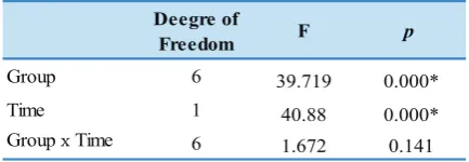
Normality and homogeneity tests showed a normally distributed and homogen data (p>0.05) therefore the statistical analysis was continued using two-way ANOVA
parametric test in order to find out whether there were differences among PDL fibroblast applied by PRP, PRP
activateed collagen and a group which was not applied by PRP activated collagen in different observation times of storage (day 1 and 3). The result is presented in Table 2. It implied that there was an effect of group and time of
observation to the proliferation cells of fibroblast in PDL
but no effect on interaction between group and time.
0,7
ti
PRP NC T1 T2 T3 T4 T5 0
0.1 0.2 0.3 0.4 0.5 0.6 0.7
Day 1 Day 3
Groups
Fibroblast Proliferation
Table 2. Result of two-way ANOVA test of periodontal ligament fibroblast proliferation.
Deegre of
Freedom F p
Group 6 39.719 0.000*
Time 1 40.88 0.000*
Group x Time 6
1.672 0.141
Figure 1. Graph of fibroblast proliferation rate on day 1 and 3. PRP: Fibroblasts + 24-hour incubation of PRP lysates; NC: Fibroblasts without stimulation; T1: Fibroblast + PRP + RT collagen lysate, 24 hours incubation; T2: Fibroblasts + PRP + RT collagen lysate, 48 hours incubation; T3: Fibroblasts + PRP + RT collagen lysate, 72 hours incubation; T4: Fibroblasts + PRP + RT collagen lysate, 168 hours incubation; T5: Fibroblasts + PRP + RT collacure lysate, 48 hours incubation.
The result in Table 2 above was subsequently
continued with Post-Hoc LSD test to see the difference of
proliferation among groups and the observation times. The
LSD test is shown in Table 3.
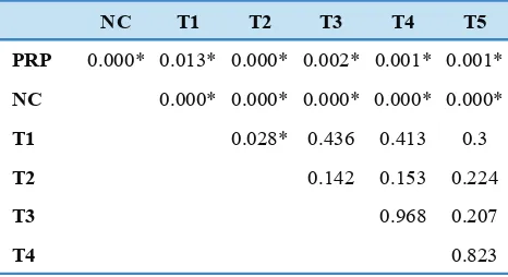
Table 3 indicated that in day 1 of observation time,
there were significant differences of PDL fibroblast
proliferation rate between group of PRP with negative control, PRP activated RT collagen in all incubations, and between negative control with all other groups. There were
no significant differences among PRP activated collagen
with different incubation times.
NC T1 T2 T3 T4 T5
PRP 0.040* 0.000* 0.000* 0.001* 0.001* 0.000*
NC 0.000* 0.000* 0.000* 0.000* 0.000*
T1 0.997 0.239 0.205 0.628
T2 0.241 0.207 0.631
T3 0.926 0.484
T4 0.428
*: significant p-value
Table 3. LSD test of periodontal ligament fibroblas proliferation among groups on day 1.
NC T1 T2 T3 T4 T5
PRP 0.000* 0.013* 0.000* 0.002* 0.001* 0.001*
NC 0.000* 0.000* 0.000* 0.000* 0.000*
T1 0.028* 0.436 0.413 0.3
T2 0.142 0.153 0.224
T3 0.968 0.207
T4 0.823
Table 4. LSD test of periodontal ligament fibroblast proliferation among all groups in day 3.
*: significant p-value
Group p-value
PRP 0.000*
NC 0.000*
T1 0.713
T2 0.050*
T3 0.002*
T4 0.010*
T5 0.470*
*: significant p-value
Table 5. Independent sample T-test of periodontal ligament fibroblast proliferation between each observed times.
Discussion
The effect of PRP to the proliferation rate of PDL fibroblast
can be seen on Table 1 with the lowest mean was observed in the negative control group. Data analyzed using
Post-Hoc LSD test is shown on Table 3 and 4. On day 1 and 3, there were significant differences between negative
control group (without PRP) compared to other groups of
PRP. It means the proliferation rate of PDL fibroblast in the
group stimulated by PRP was higher than that of without
PRP. Therefore, PRP could increase fibroblast proliferation
with observation time on day 1 and 3. This result was in line with research by Graziani, et al., that stated PRP can increase proliferation rate within 24 and 72 hours in in vitro
research.(1) Yilmaz, et al., also declared that PRP is capable to regulate biologic events such as cell proliferation, cell attachment, cell migration and extracellular matrix synthesis. (11) This may happen because PRP is supernatant with high concentration platelet that can produce various growth factors if granules in platelet are activated. Growth factor is a very important signaling molecule in various cellular processes. Each growth factor has regulatory function on cellular level and is seen in the tissue regeneration process. Table 4 implied that in day 3 of observation time, there
were significant differences of PDL fibroblast proliferation
between PRP with negative control groups, PRP-activated RT collagen in all incubation times, and between negative control groups with all other groups. There were no
significant differences among each PRP activated collagen
group with different incubation times, except between PRP-activated RT collagen group with 24 and 48 hours of incubation.
Because there was an effect of observation time to
the proliferation cells of fibroblast in PDL thus both groups
of observation times was then analyzed using independent sample T-test and the result is presented in Table 5.
Table 5 showed that there were significant differences of PDL fibroblast proliferation between day 1 and 3 of
observation times in all treatment groups. However, there
were no significant differences in groups that were applied
Growth factor is produced in platelet granulation process
including TGF-β1 to control proliferation and differentiation
of various cell types; PDGF-B to stimulate proliferation and
disrupt apoptosis; FGF inducted angiogenesis and fibroblast
proliferation; IGF acts as a mediator for other growth
factors and as chemotactic character on fibroblast of PDL;
VEGF acts in angiogenesis process. The research proved
that PRP releases TGF-β1 and PDGF-AB 3 and 2.5 times
that of whole blood respectively.(6)
Effect of collagen activation on PRP to fibroblast proliferation rate and the figure of PDL fibroblast proliferation
on observation day 1 and 3 can be observed on Table 3 and
4. There were significant differences between PRP group
(inactive PRP) compared to other groups of activated PRP. Those differences showed that adding collagen increased the
proliferation of PDL fibroblast on day 1 and 3. It meant that the proliferation of PDL fibroblast on all groups of activated
PRP with different incubation times were higher than that of the inactive PRP group. Therefore, the activation of PRP using any collagen (RT collagen or collacure sponge from
bovine’s Achilles) could increase fibroblast proliferation
with observation times of day 1 and 3. This may cause the growth factor to be released from platelet granules through molecular interaction process when platelet is activated. Enhancing platelet activation process is important in tissue regeneration process.
Collagen was used, in this research, to activate platelet granulation process. Collagen is a triple helix protein molecule with naturally involved in cascade aggregation and platelet degranulation within a body.(5,12) Platelet activation is a complex process that involved series of feedbacks and crosstalk between different pathways.
Signaling pathway in various specific receptors of platelet
simultaneously stimulated platelet form changes, growth factor secretion and other molecules from granule and at the end inducted inside-out signal process that activated
α2bβ3 integrin bond with its ligand. This bond was then
caused platelet adhesion and aggression as well as triggered outside-in signal thus caused the secretion of granule content, stabilize adhesion, aggression and platelet clot. Platelet activation process inducted by collagen started with collagen bond with glycoprotein (GP) VI receptor, which
connected to FcRγ, this bond inducted few intracellular signal series and activated integrin α2β1 which later bonded with other collagen molecules, integrin α2bβ3 as receptor of fibrinogen, integrin αvβ3 receptor of vitronectin and integrin α5β1 receptor of fibronectin. Collagen bond with
2 prominent receptors then inducted intercellular signal cascade which lead to calcium release and activation of
kynase C (PKC) protein; those were responsible to platelet activation towards series of those signals, platelet response such as aggregation was through exocytosis process.(13,14) The pattern of growth factor released from platelet granules can be different depending on the activator used. Harrison, et al., found PRP activated by collagen released growth factor continuously for 7 days and released more
TGFβ-1 in 7 days observation compared to the activation
with thrombin.(6) While the total PDGF-AB and VEGF released was relatively same on either PRP activated by
thrombin or by collagen in 7 days. TGF β-1 is a growth factor with a major role in the process of fibroblast proliferation.
Table 3 and 4 showed that there was no effect
of collagen incubation time on PRP to the fibroblast
proliferation. Activation time in this research was equal with PRP storage time. PRP was activated by collagen and kept for 24 hours (T1), 48 hours (T2), 72 hours (T3) and 168 hours (T4). After the activation, PRP lysate was
take and applied PDL culture cell. It was meant to see
whether activation or incubation time or storage time in 4°C temperature (collagen activated PRP) could be affected or
not to stimulate proliferation of PDL fibroblast. The result of Post-Hoc LSD showed comparison among group T1, T2, T3 and T4 observation on day 1 was not significantly different
(p>0.05). The similar pattern showed on observation day 3, the comparison among groups (T1, T3, and T4) did not
show any significant difference (p>0.05). This proved that PRP lysate activated with collagen and incubated for 24, 72 and 168 hours did not show differences or give the meaning
was same or equal in stimulating fibroblast proliferation.
This showed that numbers on growth factors in PRP lysate after incubation on the period of time had the same value
to stimulate proliferation of periodontal fibroblast. The
results of this study were in accordance with the conclusions obtained by Poerty, et al., that there was no different between directly and after 24 hours of long-term activation
PRP with collagen on growth factors, especially fibroblast
growth factor-2 (FGF-2).(16)
Table 5 showed there was a significant difference between proliferation figure on observation on day 1 and
3 in all groups with p<0.05. It explained that the process
of proliferation of PDL fibroblast in this research had a significant increase every day. The result is in line with prior research in which the figure of fibroblast proliferation
increased by the time or time dependent.(1,3,15) An exception to group T1 also had an increase in the mean of proliferation rate, however, using independent sample
T-test, the result was not significant. This could be caused
and treatment using MTT assay. Undissolved Formosan salt by reagent stopper (DMSO) could decrease the score of
color density by plate reader that caused absorbency figure
to be invalid.
PDL fibroblasts have the most important role in the
regeneration of periodontal tissue because of its ability to regenerate soft tissue and hard tissue. Therefore, this study used it as a representation of the periodontal tissues. PRP is a source of growth factors that can stimulate cellular activities, such as cell proliferation. PRP that is activated by collagen could also decrease the PRP clot retraction. PRP and collagen had shown to increase proliferation up
to 4 times higher. Increased fibroblast proliferation will
accelerate regeneration, thus may also clinically accelerate the healing process and improve clinical outcomes of a treatment. According to the author's knowledge, there has never been a study that examined the PRP of collagen activation in other cells.
Conclusion
In conclusion, the result of this study indicate that collagen
activation on PRP could increase periodontal fibroblast
proliferation. Incubation time of collagen activation on PRP (activated PRP storage time) was not affecting the
proliferation of PDL fibroblast, in the other hand, observation time affected the PDL fibroblast. Collacure sponge collagen had the same efficacy with rat tail collagen in inducing PDL fibroblast proliferation. We suggest for next research
to examine the effect of collagen addition on PRP at the in vivo setting, to see its effect on tissue regeneration.
References
1. Graziani F, Cei S, Ducci F, Gabriele M, Ivanovski S, Tonetti M. The in vitro effect of different PRP concentration on osteoblasts and
fibroblasts. Clin. Oral Impl. Res. 2006; 17: 212-9.
2. Careces M., Hidalgo R, Sanz A., Martinez J. Effect of
platelet-rich plasma on cell adhesion, cell migration, and myofibroblastic differentiation in human gingival fibroblast. J Periodontol. 2008;
79: 714-20.
3. Manoranjan SJ, Faizuddin M, Hemalatha M, Ranganath V. The effect of platelet derived growth factor-AB on periodontal ligament
fibroblasts: an in vitro study. J Indian Soc Periodontal. 2012; 16:
49-53.
4. Park HB, Yang JH, Chung KH. Characterization of the cytokine profile
of platelet rich plasma (PRP) and PRP-induced cell proliferation and migration: Upregulation of matrix metalloproteinase-1 and -9 in HaCaT cells. Korean J Hematol. 2011; 46; 265-72.
5. Sánchez-González DJ, Méndez-Bolaina E, Trejo-Bahena NI. Platelet-rich plasma peptides: key for regeneration. Int J Pept. 2012; 2012:5 32519. doi: 10.1155/2012/532519.
6. Harrison S, Vavken P, Kevy S, Jacobson M, Zurakowski D, Murray MM. Platelet activation by collagen provides sustained release of anabolic cytokines. Am J Sports Med. 2011; 39: 729-34.
7. Fufa D, Shealy B, Jacobson M, Kevy S, Murray MM. Activation of platelet-rich plasma using soluble type I collagen. J Oral Maxillofac Surg. 2008; 66: 684-90.
8. Anusakthien O, Webb SA, Jin QM, Giannobile WV. Platelet-derived growth factor gene delivery stimulates ex vivo gingival repair. Tissue Eng. 2003; 9: 745-56.
9. Mariner DP, Wudel JM, Miller DE. Synthetic hydrogel scaffold is an effective vehicle for delivery of INFUSE (rhBMP-2) to critical-sized calvaria bone defects in rats. J Orthop Res. 2013; 31: 401-16.
10. Naik AR, Ramesh AV, Dwarkanath CD. Use of autologous platelet rich plasma to treat gingival recession in esthetic periodontal surgery. J Indian Soc Periodontol. 2013;17: 345-53.
11. Yilmaz A, Cakar G, Ipci SD. Platelet rich plasma in reconstructive periodontal theory. In: Progress in Molecular and Environmental
Bioengineering. London: IntechOpen; 2011. p.269-83.
12. Bareil RP, Gauvan R, Berthod F. Collagen-based biomaterials for tissue engineering applications. Materials. 2010; 3: 1863-87.
13. Li Z, Delaney MK, O’Brien KA, Du X. Signaling during platelet
adhesion and activation. Artioscler Thromb Vasc Biol. 2010: 30: 2341-9.
14. Jackson EC, Ortar G, McNicol A. The effect of an inhibitor of diglyceride lipase on collagen-induced platelet activation. J Pharmacol Exp Ther. 2013; 347: 582-8.
15. Kim DH, Je YJ, Kim CD, Lee YH, Seo YJ, Lee JH, Lee Y. Can
platelet-rich plasma be used for skin rejuvenation? Evaluation of
effect of platelet-rich plasma on human dermal fibroblast. Ann
Dermatol. 2011; 23: 424-31.
16. Poerty AD, Murdiastuti K, Sudibyo. Pengaruh Lama Aktivasi