Mercury-induced oxidative stress in tomato seedlings
Un-Haing Cho *, Jung-O. Park
Department of Biology,College of Natural Science,Changwon National Uni6ersity,Changwon,Kyungsangnamdo 641-773, South Korea Received 25 August 1999; received in revised form 31 January 2000; accepted 7 February 2000
Abstract
Mercury content and distribution as well as its effects on growth and oxidative stress were investigated in 30-day-old tomato seedlings (Lycopersicon esculentum Mill.). The content of Hg increased with external Hg concentrations, and was considerably higher in roots than in shoots. Among the leaves, the mature leaves accumulated more. Excess Hg suppressed biomass production of both roots and shoots and reduced chlorophyll content in leaves. Further, substantial increases of H2O2content,
malondialde-hyde formation, and antioxidant enzyme activities such as superoxide dismutase (SOD), catalase (CAT), and peroxidase (POX) were observed in Hg-stressed plants in comparison with controls. The results suggest that the phytotoxic effects of Hg in tomato seedlings may be achieved by an enhanced production of active oxygen species (AOS) and subsequent lipid peroxidation. © 2000 Elsevier Science Ireland Ltd. All rights reserved.
Keywords:Lycopersicon esculentum; Mercury; Oxidative stress; H2O2; Lipid peroxidation; Antioxidant enzymes
www.elsevier.com/locate/plantsci
1. Introduction
The high toxicity of excessive metals has been known for a long time. The exposure of plants to metal ions causes growth inhibition or death of plants, coincidental with the alteration of mem-brane permeability of cells leading to the leakage of ions [1] and pigment destruction [2]. However, the fundamental mechanism of metal phytotoxic-ity has not yet been characterized, and little is known about the mechanisms related to absorp-tion and phytotoxicity of mercury (Hg), a cyto-toxic metal pollutant.
Active oxygen species (AOS) such as 1O 2, O2−,
OH and H2O2 are commonly generated under
stress conditions [3] and are strong oxidizing spe-cies that can rapidly attack all types of biomolecules [4], thus disrupting the normal metabolism of the cell. Meanwhile, generation of AOS, particularly H2O2has been proposed as part
of the signaling cascade leading to protection from
stresses [5]. For the protection from the oxidative stress, plant cells contain both oxygen radical detoxifying (antioxidant) enzymes such as catalase (CAT), peroxidase (POX) and superoxide dismu-tase (SOD), and non-enzymatic antioxidants such as ascorbate, glutathione and a-tocopherol [4,6].
SOD, the first enzyme in the detoxifying process, catalyzes the dismutation of O2− to H2O2 and O2
[7], CAT mediates the cleavage of H2O2 evolving
O2 [8], and POX reduces H2O2 to H2O using
several reductants available to the cells [9]. Altered activities of these antioxidant enzymes and antiox-idants commonly have been reported in plants, and are used frequently as indicators of stress [10]. In parallel to metal-induced tissue damage or cell death, alteration of both antioxidant enzyme activities [11] and antioxidant levels [12] as well as enhancement of both lipid peroxidation [13] and phytochelatin synthesis [14] have been observed. Therefore, the metal-induced phytotoxicity may be mediated by oxidative stress. However, the changes in AOS metabolism and the enzymes ac-tivities involved in scavenging AOS in response of exposing plants to metal have not been
investi-* Corresponding author. Tel.: +82-551-2797445; fax: + 82-551-2797440.
E-mail address:[email protected] (U.-H. Cho)
gated in detail. In animals, HgCl2 enhanced lipid
peroxidation in several organs, as measured by the thiobarbituric acid reaction for malondialdehyde (MDA), and reduced glutathione level [15], indi-cating that the oxidative stress-induced lipid per-oxidation may be one of the molecular mechanisms for cell injury in acute HgCl2
poison-ing. However, due to lack of data, it is difficult to assess the significance of oxidative stress induced by Hg in plants.
The objective of present study is to investigate whether Hg-induced phytotoxicity expressed as growth inhibition and chlorophyll destruction in tomato seedlings is mediated by oxidative stress. The data show that tomato seedlings exposed to toxic dose of mercury (up to 50 mM) produce
H2O2 and the activities of related antioxidant
en-zymes are altered, indicating that Hg-induced phy-totoxicity can be mediated by oxidative stress.
2. Materials and methods
2.1. Plant material
Seeds of tomato (Lycopersicon esculentum Mill. cv. Seokwang) were germinated and cultivated in pots containing perlite:vermiculite (1:1) mixture in a controlled environment chamber at 25°C with 12 h of light (250mM m−2s−1) and 70 – 80%
humid-ity. Seedlings were supplemented daily with water and twice a week with modified Hoagland solution containing the following nutrients: 28.7 mg l−1,
NH4H2PO4, 0.71 mg l−1 H3BO3, 164.1 mg l−1
Ca(NO3)2, 0.02 mg l−1 CuSO4, 2.66 mg l−1ferric
tartrate, 60.19 mg l−1 MgSO
4, 0.45 mg l−1
MnCl2, 0.004 mg l−1 MoO3, 151.65 mg l−1
KNO3, and 0.055 mg l−1ZnSO4. Thirty days after
germination, Hg was added daily to the pots as 0, 10 and 50 mM of HgCl2 in water. Plants collected
from each treatment after 10 or 20 days of Hg treatments were dried for 48 h at 70°C and weighed for biomass and Hg determination. For measurements of H2O2, MDA, chlorophyll and
antioxidant enzyme activities, fresh samples were weighed and used.
2.2. Measurement of Hg
Samples (leaves, shoots and roots) were sepa-rated and washed in deionized water two times,
and dried at 70°C for 48 h. The dried tissues were weighed and ground into a fine powder before wet ashing in HClO4:HNO3(4:1, v/v) solution. Hg was
determined by atomic absorption spectrophotome-ter equipped with vapor generative accessory (Ve-rian 200AA, Australia).
2.3. Measurement of chlorophyll and lipid peroxidation
Leaves collected at day 10 or 20 after Hg treat-ment were weighed and ground in 80% acetone. The resulting suspension was centrifuged for 10 min at 5000 rpm. The chlorophyll content of supernatant was estimated according to Arnon [16]. The level of lipid peroxides in the leaves and roots was determined as malondialdehyde (MDA) content by the thiobarbituric acid (TBA) reaction as described by Dhindsa et al. [17]. The concentra-tion of MDA was calculated based on A532–A600
(o=155 mM−1 cm−1).
2.4. Measurement of H2O2
Content of H2O2in plant tissues was determined
based on the modified method of Patterson et al. [18]. Fresh leaves or roots (150 – 300 mg) were frozen in liquid nitrogen and ground to a powder in a mortar together with frozen 5% TCA (1.5 ml) and with activated charcoal (45 mg). The ho-mogenate was centrifuged at 18 000×g for 10 min at 0°C. The supernatant was filtered through a nylon filter (45 mm, MSI) and the filtrate was
adjusted to pH 8.4 with NH4OH. After
re-filtra-tion through a nylon filter, a 500-ml aliquot was
brought to 1 ml by adding 500 ml of colorimetric
reagent. The colorimetric reagent was made daily by mixing 1:1 (v/v) 0.6 mM 4-(2-pyridy-lazo)resorcinol (disodium salt) (Sigma, St. Louis, MO) and 2% titanium (IV) chloride (diluted from 20% TiCl2 in 1 N HCl and adjusted to pH 8.4)
(Kanto Chemical, Japan) and was maintained in ice until use. The mixture was incubated for 60 min at 45°C and contents of H2O2(o=3.67mM−
1
cm−1) were determined from A
508, using H2O2
(30% Sigma) (5 – 50 mM) as a standard.
2.5. Measurement of antioxidant enzymes
by homogenization of the fresh tissue material with a mortar and pestle and a small amount of sand in a solution (5 ml g−1fresh weight)
contain-ing 50 mM KH2PO4/K2HPO4 (pH 7.0), 10 g l−1
PVP, 0.2 mM EDTA and 10 ml l−1Triton X-100.
After the homogenate was centrifuged at 12 000× g for 20 min at 4°C, the supernatant was used for immediate determination of enzyme activities. All spectrophotometric analyses were conducted on a Uvikon 922 spectrophotometer (Kontron Instru-ments, Italy). Activity of CAT was determined by monitoring the disappearance of H2O2by
measur-ing the decrease in absorbance at 240 nm of a reaction mixture containing 2 ml 29.8 mM H2O2
in KPO4 buffer (pH 7.0) and 1 ml extract [19].
Activity of SOD was assayed by the inhibition of the photochemical reduction of nitroblue tetra-zolium (NBT) according to the modified method of Becana et al. [20]. The reaction medium com-prised 0.25 ml 50 mM Na – phosphate buffer (pH 7.8) with 0.1 mM Na2EDTA, 2.73 ml O2−
-generat-ing solution and 20.45ml extract. The O2−
-generat-ing solution contained 2.2mM riboflavin, 14.3 mM
methionine, and 82.5 mM NBT. Glass cells
con-taining the mixture were placed in a cylindrical bath lined with aluminium foil at 25°C and fitted with a 22 W fluorescent lamp. The reaction was initiated by turning the light on and the reduction of NBT was followed by reading the A560 for 10
min. Blanks were run the same way but without illumination. One unit of SOD was defined as the amount of enzyme which produced a 50% inhibi-tion of NBT reducinhibi-tion under the assay condiinhibi-tions. Activity of guaiacol peroxidase (GPX) was mea-sured by monitoring the H2O2−-dependent
oxida-tion of reduced guaiacol at 470 nm [21]. One unit was defined as the enzymic amount which oxidizes 1 mM guaiacol min−1 (o=26.6 mM−1 cm−1).
Total activities (U) of enzymes and contents (nmol) of H2O2 and MDA were expressed on a
fresh weight basis.
2.6. Statistical analysis
The results are the means9S.E. of at least three independent replicates. The analyses of variance were computed on statistically significant differ-ences determined based on the appropriateF-tests. The mean differences were compared utilizing Duncan’s multiple range test.
3. Results
3.1. Hg accumulation and seedling growth
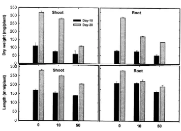
The content of Hg in tissues of tomato seedlings increased concurrently with increase in external Hg concentration and exposure time (Table 1). Hg was more accumulated in roots than in upper plant parts; Hg content in roots after 20 days was about 27-fold higher than that in shoots. The maximum accumulation of Hg was 1418.9 mg g−1
dry weight in roots treated with 50 mM Hg for 20
days. Among the leaves, the mature first leaves contained the highest Hg level whereas the younger third leaves contained the least. The ef-fects of Hg on seedling growth, expressed as dry weight and length of shoots and roots, are shown in Fig. 1. Hg-exposure induced a substantial de-pression of both root and shoot dry weights, and this effect varied as a time of the exposure and the concentration of the exogenous Hg. The growth reduction observed at the high doses of Hg ap-peared to coincide with an increased accumulation of this metal (Table 1). However, 10 mM Hg
treatment for 10 days (625.2 mg g−1dry weight of
Hg content) was not enough to suppress both the dry weight and length of roots, indicating less sensitivity of roots to initial Hg stress than that of shoots.
3.2. Chlorophyll le6els, H2O2 production and lipid
peroxidation
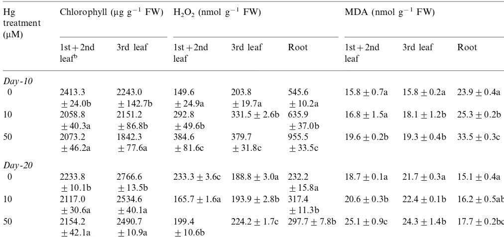
The effects of Hg on chlorophyll levels, H2O2
formation and lipid peroxidation are shown in Table 2. With a substantial amount of Hg accu-mulation (Table 1), Hg-exposure for 10 days was enough to decrease chlorophyll content particu-larly in the first and the second leaves. However, the younger third leaves were more resistant to chlorophyll destruction since 10-day treatment with 10 mM Hg was not enough to decrease
chlorophyll level.
H2O2content in roots was much higher than in
leaves. Subjecting tomato seedlings to up to 50mM
Hg for 10 days increased the level of endogenous H2O2 in comparison with control plants, and the
effect of Hg on the H2O2level measured at day-10
was much higher in leaves than in roots. However, after 20 days of exposure, H2O2 level then
To know whether lipid peroxidation was in-volved in the reduction of both seedling growth (Fig. 1) and chlorophyll level with Hg treatments,
MDA formation was investigated. A consistent increase in MDA level paralleled both an increase of Hg accumulation and a decrease of chlorophyll
Table 1
Contents of Hg in tomato seedlingsa
Hg treatment (mM) Hg content (mg g−1dry wt.)
2nd leaf 3rd leaf Shootc
1st leafb Root
Day-10
0 0
0
0 0 NDd
22.290.01e
10 20.190.03 12.890.04 ND 625.292.50
(3.5)f (3.2) (2.0)
50 25.290.01 24.490.00 20.290.01 ND 1213.797.59
(2.0) (1.7)
(2.1)
Day-20
0 0
0 0
0 0
19.490.06 16.690.3
24.790.02 819.5926.50
33.790.02 10
(3.0) (2.4)
(4.1) (2.0)
50 58.390.01 33.790.06 20.990.16 51.990.8 1418.9923.67
(2.4) (1.5) (3.7)
(4.1)
aThirty-day-old seedlings were grown in perlite:vermiculite (1:1) mixture supplemented daily with different levels of HgCl 2for
up to 20 days, and Hg content was determined at day-10 and day-20 after treatment initiation.
bLeaf number is from the bottom of the plant.
cIntact shoots containing leaves, stem and apex were used for analysis of Hg content. dND, not determined.
eValues are means9S.E. of at least three independent replicates.
fThe numbers in parenthesis indicate the relative accumulation ratio compared to root (%).
Table 2
Contents of chlorophyll, H2O2and malondialdehyde (MDA) in various parts of tomato seedlingsa
MDA (nmol g−1FW)
1st+2nd 3rd leaf 3rd leaf Root
leafb leaf leaf
50 2073.2 1842.3 384.6 379.7 955.5 19.690.2b 19.390.4b 33.590.3c
931.8c 933.5c
2490.7 199.4 17.790.2bc
2154.2
50 224.291.7c 297.797.8b 25.190.9c
942.1a 910.9a 910.6b
aThirty-day-old seedlings were grown and treated with HgCl
2 as described in Table 1, and the contents were determined at
day-10 and day-20 after treatment initiation. Note: Values are means9S.E. of at least three independent replicates. Values in a column followed by the same letter are not significantly different at the 0.05 level according to Duncan’s multiple range test.
bLeaf number is from the bottom of the plant.
level in leaves, and the MDA level observed at day-20 appeared to be related to the H2O2 level
observed at day-10. In roots, the MDA level ob-served at day-20 was much lower than that at day-10 although Hg-exposure induced a substan-tial increase of MDA at all the levels of Hg treated. Further, the pattern of MDA formation paralleled that of H2O2 formation in roots.
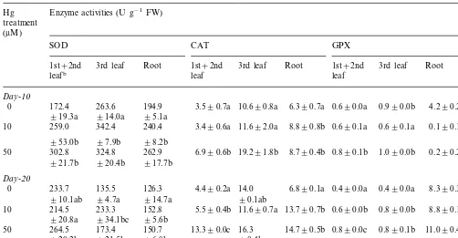
3.3. Antioxidant enzymes
The activities of SOD, CAT and GPX were investigated to determine whether Hg-exposure influenced these antioxidant enzymes (Table 3). All enzyme activities, estimated on a fresh weight basis, were substantially increased by Hg-expo-sure, depending on exposure time and treatment levels. Compared to the controls, the activity of SOD markedly increased in both leaves and roots exposed to Hg. Ten-day exposure to 10 mM Hg
was enough to increase the activity, and the in-creased SOD activities paralleled the levels of H2O2formed in leaves and roots (Table 2).
Exam-ination of two enzymes, which decompose the H2O2generated by SOD, indicated that the
activi-ties of CAT and GPX also increased in response to Hg exposure. The CAT activity in the first and the second leaves was not changed at day-10 with 10 mM Hg but increased at day-20 with 50mM Hg
compared to the controls. In the third leaves, the CAT activity increased with 50 mM Hg regardless
of exposure time. Meanwhile, when subjected to Hg stress for up to 20 days, roots maintained higher levels of activity compared to the controls. The levels of H2O2 formed in response to
Hg-ex-posure (Table 2) might be comparable to the activities of CAT particularly at day 20. The unex-pected low H2O2 levels measured at day-20 with
an increased SOD activity might be due to the increased CAT activity.
Mean GPX activity was higher in roots than in leaves. In leaves, treatment with 50 mM Hg for 10
days or all treatments with Hg for 20 days resulted in a marked increase in GPX activity. In roots, all treatments with Hg for 10 days drastically reduced the enzyme activity but further treatments up to 20 days did not change (with 10 mM) or increased
(with 50 mM) the activity. The results also
enhanced GPX activity might contribute to the reduction of H2O2 level measured at day-20 in
leaves and roots.
4. Discussion
Although a number of studies have demon-strated that metals are generally immobilized to a far greater extent at the site of metal uptake [22,23], details have not been provided with re-spect to time and concentration in specific tissues to allow for distribution in the growing plant. Since translocation will require the movement of Hg across the endodermis, membrane integrity to allow the symplastic movement might be impor-tant for the continuous Hg accumulation in shoots. However, since metal accumulation is also found in the cell wall [24] or in the apoplast [25], high Hg accumulation in roots even with substan-tial cell damage might be possible. High Hg accu-mulation in roots (Table 1) in spite of high MDA
production (Table 2) indicates the extent of cell damage which might be explained on this basis. The lowest accumulation in the upper third leaves also implies that absorbed Hg is not readily mobi-lized and redistributed in the plants, and transpira-tion [26] may not be involved in Hg translocatranspira-tion. The observed changes in the biomass of tomato seedlings were consistent with previous results ob-tained at high Hg in pea [27] and tobacco [28]. The growth reduction observed at the levels of Hg in treatments (Fig. 1) closely coincided with a consid-erable accumulation of this metal, especially in the roots. The growth reduction might be due to both the reduction in chlorophyll contents in leaves (Table 2) and membrane damage indicated as an enhanced lipid peroxidation (Table 2). It has also been suggested that heavy metals induce the defi-ciency in nutrients by reducing the uptake and transport of some mineral nutrients since metal accumulation in root may block the entry or bind-ing of the ions such as Ca, Mg and K to ion-carri-ers [29].
Table 3
Activities of antioxidant enzymes in various parts of tomato seedlingsa
Hg Enzyme activities (U g−1FW)
treatment (mM)
GPX
SOD CAT
3rd leaf Root
Root 1st+2nd
3rd leaf 3rd leaf Root 1st+2nd
1st+2nd
leafb leaf leaf
Day-10
6.390.7a 0.690.0a 0.990.0b 4.290.2b
0 172.4 263.6 194.9 3.590.7a 10.690.8a
95.1a
919.3a 914.0a
0.690.1a
10 259.0 342.4 240.4 3.490.6a 11.692.0a 8.890.8b 0.690.1a 0.190.1a
98.2b
97.9b
953.0b
324.8 0.290.2a
50 302.8 262.9 6.990.6b 19.291.8b 8.790.4b 0.890.1b 1.090.0b
920.4b 917.7b
921.7b
Day-20
0 233.7 135.5 126.3 4.490.2a 14.0 6.890.1a 0.490.0a 0.490.0a 8.390.3a
90.1ab
914.7a
94.7a
910.1ab
214.5 233.3 152.8 0.890.0b 8.890.1a
10 5.590.4b 11.690.7a 13.790.7b 0.690.0b
920.8a 934.1bc 95.6b
0.890.1b
173.4 11.090.4b
264.5
50 150.7 13.390.0c 16.3 14.790.5b 0.890.0c
96.8b
921.5b 90.4bc
920.2b
aThirty-day-old seedlings were grown and treated with HgCl
2as described in Table 1, and enzyme activities were determined
at day-10 and day-20 after treatment initiation. Note: Values are means9S.E. of at least three independent replicates. Values in a column followed by the same letter are not significantly different at the 0.05 level according to Duncan’s multiple range test.
The reduction of chlorophyll content (Table 2) observed in this study might be due to an in-creased cell or tissue damage estimated by MDA production (Table 2). Destruction of lipid compo-nents of membrane by lipid peroxidation causes membrane impairment and leakage. Meanwhile, it has also been suggested that the reduction in chlorophyll content in the presence of metal is caused by an inhibition of chlorophyll biosynthesis [30].
The present study clearly indicates that Hg-ex-posure results in an increase in H2O2 content in
plants (Table 2). Although the mechanism of Hg-induced H2O2 formation is not presently known,
heavy metals are known to be involved in many ways in production of AOS [2]. The H2O2
accumu-lation after Hg-exposure may be produced in a manner similar to H2O2 in plants cold-stressed
[31]. It is conceivable that a decrease of enzymic and non-enzymic free radical scavengers caused by heavy metals [23] may also contribute to the shift in the balance of free radical metabolism towards H2O2accumulation, and H2O2and O2− may
inter-act in the presence of certain metal ions or metal chelates to produce the highly reactive hydroxyl radical (OH). The increased H2O2 and OH
pro-duction might be involved in the lipid peroxida-tion observed in tomato seedlings (Table 3). The susceptibility to oxidative stress is a function of the overall balance between the factors that in-crease oxidant generation and those substances that exhibit antioxidant capability [9,32]. Some protective enzymes are activated in plants when production of oxygen free radicals is stimulated by stresses, and increased SOD activity may be con-sidered as circumstantial evidence for enhanced production of AOS [33]. The enhanced SOD activ-ity observed in this study (Table 3) might support the hypothesis that the H2O2resulted from oxygen
free radicals including O2−.
The increased CAT activity (Table 3) might be related to the lowered H2O2 production observed
at day 20 (Table 2), and indicated that the role of CAT might be critical to removal of H2O2induced
by Hg. Although Cd [11] inhibits CAT activity, the enzyme can take part in an efficient defense mechanism against Cu-induced oxidative stress in bean [34].
Because of a significant increase in GPX activity and strong qualitative metal-specific changes in the GPX isozyme pattern [35 – 37], the role of GPX in
removal of H2O2 might be critical in
metal-in-duced oxidative stress. GPX is a general POX which exists in both cytosol and cell wall and decomposes H2O2 [4]. The activity of GPX was
not changed in the first and the second leaves, and was reduced in both the third leaves and roots with 10 days exposure, but was increased in all organs with 20 days exposure (Table 3). Therefore, GPX activity appeared to be expressed in a long-term Hg exposure or at high Hg accumulation. It might be possible that Hg-induced GPX activity is associated with cell wall lignification and, conse-quently, with a decrease of root and stem growth (Fig. 1). POX has been postulated to stiffen the cell wall and POX-mediated lignification decreases the cell wall plasticity, and therefore reduces cell elongation, which might represent a mechanical adaptation to stress conditions [38].
Based on the present work, it can be concluded that the amount of Hg in the tissues of tomato seedlings might be associated with the reduction of both biomass (Fig. 1) and chlorophyll (Table 2). Toxic concentrations of Hg cause oxidative stress, as evidenced by the increased H2O2formation and
lipid peroxidation in leaves and roots of seedlings. The reduction of both biomass and chlorophyll concentration might result from lipid peroxida-tion-mediated cell damage in tissues. Hg-induced H2O2 formation may be associated with an
in-creased activity of SOD for O2− conversion.
Al-though parallel increases in activities of CAT and POX occur and might contribute to lower H2O2
content, the antioxidant potential in the tissues of seedlings might not be enough to block the lipid peroxidation process. The high POX activity might contribute to suppress elongation of both shoots and roots. Summing up, it was proposed that the reduced growth of tomato seedlings ex-posed to toxic levels of Hg may be induced by an enhanced production of toxic oxygen species and subsequent lipid peroxidation.
Acknowledgements
References
[1] C.H.R. De Vos, W.M. Ten Boukum, R. Vooijs, H. Schat, L.J. De Kok, Effect of copper on fatty acid composition and peroxidation of lipids in the roots of copper-tolerant and -sensitive Silene cucubalus, Plant Physiol. Biochem. 31 (1993) 151 – 158.
[2] C.M. Luna, C.A. Gonzalez, V.S. Trippi, Oxidative dam-age caused by an excess of copper in oat leaves, Plant Cell Physiol. 35 (1994) 11 – 15.
[3] B. Halliwell, J.M.C. Gutteridge, Oxygen toxicity, oxygen radicals, transition metals and disease, Biochem. J. 219 (1984) 1 – 14.
[4] K. Asada, Radical production and scavenging in the chloroplasts, in: N.R. Baker (Ed.), Photosynthesis and the Environment, Kluwer, Dordrecht, The Netherlands, 1996, pp. 123 – 150.
[5] J.F. Dat, H. Lopez-Delgado, C.H. Foyer, I.M. Scott, Parallel changes in H2O2and catalase during
thermotol-erance induced by salicylic acid or heat acclimation in mustard seedlings, Plant Physiol. 116 (1988) 1351 – 1357. [6] L.A. del Rio, G.M. Pastori, J.M. Palma, L.M. Sandalio, F. Sevilla, F.J. Corpas, A. Jimenez, E. Lopez-Huertas, J.A. Hernandez, The activated oxygen role of perox-isomes in senescence, Plant Physiol. 116 (1998) 1195 – 1200.
[7] I. Fridovich, Biological effects of the superoxide radical, Arch. Biochem. Biophys. 24 (1986) 1 – 11.
[8] J.G. Scandalios, Oxygen stress and superoxide dismu-tases, Plant Physiol. 101 (1993) 7 – 12.
[9] C.H. Foyer, M. Lelandais, K.J. Kunert, Photooxidative stress in plants, Physiol. Plant 92 (1994) 696 – 717. [10] J. Koricheva, A. Roy, J.A. Vranjic, E. Haukioja, P.R.
Hughes, O. Hanninen, Antioxidant responses to simu-lated acid rain and heavy metal deposition in birch seedlings, Environ. Pollut. 95 (1997) 249 – 258.
[11] B.V. Somashekaraiah, K. Padmaja, A.R.K. Prasad, Phy-toxicity of cadmium ions on germinating seedlings of mung bean (Phaseolus 6ulgaris): involvement of lipid peroxides in chlorophyll degradation, Physiol. Plant 85 (1992) 85 – 89.
[12] S. Sinha, M. Gupta, P. Chandra, Oxidative stress in-duced by iron in Htdrilla 6erticillata (i.f.) Royle: re-sponse of antioxidants, Ecotoxicol. Environ. Saf. 38 (1997) 286 – 291.
[13] O. Ouariti, N. Boussama, M. Zarrouk, A. Cherif, M.H. Ghorbal, Cadmium- and copper-induced changes in tomato membrane lipids, Phytochemistry 45 (1997) 1343 – 1350.
[14] S.C. Gupta, P.B. Goldsbrough, Phytochelatin accumula-tion and cadmium tolerance in selected tomato cell lines, Plant Physiol. 97 (1991) 306 – 312.
[15] Y.L. Huang, S.L. Cheng, T.H. Lin, Lipid peroxidation in rats administered with mercuric chloride, Biol. Trace Elem. Res. 52 (1996) 193 – 206.
[16] D.I. Arnon, Copper enzymes in isolated chloroplasts. Polyphenol oxidase in Beta 6ulgaris, Plant Physiol. 24 (1949) 1 – 15.
[17] R.S. Dhindsa, P. Dhindsa, T.A. Thorpe, Leaf senescence correlated with increased levels of membrane
permeabil-ity and lipid peroxidation and decreased levels of super-oxide dismutase and catalase, J. Exp. Bot. 32 (1987) 93 – 101.
[18] B.D. Patterson, E.A. McRae, I.B. Ferguson, Estimation of hydrogen peroxide in plant extracts using titaniu-m(IV), Anal. Biochem. 139 (1984) 487 – 492.
[19] R.F. Beers, I.W. Sizer, A spectrophotometric method for measuring the breakdown of hydrogen peroxide by cata-lase, J. Biol. Chem. 195 (1952) 133 – 140.
[20] N. Becana, P. Aparcio-Tejo, J.J. Irigoyen, M. Sanchez-Diaz, Some enzymes of hydrogen peroxide metabolism in leaves and root nodules of Medicago sati6a, Plant Physiol. 82 (1986) 1169 – 1171.
[21] B. Chance, A.C. Maehly, Assay of catalases and peroxi-dases, Methods Enzymol. 2 (1955) 764 – 775.
[22] D.A. Cataldo, T.R. Garland, R.E. Wildung, Cadmium distribution and chemical fate in soybean plants, Plant Physiol. 68 (1981) 835 – 839.
[23] D.E. Salt, R.C. Prince, I.J. Pickering, I. Raskin, Mecha-nisms of cadmium mobility and accumulation in Indian mustard, Plant Physiol. 109 (1995) 1427 – 1433.
[24] E. Lozano-Rodriguez, L.E. Hernandez, P. Bonay, R.O. Carpena-Ruiz, Distribution of cadmium in shoot and root tissues of maize and pea plants. Physiological dis-turbances, J. Exp. Bot. 306 (1997) 123 – 128.
[25] D. Neumann, U.Z. Nieden, W. Schwieger, I. Leopold, O. Lichtenberger, Heavy metal tolerance of Minuartia 6erna, J. Plant Physiol. 151 (1997) 101 – 108.
[26] R.T. Hardiman, B. Jacoby, Absorption and transloca-tion of Cd in bush beans (Phaseolus6ulgaris), Physiol. Plant 61 (1984) 670 – 674.
[27] W. Beauford, J. Barber, A.R. Barringer, Uptake and distribution of mercury within higher plants, Physiol. Plant 39 (1977) 261 – 265.
[28] E.M. Suszcynsky, J.R. Shann, Phytotoxicity and accu-mulation of mercury in tobacco subjected to different exposure routes, Environ. Tox. Chem. 14 (1995) 61 – 67. [29] M. Burzynski, The influence of lead and cadmium on the absorption and distribution of potassium, calcium, mag-nesium and iron in cucumber seedlings, Acta Physiol. Plant 9 (1987) 229 – 238.
[30] F. Van Assche, H. Clijsters, Effects of metals on enzyme activity in plants, Plant Cell Environ. 13 (1990) 195 – 206. [31] T.K. Prasad, M.D. Anderson, B.A. Martin, C.R. Stew-art, Evidence for chilling-induced oxidative stress in maize seedlings and a regulatory role for hydrogen peroxide, Plant Cell 6 (1994) 65 – 74.
[32] C.H.R. de Vos, H. Schat, M.A.M. de Waal, R. Vooijs, W.H.O. Ernst, Increased resistance to copper-induced damage of the root cell plasmalemma in copper-tolerant Silene cucubalus, Physiol. Plant 82 (1991) 523 – 528. [33] E.F. Elstner, G.A. Wagner, W. Schultz, Activated
oxy-gen in green plants in relation to stress situations, Curr. Top. Plant Biochem. Physiol. 7 (1988) 159 – 187. [34] J. Weckx, H. Clijsters, Oxidative damage and defense
mechanisms in primary leaves of Phaseolus6ulgarisas a result of root assimilation of toxic amounts of copper, Physiol. Plant 96 (1996) 506 – 512.
specific isozyme patterns of peroxidase in Phaseolus 6ulgaris L., Arch. Int. Physiol. Biochim. Biophys. 94 (1986) 60.
[36] S. Mazhoudi, A. Chaoui, M.H. Ghorbal, E.E. Ferjani, Response of antioxidant enzymes to excess copper in tomato (Lycopersicon esculentum, Mill), Plant Sci. 127 (1997) 129 – 137.
[37] A. Chaoui, M.H. Mazhoudi, S. Ghorbal, E.E. Ferjani, Cadmium and zinc induction of lipid peroxidation and effects on antioxidant enzyme activities in bean (Phaseo -lus6ulgarisL.), Plant Sci. 127 (1997) 139 – 147.
[38] M. Sanchez, G. Revilla, I. Zarra, Changes in peroxidase activity associated with cell walls during pine hypocotyls growth, Ann. Bot. 75 (1995) 415 – 419.