Right Heart Catheterization on Sinus Venosus type of Atrial Septal Defect with Partial Anomalous Pulmonary Vein Drainage
Pamrayogi Hutomo , Lucia Krisdinarti, Nahar Taufi q
Department of Cardiology and Vascular Medicine
Faculty of Medicine Universitas Gadjah Mada - Dr. Sardjito General Hospital, Yogyakarta, Indonesia
BACKGROUND
Atrial septal defect (ASD) is a non-cyanotic congenital heart disease characterized by the presence of defects on atrium septal wall (1). ASD is 10% of congenital heart disease (2). This defect is the third most common congenital heart disorder with an estimated incidence of 56 out of 100,000 live births. With the improved echocardiographic technique that can diagnose a defect in asymptomatic abnormalities, the estimated incidence is increased to 100 out of 100,000 live births (3). The disorder is more common in women than men with a ratio of male: female 1: 2. Sinus venosus defect itself occurs in approximately 10% of all ASD and most often located at the entrance of the superior vena cava to the right atrium which is also often associated with abnormalities on the right pulmonary venous drainage into the right atrium as in this case. The more rare disorder is at the entrance of the inferior vena cava to the right atrium where it is often associated with anomalous pulmonary venous drainage of the right lung into the inferior vena cava (Scimitar Syndrome). Anomalous pulmonary venous drainage concomitantly happen on 10% of ASD (4).
A t r i a l s e p t a l d e f e c t c a n s o m e t i m e s undiagnosed for years. Of all congenital heart defects that were newly diagnosed in adulthood, 25-30 % of which is an atrial septal defect. With increasing age, symptoms such as weakness, exercise intolerance, shortness of breath, palpitations, and manifestations of heart failure becomes more prominent and life expectancy is reduced. The onset of ischemic heart disease and other comorbid associated with decreased compliant and left ventricular (such as essential hypertension, aortic valve stenosis, aging) leading to increased flow left to right shunt through the defect, which would further exacerbate the symptoms and clinical deterioration (3).
A cardiac catheterization is rarely used only for diagnostic purposes. Most catheterization
is performed to close the defect transcatheter. Diagnostic procedure usually precedes the transcatheter defect closure, by measuring the pressures in heart chambers and comparison between pulmonary flow and systemic flow. In some patients, angiography is done to find related anomalies that are not visible from the non- invasive imaging. In patients with coronary artery disease and in pulmonary hypertension, diagnostic catheterization is indicated for further evaluation (3).
[image:1.595.309.525.534.682.2]In this case report, we reported a woman aged 21 years with a chief complaint of fatigue during activity that is already being felt since childhood. From a physical examination, electrocardiogram, chest x-ray and echocardiography, patient was diagnosed with sinus venosus type ASD with partial anomalous pulmonary venous drainage. Patient then underwent right heart catheterization procedure to determine whether it is still possible to do defect closure. The purpose of this case report is to discuss the indications and right heart catheterization techniques on ASD in particular of sinus venosus type accompanied by anomalous pulmonary venous drainage.
CASE
A woman aged 21 years was referred to Dr. Sardjito Hospital from Graha Medika Hospital with complaints of fatigue on exertion. The complaints are already being felt since childhood but never examined because it does not interfere with daily activities. Patients also said that she was susceptible to coughs almost once a month in childhood. When she was examined to the general practitioner, it is said that she had respiratory tract infection. Two years before admission, complaints of fatigue and shortness of breath on exertion increases. Patients also complained of occasional chest pain on strenuous activity. Patients comfortably lying in bed with one pillow and never woke at night because of shortness of breath. Patients went to the Graha Medika Hospital in Yogyakarta and is said to be suffering from congenital heart disease. Patients then referred to Dr. Sardjito Hospital to get further examination. A history of congenital heart disease in the family was denied.
From physical examination, the patient was fully conscious, height 155 cm, weight 48 kg, body mass index 19.98 kg/ m2. Blood pressure 105/65
[image:2.595.71.288.82.343.2]mmHg, pulse 72 beats/min, respiration rate 20 times/min, body temperature of 36.4 °C. There is no anemic conjunctiva and icteric sclera. There is no palpable neck lymph node enlargement. The chest looks symmetrical, with no retraction or lag motion. From heart examination, there is no impression of cardiomegaly. The S1 is single, there is S2 split wide fi xed, pan systolic murmur in the second spatium intercostal space in left the linea of left parasternalis. From lung examination, we found resonant percussion sounds, with normal
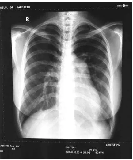
Figure 2.Chest x-ray showed the cardio-thoracic ratio of 0.47, protrusion of pulmonary segments, increased perihilar hazing as well as pruning on peripheral pulmonary vascular.
F i g u r e 3 . O v e r v i e w f r o m t r a n s t h o r a k a l echocardiography in apical view showed a sinus venosus type of ASD.
[image:2.595.307.528.85.218.2]F i g u r e 4 . O v e r v i e w f r o m t r a n s t h o r a k a l echocardiography in apical view showed the pulmonary veins empty into the right atrium.
[image:2.595.69.288.406.539.2] [image:2.595.71.290.589.721.2]Figure 6. Right heart catheterization: with the tip of a catheter in the right pulmonary vein, visible fl ow of contrast fi lling the pulmonary vein lumen and empties into the right atrium.
Figure 7. Diagram of right heart catheterization before oxygen test.
Figure 8. Diagram of right heart catheterization after oxygen test.
vesicular sound, without any additional breath sounds on auscultation. On examination of the abdomen, there is no distended abdominal wall, normal bowel sounds with timpani percussion. There is no hepatomegaly and splenomegaly. The extremities are warm with a palpable pulse, capillary refi ll time <2 seconds, with no sign of cyanosis.
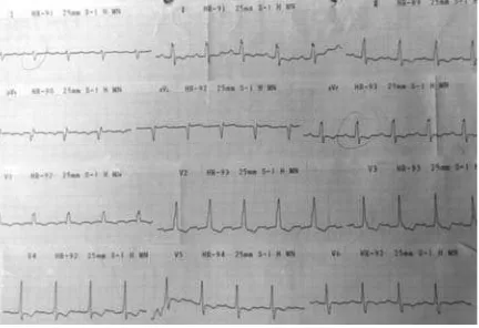
Laboratory examination are within normal limit. Investigations by electrocardiogram (ECG) showed sinus rhythm with a frequency of 91 x/min, right axis deviation. There is also right ventricular enlargement with right ventricular strain and incomplete right bundle branch block.
From posteroanterior (PA) chest x-ray examination, the cardio-thoracic ratio is 0.47 with protrusion of pulmonary segments as well as increased perihiler opacity. There is reduced pulmonary vascularization in the peripheral lung (pruning) that support the possibility of pulmonary hypertension in these patients.
Echocardiography examination revealed right atrial dilation (47 mm) and right ventricle dilation (47 mm), sinus venosus type of ASD with diameter of 1.6-1.8 cm with left to right shunt, global and segmental left ventricular systolic function is normal with ejection fraction is 78 %, normal left ventricular diastolic function, normal right ventricular systolic function, mild tricuspid valve regurgitation, mild pulmonary hypertension, and partial anomalous pulmonary venous drainage. Transesophageal echocardiography was then performed and showed sinus venosus type of ASD with a diameter of 1.6 to 1.8 cm, with left to right shunt. Both the left pulmonary veins drainage into the left atrium while both right pulmonary veins drainage into the right atrium. There is mild tricuspid valve regurgitation with a pressure gradient of 38 mmHg and moderate pulmonary hypertension.
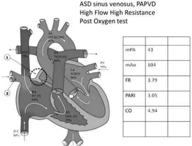
[image:3.595.87.285.572.723.2]aorta pulmonary artery pressure (mPA/mAo) is 0.52. Cardiac output is 3.39 L/min. After oxygen tests, the pulmonary artery pressure is 74/21(43) mmHg, right ventricle pressure is 69/0 mmHg, right atrial pressure is 6/7(3) mmHg, left atrial pressure is 11/8(5) mmHg, aortic pressure is 139/80(104) mmHg. The O2 saturation obtained in SVC is 72%, IVC is 84%, RA is 93%, RV is 95%, Aorta is 97%, PA is 94%, PV is 99%, LA is 96%. Flow Ratio is 3.79, PARI is 3.05 WU. The average ratio of the aorta pulmonary artery pressure (mPA/mAo) is 0.41. Cardiac output is 4.94 L/min. The conclusion of the examination is sinus venosus type of ASD with a value of fl ow ratio >1.5 and the value of PARI >8 WU and not reactive with oxygen test. There is concomitant partial pulmonary veins drainage into the right atrium (fi gure 6).
Based on the results of the examination, the patient was discharged and planned surgery to close the ASD and fix the pulmonary veins drainage back to the left atrium.
DISCUSSION
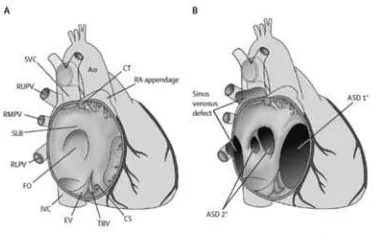
Depending on the location of defect in the atrial septum, ASD classifi ed into :
1. ASD secundum, lies in the middle of an atrial septal defect .
2. ASD sinus venosus if the location of the defect on the top near the mouth of the superior vena cava (87%) or on the bottom near the mouth of the inferior vena cava. Approximately 4-11 % of ASD is a sinus venosus type (5).
3. ASD primum, located near the tricuspid valve. This type is usually accompanied by the splitting (cleft) of the mitral valve anterior.
Nowdays ASD primum included in the group of atrio-ventricular septal defects (AVSD) 4. ASD coronary sinus, the defect is located at
[image:4.595.87.275.85.205.2]the mouth of the coronary sinus. This type is usually accompanied by a persistent left superior vena cava and unroof coronary sinus. Figure 8 shows the anatomy of the atrial septum and the types of ASD (6).
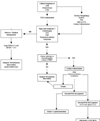
Troise et al. (7) proposed an algorithm of ASD management in fi gure 10. The ASD sinus venosus type and pulmonary hypertension in this case can be diagnosed by performing transthorakal and transesophageal echocardiography. The purpose of right heart catheterization in this case is to assess the value of pulmonary vascular resistance and reactivity towards oxygen test.
Cardiac catheterization in this case was initiated in the access of right femoral artery and vein. On the right femoral vein, we inserted 7F size sheath and 7F size Swan Ganz catheter. The same procedure was performed on the femoral artery, herein incorporated smaller sheath which is 6F sheath.
Measurement of oxygen saturation and pressure then performed. C-arm was positioned on antero-posterior view. At fi rst, we measure oxygen saturation and pressure in the abdominal aorta with a 5F size MP catheter. The obtained aortic pressure was 132/70 (94) mmHg and oxygen saturation was 97%. Then the 5F MP catheter was inserted into the femoral vein through a sheath in the femoral vein, the catheter then pushed into the inferior vena cava up (IVC) to approximately just before the diaphragm. Blood samples were taken in the IVC, the obtained oxygen saturation was 74%. Carefully, the catheter is pushed upwards by the catheter tip facing lateral to the superior vena cava (SVC) and then the tip of the catheter is positioned to the medial and pushed into the innominate vein. In the innominate vein, selective contrast agent injection should be done to know whether there is supra-cardiac shunt or not. After that, the tip of the catheter is positioned in the SVC, and blood samples was taken, the obtained oxygen saturation was 62%.
From these results, mixed venous blood saturation then calculated using the formula :
Mixed Vein (MV) = 3SVC + 1 IVC 4
The obtained MV oxygen saturation was 65%. The catheter tip is derived from the SVC to the right atrium (RA) by placing the tip of the catheter laterally to ensure that the catheter tip is in the RA. If the tip of the catheter is directed medially, it is possible that blood samples taken
[image:5.595.90.506.78.582.2]are not from the right atrium but from the left atrium (LA) through the ASD. Obtained RA pressure was 12/13(9) mmHg with O2 saturation was 85%. Here we can see the O2 saturation in the RA increase (step-up) of more than 7% compared with MV saturation which indicating that the shunt occurs at
the atrial level. In the situation where there is an O2 saturation step-up of 7% then a shunt is suspected (8). Increased O2 saturation ≥11% in the SVC-RA is required to demonstrate a shunt between SVC and RA. An increase of 7% is needed to detect shunt between the ventricle and the RA, while a 5% increase is needed to detect shunts between the right ventricle and the pulmonary artery (PA) (9). In this case, the step-up that occurred in the SVC-RA is 30% indicating a shunt between the SVC with the right atrium where one of the causes is the anomaly of pulmonary venous (PV) drainage to the RA.
In right heart catheterization, especially in the case of ASD, the RA saturation measurements should be obtained in three places (the upper, middle, and bottom of atrium) to be able to confi rm the type of ASD from the echocardiography result. An O2 saturation step-up at the top of the RA supports the diagnosis of sinus venosus type ASD. In secundum type ASD, the step-up happens in the middle of the RA. If the step-up occurs in the lower RA near the tricuspid valve and the ASD can not be bypassed by catheter, consider the possibility of anomalous pulmonary venous drainage into the coronary sinus (10) . In this case unfortunately the RA saturation measurements are not taken in three places, so the right heart catheterization can not confi rmed the type of ASD.
Measurement was continued by pointing the catheter tip to medial through the ASD to the LA, the tip of the catheter is then directed upward to the PV. The obtained PV O2 saturation was 98%. The tip of the catheter is then withdrawn into the LA. The obtained LA pressure was 13/12(8) mmHg with O2 saturation was 90%. LA systolic blood pressure was slightly higher than the RA which indicates that the shunt direction is left to right.
Subsequent measurement was intended to measure the saturation and pressure in the right ventricle (RV), PA and pulmonary capillary. For this purpose, we use the 7F size Swan Ganz catheter that were inserted through the femoral vein sheath. At fi rst, the tip of the catheter is placed in the RA. Then the balloon catheter was developed. The balloon catheter is then going to follow the fl ow of blood to the RV, the operator may need to help steer the balloon tip more easily if the balloon tip did not led to the place we want to reach. When the tip of the balloon is in the RV, operators need to consider carefully the ECG recording on the monitor. Ventricular arrhythmias often appears
in the form of ventricular premature contraction (VPC) when the catheter tip touches the ventricular wall. The pressure in the ventricles will decrease at the time of VPC, so if VPC happens too often it will lead to the patient’s hemodynamic disturbances. In addition, measurements of pressure exerted at the time of VPC became less valid assessed. In this case, the obtained RV pressure was 77/0 mmHg with O2 saturation of 84%.
The balloon catheter then directed upward to follow the fl ow of blood to the PA. Then we calculated the pressure and O2 saturation in the main pulmonary artery (MPA). The obtained pressure was 74/29(49) mmHg with O2 saturation was 80%. The balloon catheter is then directed forward to the pulmonary capillary wedge (PCW). The obtained PCW pressure was 8/11(5) mmHg. From these saturation and pressure, we calculated the value of fl ow ratio (FR), pulmonary vascular resistance (PVR) or pulmonary arteriolar resistance index (PARI), and systemic vascular resistance (SVR) using these formulas:
FR = Ao saturation – MV saturation PV saturation – PA saturation
Indexed pulmonary fl ow (Qp) = O2 Consumption (mL/min/m2).
1,36 x 10 x Hb x ((PV Sat–PA Sat)/100)
Indexed systemic fl ow (Qs) =
O2 Consumption (mL/min/m2)
1,36 x 10 x Hb x ((Ao Sat–MV Sat)/100
PVR/PARI =
Mean PA pressure – mean LA pressure Qp
SVR = Mean Ao pressure – mean RA pressure Qs
From the above formula, the fl ow ratio was 1.74, PARI was 9.95 WU and SVR was 36.8 WU. Flow ratio of more than 1.5 indicates a need for action in defect closure. While the value of PARI >8 showed pulmonary hypertension.
PARI >8 Woods Unit (WU), the patients were given 100% oxygen for 10 minutes and then the same measurement performed. If after the test oxygen FR was rise and PARI was decline, meaning that pulmonary hypertension is still reversible. But if there are no change in FR and PARI, that means pulmonary hypertension is already irreversible so the closure of ASD is contra-indicated. Oxygen test is also done if the pressure in the PA is more than two- thirds of systemic pressure (6).
Lopes and O’Leary (11) proposed a hemodynamic criteria for assessing the operability of patients with pulmonary hypertension due to congenital heart disease. Patients are expected to have good postoperative results if it has the baseline PVR <6 WU/m2 and the ratio of PVR/
SVR <0.3. If the patient has a value of 6-9 PVR WU/m2 and the ratio of PVR/SVR 0.3-0.5 then it is
recommended to test the pulmonary vasoreactivity to vasodilator agents (such as O2, Nitric Oxide) and if it meets all the criteria of which PVR index decrease by 20%, the ratio of PVR/SVR decreased 20 %, the PVR index post test <6 WU/m2, and the
PVR/SVR ratio post test <0.3 then the patient may do the ASD closure surgery.
European Society of Cardiology (ESC) in 2015 proposed a positive response criteria on acute vasodilator test which is as follow: decrease of mean PA pressure ≥10 mmHg to reach an absolute value of ≤40 mm Hg with a fixed or increased value of cardiac output (12). European Society of Cardiology (ESC) in 2010 stated that patients with signifi cant shunt (with symptoms of fl uid overload on the right ventricle) and PVR <5 WU must undergo ASD closure regardless of any symptoms (Class recommendation I, Level of Evidence B). Patients with PVR ≥5 WU, but <2/3 SVR or PA pressure <2/3 systemic pressure (baseline or after vasodilator test) with a ratio of Qp : Qs >1.5 then it could be considered for intervention procedure (Class of recommendation IIb, Level of Evidence C) (13).
In this case report, we performed vasodilator test with 100% oxygen with an oxygen mask for 10 minutes. After that, we re-measure the pressure and oxygen saturation in each heart chambers. There were decreased of PARI from 9.95 to 3.05 and SVR 29.9 WU. There is also an increase in the FR from 1.74 to 3.79. According to ESC criteria, this patient were not reactive to oxygen test because the decrease of mean PA pressure <10 mmHg and absolute PA pressure >40 mmHg,
but based on the criteria proposed by Lopes and O’Leary, patient may underwent ASD closure by surgery because there is decrease of PVR 69%, a decrease in the ratio of PVR/SVR 63%, the fi nal PVR of 3.03 WU and fi nal PVR/SVR ratio 0.1. Based on the criteria of operability from Lopes and O’Leary, the patient may underwent ASD correction surgery.
From the results obtained in cardiac catheterization, it is also necessary to consider the vasodilator agents we used. The data from INOP I Study (Inhaled Nitric Oxide as a Preoperative Test) showed that vasodilator test with oxygen and Nitric Oxide (NO) using the criteria of ratio PVR/SVR <0.33 has a sensitivity of 97% and specifi city of 90% in evaluating the operability of patients when compared with using oxygen only (sensitivity 64%, specifi city 68%) (14). This is especially important in patients who has borderline result.
CONCLUSION
In this case report, we reported a woman aged 21 years with complaints of fatigue on exertion. From the physical examination, we found single fi rst heart sound (S1), S2 wide fi xed, pan systolic murmur in the left parasternal border second intercostal space. The electrocardiogram showed sinus rhythm with heart rate frequency of 91 x/min, right axis deviation, right ventricular enlargement with right ventricular strain and incomplete right bundle branch block. Patients then underwent transthorakal and then transesophageal echocardiography, the results indicate the presence of sinus venosus type ASD with partial anomalous pulmonary venous drainage to the right atrium. The right heart catheterization was performed, the FR was >1.5 and the PARI >8 WU and not reactive to oxygen test.
Right heart catheterization in ASD can be addressed as invasive diagnostic procedures and interventions. In this case, where the type of ASD is sinus venosus, the closure of the defect can only be done with surgery, so the purpose of right heart catheterization was to assess the pulmonary artery reactivity with oxygen test as an indication to correct the defect.
The right heart catheterization in the case of ASD with partial anomalous pulmonary venous drainage can be differentiated with the case of ASD without abnormalities anomalous pulmonary venous drainage in which the step-up value of O2 oxygen saturation in the RA was higher in the former than the latter.
REFERENCES
Porter CBJ, Edwards WD. 2008.
1. Atrial septal
defect. In: Allen HD, Driscoll DJ, Shaddy RE, Feltes TF. Moss and Adam heart disease in infants, children, and adolescents, including the fetus and young adult. 7th edition. Philadelphia:
Lippincott William & Wilkins. h.632-45. Pedra CAC, Pedra SRF. 2012.
2. Atrial level
shunts including partial anomalous pulmonary venous connection and Scimitar syndrome. In: Moller JH, Hoffman JIE, Benson W, Van Hare GF, Wren C, editor. Pediatric cardiovascular medicine. Second Edition. Oxford: Blackwell Publishing. h.289-307.
Geva T, Martins JD, Wald RM. 2014. Atrial 3.
septal defects. Lancet. 383: 1921-32. Park MK. 2008.
4. Pediatric cardiology for
practitioners. Fifth edition. Philadelphia: Mosby Elsevier.
Attenhofer JCH, Connolly HM, Danielson 5.
GK, Bailey KR, Schaff HV, Shen WK, Warnes CA, Seward JB, Puga FJ, Tajik AJ. 2005. Sinus venosus atrial septal defect: long-term postoperative outcome for 115 patients.
Circulation. 112: 1953–58. Djer MM. 2014.
6. Penanganan penyakit jantung
bawaan tanpa operasi (kardiologi intervensi)-petunjuk praktis menangani pasien dan mengedukasi keluarga. Jakarta: CV Sagung Seto.
Troise D, Ringel RE, Vricella LA, Arciprete 7.
P. 2006. Atrial Septal Defects and Partial
Anomalous Pulmonary Venous Connection. Techbooks: PPG-Quark. p.1057-75.
Watson S, Gorski KA. 2011.
8. Invasive Cardiology
A Manual for Cath Lab Personnel. Kanada: Jones & Bartlett Learning.
Moscucci M. 2014.
9. Grossman & Baim’s
Cardiac Catheterization, Angiography, and Intervention. Eight Edition. Philadelphia: Lippincott Williams & Wilkins.
Swanton RH, Banerjee S. 2008.
10. Swanton’s
Cardiology: a concise guide to clinical practice. Sixth Edition. Massachusetts: Blackwell Publishing.
Lopes AA, O’Leary PW. 2009. Measurement, 11.
interpretation and use of haemodynamic parameters in pulmonary hypertension associated with congenital cardiac disease.
Cardiol Young. 19: 431 – 435.
Galie N, Humbert M, Vachiery J, Gibbs S, 12.
Lang I, Torbicki A, Simonneau G, Peacock A, Noordegraaf AV, Beghetti M, Ghofrani A, Sanchez MAG, Hansmann G, Klepetko W, Lancellotti P, Matucci M, McDonagh T, Pierard LA, Trindade PT, Zompatori M, Hoeper M. 2015. 2015 ESC/ERS Guidelines for the diagnosis and treatment of pulmonary hypertension. European Heart Journal. 1-58. Baumgartner H, Bonhoeffer P, DeGroot NMS, 13.
de Haan F, Deanfi eld JE, Galie N, Gatzoulis MA, Gohlke-Baerwolf C, Kaemmerer H, Kilner P, Meijboom F, Mulder BJM, Oechslin E, Oliver JM, Serraf A, Szatmari A, Thaulow E, Vouhe PR, Walma E. 2010. ESC Guidelines for the management of grown-up congenital heart disease (new version 2010). European Heart Journal. 1-43.
Balzer DT, Kort HW, Day RW, Corneli HM, 14.