Antiviral activity of NMSO3 against respiratory syncytial
virus infection in vitro and in vivo
Kazufumi Kimura
a, Shuichi Mori
a, Kiyoko Tomita
a, Kouei Ohno
a,
Kazuo Takahashi
a, Shiro Shigeta
a,*, Masaki Terada
b,1aDepartment of Microbiology,Fukushima Medical Uni6ersity School of Medicine,1,Hikarigaoka,Fukushima,960-1247, Japan bNissin Molecular Biology Institute,20 O6erland Street,Boston,MA 02215, USA
Received 18 November 1999; accepted 21 March 2000
Abstract
NMSO3, a sulfated sialyl lipid was evaluated for its efficacy against respiratory syncytial virus (RSV) and other myxovirus infections in cell culture. The median effective concentration (50% effective concentration, EC50) of
NMSO3 against replication of the Long strain of RSV in HEp-2 cells was 0.2 and 0.32mM by optical ELISA and the plaque reduction method, respectively. On the other hand, the corresponding values for ribavirin were 10.5 and 11.2mM, respectively. NMSO3 showed potent activity against other laboratory strains as well as fresh clinical isolates of RSV, and the average EC50was similar to that for Long strain. NMSO3 exhibited minimal cytotoxicity against
HEp-2, MDCK, HMV-2 and Vero cells for which the median cytotoxic concentration (CC50) was more than 685mM.
The selectivity index [SI=(CC50for HEp-2/EC50)] of NMSO3 for RSV exceeded 2978 and that of ribavirin was 6.
The EC50of NMSO3 against influenza virus (FluV) A (H3N2) was 23.8 by the MTT method using HMV-2 cells, and
17.8 mM by the TCID50 method using MDCK cells. NMSO3 did not inhibit replication of influenza B virus,
parainfluenza virus type 2 and canine distemper virus at 103mM. NMSO3 inhibited RSV infection of HEp-2 cells when it was added between 0 and 1.5 h after virus infection. By a temperature shift experiment during the period of contact between the virus and cells, NMSO3 inhibited both the binding of RSV to the cells and its penetration into the cells. Prophylactic and therapeutic efficacy of NMSO3 against RSV infection in cotton rats was examined. Intraperitoneal administration of 100 mg/kg per day of NMSO3 to cotton rats from 1 day before or 1 h after to 3 days after the RSV infection, once a day every day, decreased the RSV titer in lungs to 10−1.26to 10−1.63compared
to the control rats which were infected with RSV and left untreated. © 2000 Elsevier Science B.V. All rights reserved.
Keywords:Cell culture infectious dose; RSV infections; NMSO3
www.elsevier.com/locate/antiviral
1. Introduction
Respiratory syncytial virus (RSV) causes severe lower respiratory tract infections in infants and immunocompromised adults (MacDonald et al.,
* Corresponding author. Tel.:+81-24-5482111; fax:+ 81-24-5485072.
1Present address: Central Research Institute, Nissin Food Products Co. Ltd., 2247 Noji, Kusatsu, Shiga 525-0055, Japan.
1982; Harrington et al., 1992). No vaccines are currently available for the prevention of RSV infections. As a chemotherapeutic drug, only rib-avirin is approved for use in RSV infections. Ribavirin is reported to be myelocytotoxic when it is administered by the intravenous route and is therefore licensed only for delivery to the respira-tory tract as a small particle aerosol (Smith et al., 1991; Lewinsohn et al., 1996). Aerosol administra-tion of drugs to patients, especially infants and children, is rather difficult to manipulate and con-trol at home and the patients must visit or have to be admitted to hospitals for the chemotherapy. Thus, the development of novel anti-RSV drugs that can be administered orally or parenterally is strongly demanded.
NMSO3 is a sulfated sialyl lipid and was re-ported to be nontoxic to LCR male mice following single intraperitoneal injection at 1500 mg/kg and by 14 daily injections of 350 mg/kg per day (M. Terada, personal communication). In this study, we examined the antiviral activity of this com-pound against various RSV and other paramyx-ovirus and orthomyxparamyx-ovirus infections in cell culture and found that it was inhibitory to antigen production and plaque formation of RSV in HEp-2 cells. We also examined the prophylactic and therapeutic activity of NMSO3 against RSV infec-tion in cotton rats by intraperitoneal administra-tion and found a significant decrease of RSV titer in lungs of the infected rats.
2. Materials and methods
2.1. Cell cultures and 6iruses
HEp-2, HeLa, MDCK, HMV-2 and Vero cells were serially passaged in Eagle’s minimum essen-tial medium (MEM) supplemented with 5% fetal calf serum (FCS), 100 units of penicillin G, 100
mg/ml of streptomycin, 0.2% sodium bicarbonate and 2 mM L-glutamine. The source of the Long
strain of RSV and preparations of stocks of the virus have been reported previously (Kawana et al., 1987). FM58-8 (RSV, type A) and SM61-48 (RSV, type B) strains were provided by the De-partments of Pediatrics at the Fukushima Medical
University and Sapporo Medical University, re-spectively, and passaged more than 10 times in our laboratory in HEp-2 cells (Shigeta et al., 1992a). K686, K656, N869 and Y751 are fresh RSV strains isolated from patients with acute respira-tory infections by Dr K. Mizuta of National Sendai Hospital. The serotype of these clinical strains had not been determined. All the RSV strains were passaged in HEp-2 cells and virus stocks were stored at −80°C. Influenza virus (FluV) type A, Ishikawa/7/82//H3N2 and FluV type B, Singapore/222/79 were passaged more than ten times in embryonated eggs and three times in MDCK cells in the presence of 2mg/ml of trypsin in our laboratory and the virus stocks were stored at −80°C. Parainfluenza virus (PfluV) type 2 (Greer) and canine distemper virus (CDV) (On-derstepoort) strains were obtained from Dr K. Mizuta of National Sendai Hospital and Dr C. Kai of Tokyo University, respectively. Both viruses were passaged more than three times in Vero cells in our laboratory and the virus stocks were stored at −80°C.
2.2. Chemicals and reagents
NMSO3 was synthesized at The Central Re-search Institute of Nissin Food Products Co. Ltd., Kusatsu, Shiga, Japan. The chemical name of NMSO3 is sodium [2,2-bis(docosyl-oxymethyl) propyl-5-acetoamido-3,5-dideoxyl-4,7,8,9-tetra-O-(sodium-oxysulfonyl) -D-glycero -a-D galacto 2
-nonulopyranosid]onate with a molecular weight of 1478.7. The structural formula is shown in Fig. 1. NMSO3 is soluble in water (more than 10% at 60°C and more than 20% at 80°C). Ribavirin was used as the positive anti-RSV drug control and was provided by Yamasa Cooperation, Choshi, Chiba, Japan. Dextran sulfate (Mw, 8000) was
from Denka Seiken Co. (Niigata, Japan) and rabbit anti-mouse IgG antibody conjugated with peroxidase from Dako Japan (Kyoto, Japan). Mouse anti-RSV monoclonal antibody was pur-chased from Chemicon International Inc. (Temec-ula, CA, catalog number, MAB-858-1, isotype is IgG2a,k). This antibody is specific for the fusion protein of RSV with a molecular weight of 47 – 49 kD.
2.3. Titration of RSV infecti6ity
Titration of RSV infectivity was performed with a 50% cell culture infectious dose (CCID) method. HEp-2 cells were seeded in a Falcon 3072 plate (96 wells, Becton Dickinson Co.) and cul-tured with 200 ml of MEM plus 5% fetal calf serum (FCS) for 2 days at 37°C in 5% CO2. When
cell sheets became confluent, the culture medium was changed with 100 ml of MEM plus 2% FCS and 100ml RSV suspension at serial 10-fold dilu-tions was added. The virus-inoculated cultures were further incubated at 35°C in 5% CO2 and
characteristic cytopathic effects (CPE) of RSV were observed. The culture medium was replaced every 2 days and the median CCID (CCID50) was
determined at 6 days after infection by the method of Reed and Muench (1938).
2.4. E6aluation of anti6iral acti6ity in 6itro
The antiviral activity for RSV was assessed using both enzyme-linked immunoadsorbent as-say (ELISA) and plaque reduction (PR) methods. For ELISA, NMSO3 or ribavirin was added to
eight wells in the first line of a Falcon 3072 plate (96 wells) at a concentration of 1000mg/ml. Serial 5-fold dilutions were performed in MEM plus 2% FCS and antibiotics cited above (maintenance medium), and 100ml dilutions/well were prepared. To all wells except those of the control cells, 50ml of maintenance medium containing 100 TCID50
of RSV was added. Approximately 8×10³cells in
50 ml of MEM plus 2% FCS were then added to each well. As controls, the wells containing cells and virus but no compound (virus control), and those containing only cells and neither virus nor compound (cell control) were prepared.
The enzyme immunoassay technique used for RSV is a slight variation of the method of Ander-son et al. (1985). Briefly, at 5 days after infection, the cells in the wells were washed three times with PBS containing 0.5% Tween 20 (washing medium) and fixed with an 80% (vol./vol.) solution of acetone-PBS at 4°C for 15 min. After fixation, the cells were air-dried, precoated with 200 ml of PBS with 10% FCS for 30 min at 35°C, washed with the washing medium, and after the addition of 40
ml of mouse anti-RSV monoclonal antibody di-luted in PBS with 10% FCS, the cells were incu-bated at room temperature for 1 h. After the incubation, the cells were washed three times with the washing medium and 40ml of peroxidase-con-jugated rabbit anti-mouse immunoglobulin G (IgG) diluted in PBS with 10% FCS was added. The plates were incubated at room temperature for 1 h and the cells were washed three times with washing medium. Following the addition of 0.4 mg/ml of O-phenylendiamine dihydrochloride plus 0.015% H2O2 in 0.15 M citrate phosphate
buffer (Ph 5.5), the cells were incubated at room temperature for 30 min. The reaction was stopped with 3.5 M HCl and the color of the product was read at an absorbance of 490 nm wavelength using a computer-controlled microplate reader (Model 3550, Bio Rad). Thus, the viral antigen production of mock-infected and infected HEp-2 cells was evaluated by absorbance of the product oxidized by peroxidase and H2O2.
The EC50 of a compound was defined as the
concentration at which 50% decrease of viral anti-gen synthesis was achieved. The percent protec-tion was calculated using the following formula:
[(ODc)V−(ODt)V] /[(ODc)V−(ODc)M]×100, where (ODc)V, (ODt)V, (ODc)M indicate the ab-sorbance (optical density) of the virus-infected control (no compound), test sample, and the mock-infected control (no virus and no com-pound), respectively.
The plaque reduction method for RSV and CDV was described previously (Shigeta et al., 1992b). HEp-2 cells were used for RSV, and Vero cells for CDV. Each one ml of the HEp-2 cell or Vero cell suspensions with 1×10⁴
cells/ml in MEM plus 5% FCS were seeded in a Falcon 3047 tray (24 wells per tray), and incubated at 35°C for 2 days. When a monolayer of cells was obtained after 2 days incubation, approximately 50 plaque-forming units (pfu) of RSV or CDV in 100ml of maintenance medium were inoculated into the cells, immediately followed by the addition of 900
ml of serially 4-fold diluted compound in the maintenance medium and the culture was incu-bated at 35°C for 90 min. After the incubation, the culture medium was refed with the overlay medium which contained the same concentrations of the compound as in the maintenance medium plus 0.6% methylcellulose (Methocel A-4M Pre-mium, Dow Chemical Co. Midland, MI). After 4 days incubation at 35°C, the medium was aspi-rated and the cells were fixed with 5% formalin in PBS for 60 min and stained with 0.02% crystal violet for 30 min at room temperature. The num-ber of plaques was counted under 40× magnifi-cation by light microscopy and the concentration of the compounds which reduced the number of plaques to 50% of the control was determined as the EC50.
The activities of the compounds against FluV-A, B and PfluV-2 were examined by the MTT method. The precise method of MTT antiviral assay for FluV and PfluV was reported previously (Watanabe et al., 1994; Mori et al., 1995).
2.5. Inhibition assay for 6irus adsorption and penetration
At 4°C RSV binds to cells but does not pene-trate through the cell membrane. When virus-bound cells are exposed to a 35°C condition, the virus starts to penetrate the cell membrane. The
inhibition assay basically followed that described by Hosoya et al. (1991); in brief, HEp-2 mono-layer cells were prepared in two 24-well Falcon tissue culture trays. Approximately 100 pfu of RSV in 100 ml of the maintenance medium were inoculated into the cells and incubated at 4°C for 90 min with or without NMSO4 or the other compounds. After 90 min adsorption of virus at 4°C, the cells in one tray containing the virus and the compound were washed three times with the maintenance medium, and 1 ml of overlay medium was added before incubation at 35°C. The cells in the other tray with the virus only were washed three times with the maintenance medium after 90 min adsorption, and the test compound was added to the cells before incubation at 35°C for 90 min. After incubation, the cells were washed three times with the maintenance medium and the overlay medium was then added. Both trays were incubated for 5 days at 35°C and the cells were fixed with 10% formalin, stained with 0.02% crystal violet and examined for the number of plaques. The former tray was used for the evaluation of inhibition of adsorption (binding) and the latter for the evaluation of inhibition of penetration.
2.6. Inhibition of syncytium formation and immunofluorescent staining
The inhibitory effect of the test compounds against syncytium formation was monitored by counting the number of antigen-positive foci and cells in a syncytium after immunofluorescence staining of the infected cells. HeLa cells were seeded in a Lab-Tek chamber slide (eight cham-bers, Nunc, Naperville, IL) and incubated at 37°C in an atmosphere containing 5% CO2. When the
cell sheets became confluent, approximately 50 pfu of RSV were inoculated into the cells in each chamber. After adsorption and penetration of the cells by the virus following incubation of the cells at 37°C for 90 min, serial 4-fold dilutions of the compound in the maintenance medium containing 0.4% methylcellulose were added to the chamber (in triplicate) and the cultures were incubated at 35°C in a CO2 incubator. At 36 h after infection
washed twice with PBS and fixed with acetone for 10 min at room temperature. The fixed cells were stained with FITC-conjugated rabbit antibody against RSV (Denka Seiken, Niigata, Japan) for 30 min at 37°C. After staining, the cells were washed with PBS and mounted with 20% glycerol in PBS. Immunofluorescence was analyzed under a fluorescent microscope (Nikon Opiphot, Nikon Industrial, Tokyo, Japan).
2.7. Cytotoxicity
The cytotoxicity of the compounds was deter-mined using quantitative colorimetric MTT assay (Mosmann, 1983). Compounds were diluted seri-ally in 2-old using MEM plus 6% FCS. The dilutions were transferred to the wells of a flat-bottomed cell culture plate (Falcon 3072, 96 wells) containing the cultured cells at 3×10³ cells per
well (approximately 30% confluent). Duplicate control wells containing the same number of cells in the same medium but no compound were in-cluded in each assay. The culture tray was incu-bated at 35°C and when the cells in the control reached the stage of confluent sheets (1×10⁵
cells on the 4th day of culture), the cells were examined for morphological changes and the number of viable cells. For evaluation of the viable cell num-ber, 50ml of MTT at a concentration of 5 mg/ml in PBS was added to each well containing the cells and 200 ml of the culture medium. After 3 – 6 h incubation of the cells at 37°C, when the cultures in the control wells turned blue, acidified iso-propanol and triton X were added to each well. The optical density (OD) in each well of each plate was determined using a 96-well plate reader (Model 3550, Bio Rad) with double wavelengths of 540 and 690 nm. To ensure that the OD reading was proportional to the cell number, the ODs obtained from a linearly decreasing number of cells were plotted for each assay and only the assays in which the resulting correlation coeffi-cients were 0.90 or more were employed in the analyses. The median cytotoxic concentration (CC50) was calculated as the concentration of the
compounds which decreased the number of viable cells to 50% of the cell control at 4 days.
2.8. Infection of cotton rats with RSV and assessment of prophylactic effect of NMSO3
Ten-week-old female cotton rats were grouped into three separate cages and infected with 1000 CCID50 of RSV Long strain intranasally. One
hundred mg/kg each of NMSO3 and ribavirin was introduced intraperitoneally to two groups with five rats each. The diluent of the compounds (PBS, pH 7.2) was introduced to the third group. The compounds were injected to rats every 24 h, from 1 day before to 3 days after the RSV infec-tion. At 24 h after the last injection of compound, the rats were anesthetized and sacrificed for the titration of RSV in the lungs. The same ment was repeated twice and in the third experi-ment, the infected rats were grouped into four and treated with 100, 20, and 4 mg/kg NMSO3, re-spectively. Four rats each were used per group, including the control which received PBS instead of NMSO3. The compound was administered in-traperitoneally from 1 h after the RSV infection and same doses of NMSO3 were administered every 24 h to 3 days after infection. Lungs were removed at 24 h after the last administration of NMSO3. All lungs were weighed, minced in equal volume (wt./wt.) of MEM plus 2% FCS and homogenized with an Hitachi homogenizer (HG-30) for 30 s in ice. After the homogenization, the suspension was centrifuged at 3000 rpm for 5 min and the supernatant was used for the titration of virus infectivity.
2.9. Statistic analysis
The difference between RSV titer in the lungs of three experimental groups was analyzed by a nonparametric analysis using the Kruskal – Wallis method.
3. Results
3.1. Anti6iral acti6ity of NMSO3 against RSV, FluV, PfluV-2 and CDV
Table 1
Antiviral activities of NMSO3 and ribavirin against RSV and FluV A
FluV A(Ishikawa strain)
23.896.3
MTTc 11.297.7
7.797.7 17.894.9
CCID50d
aAverage for three independent experiments. bHEp-2 cells were used.
cHMV-2 cells were used. dMDCK cells were used.
values were obtained. The EC50 values for RSV
obtained by the ELISA and PR methods were similar or slightly lower in those determined by ELISA than by PR. Those of MNSO3 for RSV were 0.20 and 0.32mM and those of ribavirin for RSV were 10.5 and 11.1 mM, respectively. NMSO3 showed a 35 – 53 times lower EC50 than
ribavirin for the Long strain of RSV. The EC50s
of NMSO3 for FluV A was examined by the MTT and TCID50methods and the EC50s (24 and
18 mM) were twice higher than those of ribavirin (Table 1). NMSO3 did not show any antiviral activity against FluV-B, PfluV-2 and CDV at 103
mM (data not shown). Because the ELISA method is more sensitive and objective than the PR method for the evaluation of anti-RSV activity, we employed ELISA for further experiments on the anti-RSV activity of NMSO3.
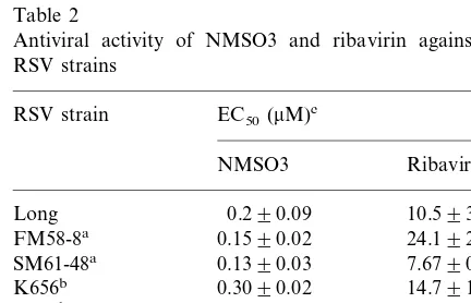
3.2. Anti6iral acti6ity of NMSO3 against se6eral RSV strains
The antiviral activities of MNSO3 and ribavirin against a standard RSV strain (Long), laboratory strains (FM58-8 and SM61-48), and newly iso-lated clinical strains (K656, K686, Y751 and N689) of RSV were examined. Both MNSO3 and ribavirin exhibited significant antiviral activities against the standard, laboratory and clinical RSV strains. The antiviral activity of MNSO3 against all the RSV strains was much stronger than that of ribavirin (Table 2). The average EC50 of
MNSO3 for all the RSV strains was 0.23mM and that of ribavirin was 12.3 mM. Thus MNSO3 possessed 53.5 times more potent inhibitory activ-ity than ribavirin against RSV replication in HEp-2 cells.
3.3. Cytotoxicity
The cytotoxicities of NMSO3 and ribavirin against HEp-2, HMV-2, MDCK and Vero cell lines were examined. No significant cytotoxicity of NMSO3 was observed against any of the cell lines both by MTT assay and morphological observa-tion, even at the highest concentration of the compound examined (685 mM for MTT assay, and 1028mM for morphological observation). On
Table 2
Antiviral activity of NMSO3 and ribavirin against several RSV strains
cAverage for three independent experiments.
the other hand, the CC50s of ribavirin for four cell
lines were 74 – 97mM by MTT assay and 2.5 – 5.5 times these concentrations by morphological obser-vation. The selectivity indices (SI: CC50for HEp-2
by MTT assay/EC50for RSV by ELISA assay) of
NMSO3 and ribavirin were calculated as being more than 2978 and 6, respectively.
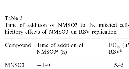
3.4. Time of addition experiment
In order to explore the mechanism of antiviral activity of NMSO3 against RSV, the compound was added to HEp-2 cells before, during and after virus adsorption, and its inhibitory effect on virus replication was examined. When NMSO3 was added before the viral infection or 1.5-h after viral infection, it was less inhibitory against RSV replication. When it was added during 0 – 1.5 h of the infection, that is at the time of virus adsorption, it was found to be inhibitory against RSV replication. However, when MNSO3 was added from 1.5 to 120 h after infection it was still inhibitory to some extent against virus replication (Table 3). Thus we concluded that NMSO3 primarily inhibited the adsorption of virus on the cells and some other later processes of RSV infection.
3.5. Inhibitory acti6ity of NMSO3 against RSV adsorption on and penetration of the cell membrane
The inhibitory activity of NMSO3 against RSV adsorption to the cells and penetration through the cell membrane was examined shifting the tempera-ture during the contact period of the virus with the cells. Dextran sulfate (Mw 8000) was used as a
typical adsorption inhibitor of RSV, and anti-RSV (F) monoclonal antibody as the inhibitor of pene-tration/fusion. Both NMSO3 and anti-RSV (F) monoclonal antibody inhibited adsorption as well as penetration of the virus at similar concentrations of the compound, whereas dextran sulfate inhibited the adsorption of the virus at a ten times lower concentration than that required for the inhibition of penetration of the virus (Fig. 2).
3.6. Inhibitory acti6ity of NMSO3 against syncytium formation of RSV-infected cells
After the adsorption and penetration of RSV following a by 90 min incubation of virus and cells at 37°C, the cells were incubated with or without NMSO4 and monitored for syncytium formation of RSV-infected cells. NMSO3 inhibited the ap-pearance of RSV-antigen-bearing cells at 2.74mM whereas it inhibited syncytium formation at a 0.11
mM concentration (Fig. 3).
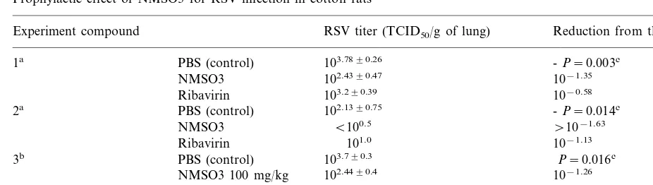
3.7. Prophylactic and therapeutic effects of RSV infection in cotton rats
One hundred milligrams per kg each of NMSO3 and ribavirin were injected to cotton rats intraperi-toneally every 24 h from 1 day before to 3 days after the intranasal infection of RSV. At 24 h after the last injection of the compounds, the rats were sacrificed, lungs were removed and RSV titer in the lungs was quantified. Two experiments indicated that the infectious virus titer in lungs of RSV-in-fected and NMSO3 injected rats decreased by 10−1.35 or 10−1.63 of the controls which were
RSV-infected and PBS-injected. The decrease of virus in lungs was greater in the NMSO3-treated group than in the group treated by ribavirin (Table 4).
Table 3
Time of addition of NMSO3 to the infected cells and in-hibitory effects of NMSO3 on RSV replication
Compound Time of addition of EC50(mM) for
NMSO3a(h) RSVb
−1–0
MNSO3 5.45
0.53 −1–1.5
0.46 −1–120
0–1.5 0.26
6.08 1.5–3
1.5–120 1.09
Ribavirin 0–1.5 234.7
1.5–120 12.0
Fig. 2. Inhibitory effects of NMSO3 (panel A), anti-RSV (F) monoclonal antibody (panel B) and dextran sulfate (panel C) on adsorption onto and penetration of RSV into HEp-2 cells. HEp-2 cell monolayers were infected with 100 pfu of RSV in 100ml of MEM plus 2% FCS (maintenance medium) per well, and incubated at 4°C for 90 min with or without the test compounds. (1) Inhibition of adsorption ("---"). The cells infected with virus and treated with the compound were washed with maintenance medium, and maintenance medium containing 0.6% methylcellulose (overlay medium) was added. (2) Inhibition of penetration (---). The cells infected with virus without the compound were washed and the compound was then added to the cells. The cells were incubated at 35°C for 90 min and washed again and then the overlay medium was added. All cells were incubated at 35°C for 5 days and examined for the number of plaques. The data give average values for three independent experiments. The bars indicate standard deviations.
In the third experiment, NMSO3 injection into the peritoneal cavity started from 1 h after the RSV infection and was repeated every 24 h to 3 days after infection. Lungs were removed at 24 h after the last injection of compound and virus infectivity was determined. A significant reduction of RSV titer in lungs was observed in the three groups that received different doses of NMSO3, that is, 100, 20 and 4 mg/kg, respectively. The most prominent therapeu-tic effect was observed in the group of rats that received 20 mg/kg of NMSO3.
4. Discussion
Acute respiratory infections caused by RSV are known to be most prevalent in infants and children. The mortality of RSV infection is usually low, whereas it increases to 37 – 73% in infants with heart or pulmonary failure, and to 36 – 45% in recipients of a bone marrow transplant (MacDonald et al. 1982; Englund et al., 1988; Harrington et al., 1992). A novel anti-RSV compound with better therapeu-tic efficacy and safety than ribavirin has been searched for several years. Nucleoside analogues, antisense oligonucleotides, polyoxometalates, and benzodithiin derivatives have been reported to be inhibitory against RSV replications in cell culture
Table 4
Prophylactic effect of NMSO3 for RSV infection in cotton rats
Experiment compound RSV titer (TCID50/g of lung) Reduction from the control
103.7890.26
PBS (control) -P=0.003c
1a
102.4390.47 10−1.35 NMSO3
103.290.39
Ribavirin 10−0.58
2a PBS (control) 102.1390.75 -P=0.014c
B100.5
NMSO3 \10−1.63
101.0 10−1.13
Ribavirin
103.790.3
PBS (control) P=0.016c
3b
NMSO3 100 mg/kg 102.4490.4 10−1.26 102.2890.2
NMSO3 20 mg/kg 10−1.42
NMSO3 4 mg/kg 102.8490.5 10−0.86
aNMSO3 and ribavirin were injected into the peritoneal cavity of RSV-infected rats at a dose of 100 mg/kg once a day from 1 day before to 3 days after the infection. Five rats were used per group.
bNMSO3 was injected into the peritoneal cavity of RSV-infected cotton rats at doses indicated in this table once a day from 1 h to 3 days after the infection. Four rats were used per group.
cPvalues were examined for the different groups by Kruskal–Wallis analysis.
(Kawana et al., 1987; Shigeta et al., 1992a; Jairath et al., 1997; Barnard et al., 1997; Watanabe et al., 1998; for a review see De Clercq, 1996). A few reports of prophylaxis for RSV infection in mon-keys and humans with monoclonal RSV anti-body and therapeutic efficacy in RSV-infected cotton rats with the natural polyphenol polymer, SP303 have appeared (Wyde et al., 1993; Weltzin et al., 1996; Impact RSV Study Group, 1998).
NMSO3 showed prominent inhibitory activity against RSV and marginal activity against FluV-A, but no activity against FluV B, PfluV-2 or CDV. NMSO3 inhibited not only the replication of the standard Long strain but also other laboratory and clinical strains of RSV at lower concentrations than ribavirin. Its EC50 in vitro for RSV was 53 times
lower than that of ribavirin. In general, negatively charged polysaccharides inhibit the adsorption of viruses on the cell membrane by interference of static electric binding between the viral envelope and cell membrane. NMSO3 has four sulfate residues in one molecule, is negatively charged and may inhibit the binding of RSV to the cell mem-brane. According to the time of addition, NMSO3 inhibited RSV replication when it was added during the time of virus adsorption and penetration (0 – 1.5 h). On the other hand, the inhibitory activity to RSV replication was reduced when added after the virus adsorption or before the viral infection.
inhib-ited adsorption of RSV to cells. Probably binding of antibody to F may interfere with the interac-tion between RSV G-protein and the cellular re-ceptor. The reason why NMSO3 did not inhibit CDV and PfluV-2 infections is not known, how-ever. Dextran sulfate was also reported to inhibit RSV but not measles virus and PfluV type 3 replications (Hosoya et al., 1991). Probably, the binding site on the RSV viral envelope is specific for NMSO3. When NMSO3 was added to the culture after adsorption/penetration of the virus, it inhibited any increase in antigen-bearing cells in the syncytia. This result implies that NMSO3 inhibits the incorporation of uninfected cells by fusion with the virus-infected syncytium. Thus, we conclude that the mechanism of anti-RSV activity of MNSO3 is inhibition of virus adsorption, pene-tration and syncytium formation.
Prophylactic/therapeutic study of NMSO3 for RSV infection in cotton rats was performed thrice and intraperitoneal administration of 100 mg/kg of NMSO3 to rats from 1 day before to 3 days after the RSV infection was shown to inhibit RSV growth in the lungs of the infected rats. In the first experiment, administration of NMSO3 one day before RSV inoculation did not prevent RSV infection, but significant reduction of RSV titer in lungs of infected rats was observed. The therapeu-tic effect of NMSO3 for RSV growth in lungs of the rats was greater than that of ribavirin. In the second experiment, it is not clear whether NMSO3 prevented the infection or decreased RSV titer to undetectable level. In the third exper-iment, NMSO3 was administered to rats at 1 h after the RSV infection. Therapeutic efficacy was observed at three different doses of NMSO3, that is, 100, 20 and 4 mg/kg. Twenty milligrams per kg may be optimal dose of NMSO3 for the therapeu-tic administration to RSV-infected cotton rats because the most pronounced reduction of virus titer in the lungs was observed at this dose. The carry over of NMSO3 in lungs of rats to cell culture systems should not occur because the in-fected lungs were removed at 24 h after the last injection of the compound. In addition, a higher therapeutic efficacy was observed in the group of mice which received 20 mg/kg of NMSO3 than those that received 100 mg/kg of the compound
(Table 4). The rats tolerated well the administra-tion of the compound. Further in vivo therapeutic experiments on RSV-infected cotton rats by aero-sol or peroral administration are underway.
In conclusion, NMSO3 proved to be potent and selective anti-RSV compound in vitro and in vivo. NMSO3 is not toxic to tissue culture cells and cotton rats at high concentrations. NMSO3 is also inhibitory to FluV A replication at a higher con-centration than those effective against RSV. From the non toxic nature of this compound to small animals, NMSO3 may be worth investigating for the treatment of both RSV and FluV-A.
References
Anderson, L.J., Hierholzer, J.C., Bingham, P.G., Stone, Y.O., 1985. Microneutralization test for respiratory syncytial virus based on an enzyme immunoassay. J. Clin. Micro-biol. 22, 1050 – 1052.
Barnard, D.L., Hill, C.L., Gage, T., Matheson, J.E., Huffman, J.H., Sidwell, R.W., Otto, M.I., Schinazi, R.F., 1997. Potent inhibition of respiratory syncytial virus by polyox-ometalates of several structural classes. Antivir. Res. 34, 27 – 37.
De Clercq, E., 1996. Perspectives for the chemotherapy of respiratory syncytial virus (RSV) infections. Int. J. Antimi-crob. Agents 7, 193 – 202.
Englund, J.A., Sullivan, C.J., Jordan, M.C., Dehner, L.P., Vercellotti, G.M., Balfour, H., 1988. Respiratory syncytial virus infection in immunocompromised adults. Ann. Int. Med. 109, 203 – 208.
Harrington, R.D., Hooton, T.M., Hackman, R.C., Storch, G.A., Osborne, B., Gleaves, C.A., Benson, A., Meyers, J.D., 1992. An outbreak of respiratory syncytial virus in bone marrow transplant center. J. Infect. Dis. 165, 987 – 993.
Hosoya, M., Balzarini, J., Shigeta, S., De Clercq, E., 1991. Differential inhibitory effects of sulfated polysaccharides and polymers on the replication of various myxoviruses and retroviruses, depending on the composition of the target amino acid sequences of viral envelope glyco-proteins. Antimicrob. Agents Chemother. 35, 2515 – 2520. Impact RSV Study Group, 1998. Palivizumab, a humanized
respiratory syncytial virus monoclonal antibody, reduces hospitalization from respiratory syncytial virus infection in high-risk infants. Pediatrics 102, 531 – 537.
Jairath, S., Vargas, P.B., Hamlin, H.A., Field, A.K., Kilkuskie, R.E., 1997. Inhibition of respiratory syncytial virus replication by antisense oligodeoxyribonucleotides. Antivir. Res. 33, 201 – 213.
respiratory syncytial virus replication in vitro. Antimicrob. Agents Chemother. 31, 1225 – 1230.
Lewinsohn, D.M., Bowden, R.A., Mattson, D., Crawford, S.W., 1996. Phase I study of intravenous ribavirin treat-ment of respiratory syncytial virus pneumonia after mar-row transplantation. Antimicrob. Agents Chemother. 40, 2555 – 2557.
MacDonald, N.E., Hall, C.B., Suffin, S.C., Alexson, C., Har-ris, P.J., Manning, J.A., 1982. Respiratory syncytial virus infection in infants with congenital heart disease. New Engl. J. Med. 307, 397 – 400.
Mori, S., Watanabe, W., Shigeta, S., 1995. A colorimetric LDH assay for the titration of infectivity and the evalua-tion of anti-viral activity against ortho- and paramyx-oviruses. Tohoku J. Exp. Med. 177, 315 – 325.
Mosmann, T., 1983. Rapid colorimetric assay for cellular growth and survival: application to proliferation and cyto-toxicity assay. J. Immunol. Meth. 65, 55 – 63.
Reed, J.L., Muench, H., 1938. A simple method estimating fifty percent endpoints. Am. J. Hyg. 27, 493 – 497. Shigeta, S., Mori, S., Baba, M., Ito, M., Hanzumi, K.,
Naka-mura, K., Oskitani, H., Numazaki, Y., Matsuda, A., Obara, T., Shuto, S., DeClercq, E., 1992a. Antiviral activi-ties of ribavirin, 5-ethyl-1-b-D -ribofuranosylimidazole-4-carboxmide, and 6%-(R)-6%-C-methylneplanocin A against several ortho- and paramyxoviruses. Antimicrob. Agents Chemother. 36, 435 – 439.
Shigeta, S., Mori, S., Baba, M., Hosoya, M., Mochizuki, N., Chiba, T., De Clercq, E., 1992b. Inhibitory effect of pyri-dobenzoazoles on orthomyxo- and paramyxovirus replica-tion in vitro. Antivir. Chem. Chemother. 3, 171 – 177. Smith, D.W., Frankel, L.R., Mathers, L.H., Tang, A.T.S.,
Ariagno, R.L., Prober, C.G., 1991. A controlled trial of aerosolized ribavirin in infants receiving mechanical venti-lation for severe respiratory syncytial virus infections. New Engl. J. Med. 325, 24 – 29.
Watanabe, W., Konno, K., Ijichi, K., Inoue, H., Yokota, T., Shigeta, S., 1994. MTT colorimetric assay system for the screening of anti-orthomyxo- and anti-paramyxoviral agents. J. Virol. Meth. 48, 257 – 265.
Watanabe, W., Sudo, K., Sato, R., Kajiyashiki, T., Konno, K., Shigeta, S., Yokota, T., 1998. Novel anti-respiratory syncytial (RS) viral compounds: benzodithiin derivatives. Biochem. Biophys. Res. Commun. 249, 922 – 926. Weltzin, R., Traina-Dorge, V., Soike, K., Zhang, J.-Y., Mack,
P., Soman, G., Drabik, G., Monath, T.P., 1996. Intranasal monoclonal IgA antibody to respiratory syncytial virus protects rhesus monkeys against upper and lower respira-tory tract infection. J. Infect. Dis. 147, 256 – 261. Wyde, P.R., Ambrose, M.W., Meyerson, L.R., Gilbert, B.E.,
1993. The antiviral activity of SP-303, a natural polyphe-nolic polymer, against respiratory syncytial and parainflu-enza type 3 viruses in cotton rats. Antivir. Res. 20, 145 – 154.
![Fig. 1. Chemical structure of NMSO3. The chemical name ofNMSO3glycero-issodium[2,2-bis(docosyloxymethyl)propyl-5-ace-toamido-3,5-dideoxy-4,7,8,9-tetra-O-(sodiumoxysulfonyl)-D-�-D-galacto-2-nonulopyranosid]onate.](https://thumb-ap.123doks.com/thumbv2/123dok/3157020.1385646/3.612.61.240.487.572/chemical-structure-chemical-issodium-docosyloxymethyl-dideoxy-sodiumoxysulfonyl-nonulopyranosid.webp)