www.elsevier.com / locate / bres
Short communication
Depletion of brain norepinephrine does not reduce spontaneous
ambulatory activity of rats in the home cage
James W. Murrough, Katherine A. Boss-Williams, Milburn S. Emery, Robert W. Bonsall,
*
Jay M. Weiss
Department of Psychiatry and Behavioral Sciences, Emory University School of Medicine, Emory West Campus, 1256 Briarcliff Road, NE Atlanta,
GA30322, USA Accepted 15 August 2000
Abstract
6-Hydroxydopamine (6-OHDA) lesions of brain noradrenergic neurons and terminals were made in rats to assess the importance of forebrain norepinephrine (NE) for mediating circadian patterns of spontaneous ambulatory activity that rats show in the home cage. 6-OHDA was injected intracranially into the fibers of the ascending noradrenergic dorsal and ventral bundle pathways or infused into the lateral ventricle or both. Rats living in a 12 / 12 h light / dark cycle exhibit a marked increase in ambulatory activity during the dark period in comparison to the light period and a ‘W-shaped’ pattern of activity during the 12 h of the dark phase. Results showed that near-total depletion of brain NE did not impair the capacity to generate normal patterns of spontaneous ambulatory activity that occur in the home cage. In the animals that sustained the most complete NE lesions, the amounts of activity generated at times of peak activity were exaggerated in comparison to the control animals, which is consistent with the possibility that NE in the brain exerts a moderating influence on behavior. 2000 Elsevier Science B.V. All rights reserved.
Theme: Neural basis of behavior
Topic: Monoamines and behavior
Keywords: Motor activity; Norepinephrine; 6-hydroxydopamine; Ambulation; Circadian rhythm
Altered activity of noradrenergic neurons in the brain activity has been of considerable interest since the cat-has long been thought to be critically involved in the echolamine hypothesis of depression was proposed. pathophysiology of depression. The ‘catecholamine hy- Despite the close connection between brain NE and pothesis of depression,’ first put forth over 30 years ago, motor activity suggested by the catecholamine hypothesis proposed that depression arose from a deficiency of of depression, basic research has by no means provided norepinephrine (NE) in the brain [4,22,23]. Although unambiguous evidence showing that alteration of brain NE shortcomings of this formulation became evident soon will affect motor activity. Experimental manipulation of after it was proposed, evidence nevertheless has continued brain NE, most commonly by making lesions of brain to accumulate indicating that brain NE is important in noradrenergic neurons, often has failed to produce a depression (reviewed in Grant and Weiss [12]). The change in motor activity of animals [7,19,27]. A recent present study examined the effect of reducing brain NE on study using a genetic manipulation to investigate the role spontaneous motor activity of rats. Because altered motor of NE (i.e., elimination of the gene for dopamine beta-activity is one of the most common changes seen in hydroxylase) also reported no significant effect on sponta-depression, the relationship between brain NE and motor neous motor activity of mice unable to synthesize NE [28]. On the other hand, other studies have reported that NE depletion leads to a decrease in motor activity in a novel *Corresponding author. Tel.: 11-404-712-9771; fax: 1
1-404-712-environment, suggesting a role for brain NE with respect to 9755.
E-mail address: [email protected] (J.M. Weiss). motor activity under these conditions [3,16,21]. In
tion, early studies found that catecholamine-depleting delivered through the cannula into each side of the brain. A drugs could reduce behavioral responses that are highly cannula was first lowered into one side of the brain to dependent on motor activity in rats (e.g., [17,18]). And, 28.0 mm, targeting axons of the VNB and 1 ml of finally, a strong correlation between regional brain NE 6-OHDA (4mg /ml) was injected over a period of half a depletion and decreased motor activity has been shown in minute. After a wait of 1 min, the cannula was raised to the context of a widely-studied animal model of depression 27.0 mm, targeting axons of the DNB and a second 1ml [24,25,29,30]. injection of 6-OHDA was performed, followed by a wait In the present study, we investigated the influence of of 1 min and then the cannula was removed. Thirty brain NE on diurnal changes in spontaneous ambulatory minutes after completion of the injection on one side of the motor activity of rats. Freely-behaving rats, living in a 12 h brain, the procedure was repeated on the other side of the on / 12 h off light / dark cycle, exhibit a pattern of sponta- brain (the 30-min delay between injections was imposed to neous motor activity such that the animals’ ambulatory promote animal survival). The ‘ventricular’ lesion group activity is markedly elevated during the dark period of the received a microinfusion of 6-OHDA into the lateral day in contrast to the light period when animals are much ventricle. For this group, the surgical procedure was the less active. Moreover, animals show a characteristic pat- same as described above except that a single hole was tern of activity during the ‘active’ dark phase. This pattern drilled in the skull for insertion of a single 26-gauge shows an upward spike in ambulatory activity at the cannula. Stereotaxic coordinates used for this condition beginning of the dark phase, after which activity then were as follows: with nose piece level with skull (23.38), declines, only to increase somewhat around the middle of AP21.0 mm from bregma; ML61.5 mm; DV23.5 mm the dark period, after which it declines again, followed by from skull (variable). After the cannula was lowered at this marked increase leading up to the end of the dark period location, placement in the ventricle was confirmed by the (i.e., a ‘W-shaped’ pattern). In this study, we examined observation of cerebrospinal fluid rising in a small length whether destruction of NE neurons and terminals in the of silastic tubing attached to the cannula. 6-OHDA intro-brain would affect these patterns of spontaneous ambulat- duced into the ventricle normally would be taken up into ory activity. dopamine (DA)-containing cells and terminals as well as To alter brain NE, lesions of the ascending noradrener- NE-containing neurons and would also destroy DA cells gic pathways — the dorsal noradrenergic bundle (DNB) and terminals. To prevent lesioning of DA terminals and and ventral noradrenergic bundle (VNB) — were made cells, the DA uptake blocker GBR 12909 (Sigma) was using the neurotoxin 6-hydroxydopamine (6-OHDA). Sub- infused through the ventricular cannula just prior to jects were male Sprague–Dawley rats weighing approxi- infusion of the 6-OHDA. GBR 12909 was dissolved in mately 400 g at time of the study. Sixteen animals were warm (378C) aCSF (1 mg /ml) and 6 ul was infused at a assigned to one of three lesion groups (i.e., the axonal, rate of 2ml / min. Following a wait of 15 min to allow time ventricular, or compound lesion group) or the control for the drug to bind to DA transporters, 4ml of 6-OHDA group (n54 per group). Animals were anesthetized with (40 mg /ml) was then infused at a rate of 2 ml / min. to 1–3% halothane mixed with oxygen and placed into a complete the ventricular infusion procedure. The ‘com-stereotaxic instrument where surgery was performed. The pound’ lesion group underwent both the axonal lesion ‘axonal’ lesion group received bilateral microinjections of procedure and the ventricular lesion procedure. For this 6-OHDA hydrochloride (Sigma) aimed at fibers of the group, first the axonal lesion was carried out and then, DNB and VNB just anterior to the locus coeruleus (LC). after a recovery period of 2 weeks, the ventricular lesion Axons arising from NE-containing cells of the LC com- procedure was conducted. All doses and surgical pro-prise the DNB, which provides over 70% of the NE in the cedures used for the compound lesion group were the same rat brain including all the NE in the neocortex and as described above for the axonal and ventricular lesion hippocampus [9,13,15]. The VNB arises from noradrener- groups. For the control group, these animals were anes-gic neurons caudal to the LC and terminates in various thetized with halothane for the amount of time that surgery regions of the diencephelon, providing particularly rich normally required but no surgery was performed (control innervation to the hypothalamus [5,11]. After an incision animals were subjected to minimal manipulation so that was made in the skin covering the skull and the skin their subsequent ambulatory behavior would not be affect-retracted, two small holes were drilled into the skull to ed by any experimental procedure and thus would consti-introduce a 26-gauge cannula bilaterally into the brain. tute normal activity for a rat).
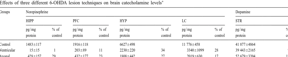
and tabulated the number of interruptions of the photocells the axon bundles, although a difference between these by each animal. Animals were housed directly on bedding techniques was not observed in the hypothalamus. Hypo-and received ad libitum food Hypo-and water. Five days was thalamic NE proved somewhat more resistant to neurotoxic allowed for acclimation to individual housing, followed by depletion than did forebrain NE, consistent with previous 7 days of monitoring the animals’ nocturnal spontaneous reports [20]. The compound lesion group showed the motor activity. On each day, the recording apparatus was largest reduction of NE, indicating that the ventricular and activated 2 h before onset of the dark phase and recording axonal lesion techniques had cumulative effects. In all ended 5 h after lights on. Ambulatory motor activity was animals of the compound lesion group, NE was undetect-expressed as ambulation counts per hour, an ambulation able in the hippocampus and cortical NE was almost count being defined as interruption of a photocell beam equally reduced. The compound lesion technique also was that had not been broken in the previous four beam breaks able to deplete hypothalamic NE to an average of 10% of and thus represented horizontal locomotion by the animal. control, with two animals of the group found to have less At the conclusion of the study, the animals were than 10% of the control level of NE remaining. DA levels sacrificed and brains removed. The following brain regions were not reduced by either the axonal or ventricular lesion were dissected and retained for analysis: LC region of techniques as estimated by DA content in striatum / nucleus brain stem (LC), hypothalamus (HYP), hippocampus accumbens, while the compound lesion technique pro-(HIPP), prefrontal cortex (PFC) and striatum / nucleus duced a small reduction in DA (i.e., 30%) that is not accumbens (STR). Brain samples were frozen at 2858C considered to be functionally effective [6,14].
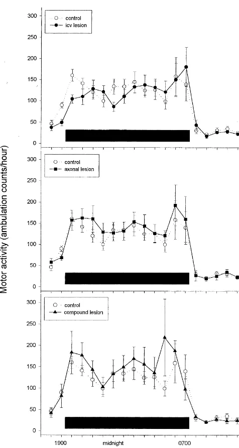
for analysis of catecholamine content using high pressure Mean (6standard error) hourly ambulatory activity of liquid chromatography (HPLC). Tissue samples were the different groups during the 19 h of the day when analyzed for NE content to assess the extent of the lesion; activity was recorded is shown in Fig. 1, with the activity striatal tissue was principally analyzed for DA to assess of each lesion group plotted against the control group. whether DA-containing terminals had been protected from These data were analyzed by analysis of variance (two-destruction by infusion of GBR 12909 prior to icv infusion way [group3h] with repeated measures across the hour of 6-OHDA. Tissues were homogenized in 0.1 M per- factor) which was carried out for each of the three panels chloric acid containing an internal standard. Catechol- shown in the Fig. 1. No analysis generated an effect of amines were separated using reverse-phase, ion–pair group that approached significance, thereby indicating that HPLC and were quantified by an electrochemical detector depletion of brain NE by these lesion techniques did not (ESA Coulochem II) attached to a computerized data cause a change in spontaneous motor activity in com-acquisition system (Perkin Elmer Turbochrom 4). Protein parison to control animals. The total nocturnal ambulatory content of each sample was measured by the Lowry counts for each of the lesion groups, which was compared technique so that catecholamine content could be ex- to the control group by t-test, showed no difference that pressed as pg NE (or DA) per mg tissue protein. approached significance. Moreover, inspection of Fig. 1 As expected, all lesion groups (axonal, ventricular, shows that even the ‘W-shaped’ pattern of dark phase compound) showed marked depletion of NE in brain activity that occurs in normal rats was intact in the regions known to be major targets of the ascending lesioned animals. The only potential departure from this noradrenergic pathways, i.e., cortex and hippocampus for was seen in the ventricular lesion condition, where a the DNB and hypothalamus for the VNB. The results are somewhat reduced elevation in the first hour of activity shown in Table 1. 6-OHDA administered icv was found to after onset of the dark period was seen. However, the lack be somewhat more effective for depleting NE in the of an overall effect of group in the analysis of variance that terminal regions of the DNB than was 6-OHDA aimed at compared this group with the control condition, which was
Table 1
a
Effects of three different 6-OHDA lesion techniques on brain catecholamine levels
Groups Norepinephrine Dopamine
HIPP PFC HYP LC STR
pg / mg % of pg / mg % of pg / mg % of pg / mg % of pg / mg % of protein control protein control protein control protein control protein control
Control 14836117 19166118 66276498 11 7766458 41 87764864
Ventricular 15615 1 203689 11 22306220 34 334061099 28 39 44362165 94 Axonal 4286157 29 4326177 23 18086442 27 20196630 17 52 67963394 125 Compound 0 0 1267 1 6926165 10 9836251 8 29 27063806 70
a
any overall difference in ambulatory activity from control rats but they also showed an unchanged pattern of noctur-nal activity; i.e., like the control (normal) rats, lesioned rats showed a marked increase in activity at the onset of the dark period, followed by a decline and then another smaller peak in activity toward the middle of the dark period, followed finally by another large increase in activity leading up to onset of the light period. Finally, ambulatory activity during hours of light also was similarly low in all groups.
What is indicated by these data is that forebrain NE is not necessary for the rat to generate a normal pattern of ambulatory activity in a home cage situation during light and dark phases of the day. Lesions of NE neurons and terminals that virtually eliminated NE from the forebrain neither reduced total spontaneous motor activity nor impaired ability to generate increases in nocturnal ambulat-ory activity that characterizes normal rat behavior. But insofar as the technique used for affecting NE in the present study was to make a lesion that would reduce brain NE, an important issue to consider is whether compensat-ory increases in the function of noradrenergic systems in the brain (such as upregulation of postsynaptic adrenergic receptors) might have overcome effects of decreased transmission of brain NE. Thus it might be argued that, despite reduced NE levels in the brain, noradrenergic neurotransmission in the lesioned animals nevertheless was functioning well enough to mask motor deficits that will result from the depletion of NE. With regard to this issue, studies that have assessed compensation after experimen-tally-produced reduction of catecholamine content in brain agree that compensation indeed occurs but also indicate that compensation sufficient to return functional responses to normal generally requires 10% or more of the normal amount of NE or DA to be present [6,8,10,14]. In regard to the present experiment, the study of Curet and Montigny [8] is particularly relevant. These investigators examined electrophysiological responses of hippocampal neurons Fig. 1. Mean (6S.E.M.) hourly ambulatory activity of animals with
following electrical stimulation of the locus coeruleus lesions of the noradrenergic neurons in the brain in comparison to
(which will cause NE to be released in the hippocampus). non-lesioned animals. Activity data was collected throughout a 12 / 12 h
In part of the study, responses were assessed in rats whose light / dark cycle over a period of 7 days; monitoring began 2 h prior to
onset of the dark phase and ended 5 h after onset of the light phase. Each NE had been depleted by intraventricular infusion of 6-panel compares the non-lesioned animals (control group) to one of three OHDA; electrophysiological measurement was made 14– lesion groups (n54 / group). The top panel shows animals who received
21 days after the NE lesioning procedure had been carried microinfusion of 6-OHDA into the lateral ventricle (ventricular lesion
out. In comparison to naturally-occurring physiological group); the middle panel shows animals who received microinjections of
stimuli that activate LC neurons, electrical stimulation of 6-OHDA into the fibers of ascending noradrenergic projections (axonal
lesion group); the bottom panel shows animals who were subjected to LC is a very strong stimulus for NE release, so that this both lesion procedures (compound lesion group). No statistically signifi- technique is likely to ‘test the limits’ by recruiting what-cant differences were found. The horizontal black bar designates duration
ever NE is still available for release. In animals that had of the dark phase.
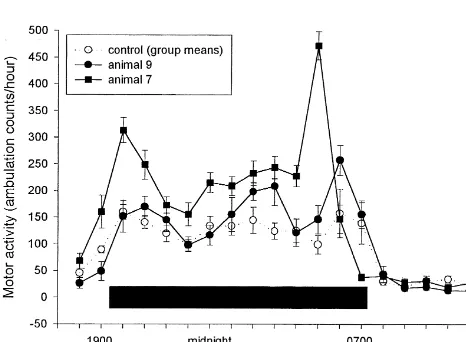
being total (i.e., 100%). Thus, Curet and Montigny re- considered that the data shown for the two lesioned ported that substantial depletions of NE (at least 90%) animals in Fig. 2 are means (6SEs) calculated from were needed to alter the influence of NE on hippocampal recordings made over 7 days, so that elevated activity at a cells that was produced by electrically stimulating the LC, particular hour was not an isolated occurrence on a single but even a strong stimulus for release such as this had no day but, instead, represents a consistent daily pattern. effect at all when NE depletion was judged to be complete. Therefore, animal[7 consistently showed (a) an exagger-In the present study, NE lesions were quite extensive, with ated response to the onset of darkness, as well as (b) a the compound lesion technique producing near-total deple- large increase in activity near the end of the dark period tion of NE in the forebrain. In this regard, Fig. 2 shows the which also occurred slightly earlier in time than did the individual activity data of the two animals in the com- usual pre-light increase in activity shown by normal pound lesion group that were found to have the most animals. Animal[9 also showed a consistent exaggeration complete lesions in the study. For these two subjects of ambulatory activity peaks in producing the ‘W-shaped’ (animals [7 and [9), no NE at all was detected in pattern that characterizes dark-phase activity. Thus, prefrontal cortex and hippocampus (assay sensitivity for eliminating forebrain NE in these animals seems to have these regions was less than 3.0 pg / ml protein, which was removed a moderating influence on ambulatory activity; as less than 0.2% of control levels) and the reduction in a result, the animals responded in an exaggerated manner hypothalamic NE was 95% and 92% respectively. Exami- to the excitatory stimuli, both internal and external, that nation of Fig. 2 reveals that these animals showed undi- give rise to ambulatory activity at the time points where minished elevation of ambulatory activity in response to the exaggerated responses were seen. In a comprehensive the onset of darkness and also showed a similar ‘W’ review of basic research relating to the function of dorsal pattern of ambulatory activity during the dark period as bundle noradrenergic pathway that originates in the LC, was shown by control animals. These results in animals Robbins, Everitt and colleagues [19] concluded that the whose NE depletions were so large as to preclude the functioning of this system allows an animal to respond in a possibility of any significant compensation indicate, to- discriminating way to the environment (pg 146). While the gether with the other findings shown in Fig. 1, that theoretical formulation of Robbins and Everitt emphasizes forebrain NE is not necessary for the ambulatory behavior how NE influences attention and stimulus processing, their assessed in this study. formulation seems consistent with the possibility that The results shown in Fig. 2 also may shed some light on forebrain NE serves to moderate behavioral responses in the function of forebrain NE. Rather than showing a failure many situations.
to respond, the two lesioned subjects whose data are shown When evaluating the results described here, these find-in Fig. 2 can be seen to manifest exaggerated ambulatory ings should not be interpreted as demonstrating that action responses at times when nocturnal ambulatory activity of NE in the brain does not affect motor activity. First, the normally increases. In evaluating these results, it should be present study assessed spontaneous ambulatory activity in the home cage and did not examine motor responses that occur within brief periods of time and / or in reaction to challenging or novel events. Second and perhaps most important, the study described here utilized lesion tech-niques to assess the influence of forebrain NE on behavior. While this methodology has a long history with respect to the question of how NE is related to motor activity, it has distinct drawbacks. Long-term reduction or removal of brain NE, which is what the technique brings about, does not reproduce how a change in NE normally occurs to exert its influence; in physiological function, transient increases or decreases in synaptic NE concentration are what occur to affect function. As a consequence, such reduction or removal of a substance that acts acutely, particularly one that appears to exert a modulatory in-fluence, ultimately may not prove to be a good method for assessing its effects. This may explain why techniques that Fig. 2. Hourly ambulatory activity of two individual rats with virtually acutely perturb NE neurotransmission (e.g., [1,2,26,31]) complete ablation of central NE compared with the mean (6S.E.M.) have observed effects on motor behavior with more activity of the control group (n54). In the case of the individual animals, regularity than studies in which brain NE has been error bars represent variation in hourly activity recorded on 7 days; the
removed by lesion techniques. Such observations argue small magnitude of error bars indicates that the pattern of activity shown
strongly against overgeneralization of the present findings was stable from one day to the next. The horizontal black bar designates
location and destruction of the locus coeruleus in the stumptail But regardless of the possibility that making acute
per-macaque (Macaca arctoides), Brain Res. 100 (1975) 157–162. turbations of NE action in the brain may reveal appropriate
[14] D. Kirik, C. Rosenblad, A. Bjorklund, Characterization of behavioral physiological functions of brain NE that are not seen when and neurodegenerative changes following partial lesions of the NE is chronically removed from the brain, the present nigrostriatal dopamine system induced by intrastriatal
6-hydroxy-dopamine in the rat, Exp. Neurol. 152 (1998) 259–277. results do indicate that the presence of NE in the forebrain
[15] R.M. Kobayashi, M. Palkovits, I.J. Kopin, D.M. Jacobowitz, Bio-is not required for the rat to generate normal diurnal
chemical mapping of noradrenergic nerves arising from the rat locus patterns of ambulatory activity. coeruleus, Brain Res. 77 (1974) 269–279.
[16] S.T. Mason, H.C. Fibiger, Altered exploratory behaviour after 6-OHDA lesion to the dorsal noradrenergic bundle, Nature 269 (1977) 704–705.
Acknowledgements
[17] E.W. Maynert, R. Levi, Stress-induced release of brain norepineph-rine and its inhibition by drugs, J. Pharmacol. Exp. Ther. 143 (1964) The authors gratefully acknowledge the assistance of 90–95.
[18] R.H. Rech, H.K. Borys, K.E. Moore, Alterations in behavior and Sandra Parks in preparation of this manuscript. This
brain catecholamine levels in rats treated with a-methyltyrosine, J. research was supported in part by grant MH56602 from the
Pharmacol. Exp. Ther. 153 (1966) 412–419.
National Institute of Mental Health. [19] T.W. Robbins, B.J. Everitt, B.J. Cole, T. Archer, A. Mohammed,
Functional hypotheses of the coeruleocortical noradrenergic projec-tion: A review of recent experimentation and theory, Physiol. Psychol. 13 (1985) 127–150.
References
[20] B.J. Sahakian, P. Winn, T.W. Robbins, R.J. Deeley, B.J. Everitt, L.T. Dunn, M. Wallace, W.P.T. James, Changes in body weight and [1] C.W. Berridge, S.L. Foote, Effects of locus coeruleus activation on food-related behaviour induced by destruction of the ventral or electroencephalographic activity in neocortex and hippocampus, J. dorsal noradrenergic bundle in the rat, Neuroscience 10 (1983)
Neurosci. 11 (1991) 3135–3145. 1405–1420.
[2] C.W. Berridge, M.E. Page, R.J. Valentino, S.L. Foote, Effects of [21] S.J. Sara, C. Dyon-Laurent, A. Herve, Novelty seeking behavior in locus coeruleus inactivation on electroencephalographic activity in the rat is dependent upon the integrity of the noradrenergic system, neocortex and hippocampus, Neuroscience 55 (1993) 381–393. Cognitive Brain Res. 2 (1995) 181–187.
[3] D.R. Britton, C. Ksir, K. Thatcher-Britton, D. Young, G.F. Koob, [22] J.J. Schildkraut, The catecholamine hypothesis of affective dis-Brain norepinephrine depleting lesions selectively enhance be- orders: a review of supporting evidence, Am. J. Psychiatry 122 havioral responsiveness to novelty, Physiol. Behav. 33 (1984) 473– (1965) 509–522.
478. [23] J.J. Schildkraut, S.S. Kety, Biogenic amines and emotion, Science [4] W.E. Bunney, J.M. Davis, Norepinephrine in depressive reactions, 156 (1967) 21–37.
Arch. Gen. Psychiatry 13 (1965) 483–494. [24] P.E. Simson, J.M. Weiss, Altered activity of the locus coeruleus in [5] V. Castagne, J.-M. Rivet, P. Mormede, The integrity of the ventral an animal model of depression, Neuropsychopharmacology 1 (1988)
noradrenergic bundle (VNAB) is not necessary for a normal 287–295.
neuroendocrine stress response, Brain Res. 511 (1990) 349–352. [25] P.G. Simson, J.M. Weiss, M.J. Ambrose, A. Webster, Infusion of a [6] E. Castaneda, I.Q. Whishaw, T.E. Robinson, Changes in striatal monoamine oxidase inhibitor into the locus coeruleus can prevent dopamine neurotransmission assessed with microdialysis following stress-induced behavioral depression, Biol. Psychiatry 21 (1986) recovery from a bilateral 6-OHDA lesion: Variation as a function of 724–734.
lesion size, J. Neurosci. 10 (1990) 1847–1854. [26] P.G. Simson, J.M. Weiss, L.J. Hoffman, M.J. Ambrose, Reversal of [7] T.J. Crow, J.F.W. Deakin, S.E. File, A. Longden, S. Wendlandt, The behavioral depression by infusion of alpha-2 adrenergic agonist into
locus coeruleus noradrenergic system — evidence against a role in the locus coeruleus, Neuropharmacology 25 (1986) 385–389. attention, habituation, anxiety and motor activity, Brain Res. 155 [27] K. Taghzouti, H. Simon, D. Herve, G. Blanc, J.M. Studler, J. (1978) 249–261. Glowinski, M. LeMoal, J.P. Tassin, Behavioural deficits induced by [8] O. Curet, C. de Montigny, Electrophysiological characterization of an electrolytic lesion of the rat ventral mesencephalic tegmentum are adrenoceptors in the rat dorsal hippocampus. II. Receptors mediating corrected by a superimposed lesion of the dorsal noradrenergic the effect of synaptically released norepinephrine, Brain Res. 475 system, Brain Res. 440 (1988) 172–176.
(1988) 47–57. [28] S.A. Thomas, R.D. Palmiter, Disruption of the dopamineb -hydroxy-[9] A. Dahlstrom, K. Fuxe, Evidence for the existence of monoamine- lase gene in mice suggests roles for norepinephrine in motor containing neurons in the central nervous system. I. Demonstration function, learning, and memory, Behav. Neurosci. 111 (1997) 579– of monoamines in the cell bodies of brainstem neurons, Acta 589.
Physiol. Scand. 62 (1964) 1–55. [29] J.M. Weiss, W.H. Bailey, L.A. Pohorecky, D. Korzeniowski, G. [10] D.J. Dooley, G.H. Jones, T.W. Robbins, Noradrenaline- and time- Grillione, Stress-induced depression of motor activity correlates dependent changes in neocortical a - and b -adrenoceptor binding in2 1 with regional changes in brain norepinephrine but not dopamine, dorsal noradrenergic bundle-lesioned rats, Brain Res. 420 (1987) Neurochem. Res. 5 (1980) 9–22.
152–156. [30] J.M. Weiss, P.A. Goodman, B.G. Losito, S. Corrigan, J.M. Charry, [11] A.H. Engelbrecht, V. Russell, M.E. Carstens, A.S. De Villiers, A. W.H. Bailey, Behavioral depression produced by an uncontrollable Searson, A. Jaffer, J.J.F. Taljaard, Evidence that noradrenergic stressor: relationship to norepinephrine, dopamine, and serotonin neurons in the A1 ands A2 nuclei are lesioned by low doses of levels in various regions of the rat brain, Brain Res. Rev. 3 (1981) 6-OHDA injected into the locus coeruleus, J. Neurosci. Met. 52 167–205.