Malaysian Journal of Pharmaceutical Sciences, Vol. 4, No. 1, (2006)
*Corresponding author: Zullies Ikawati, e-mail: [email protected]
CYTOTOXICITY AGAINST TUMOR CELL LINES OF A
RIBOSOME-INACTIVATING PROTEIN (RIP)-LIKE PROTEIN ISOLATED
FROM LEAVES OF MIRABILIS JALAPA L
ZULLIES IKAWATI1*, SUDJADI1 AND SISMINDARI1,2
1Faculty of Pharmacy, Gadjah Mada University, Jogjakarta, Indonesia 2Life Science Laboratory, Gadjah Mada University, Jogjakarta, Indonesia
The 30 kD protein fraction with properties like ribosome-inactivating protein (RIP) was isolated from the leaves of Mirabilis jalapa L and named MJ-30. This study investigated the cytotoxic effect of MJ-30 on normal and malignant cells. MJ-30 was isolated from the leave extract of M. jalapa L using cation-exchange chromatography with CM Sepharose CL-6B column to obtain MJ-30. The fraction contain DNA cleaving ability were pooled and checked for cytotoxicity against breast cancer T47D cell line, cervical cancer SiHa cell line, and human mononuclear cells derived from peripheral blood of healthy volunteers. Results showed that the MJ-30 produced cytotoxic effect against T47D and SiHa cell line to different extent. The LC50 of the
MJ-30 on T47D cell line and SiHa cell line were 0.36 µg/mL and 5.6 µg/mL, respectively. While in normal cells, represented by human mononuclear cells, MJ-30 was considerably less toxic, with LC50 of 21.04 µg/mL. The results suggest that MJ-30 produced more cytotoxic activity toward breast and cervical cancer cells (58-fold and 4-fold, respectively) as compared to normal mononuclear cells.
Keywords: Ribosome-inactivating protein, Mirabilis jalapa L, cytotoxicity, T47D and SiHa cell line,
human mononuclear cells
INTRODUCTION
Ribosome-inactivating proteins (RIPs)are a group of plant enzymes that possess
capability to inactivate ribosomes by modifying the 28S rRNA through their N-glycosidase activity, which is manifested by cleavage of the N-glycosidic bond at a specific adenine. The adenine at position 4324 of rat liver 28S rRNA and corresponding adenine on other eukaryotic or prokaryotic models are the target
sites (Stirpe et al. 1992). Through this mechanism, the binding of elongation
factor 2 is prevented, with the consequent arrest of protein synthesis leading to
autonomous cell death (Osborn and Hartley 1990). Such properties might
Zullies Ikawati et al.
2
acid cleavage of RIP has been used to identify the presence of this activity of extract preparation of plant origin (Sismindari and Lord 2000).
RIPs are currently under study as therapeutic agents against cancer
(Kreitman and Pastan, 1998). The present study is a part of our continuing research to isolate active compounds from plant for anti-cancer activity. We have previously reported that crude protein fraction extracted from the leaves of Mirabilis jalapa L displayed potential to cleave supercoiled DNA like RIP, and was able to induce cell death through the induction apoptosis and necrosis
in different cancer cell-line models (Ikawati et al. 2003). However, this being in
crude form, we carried out further purification of the protein extract using cation-exchange chromatography. In the present study, a protein fraction identified to have RIP-like activity has a molecular weight of 30 kD, and,
therefore, was designated as MJ-30 (MJ for Mirabilis jalapa, and 30 for 30 kD).
Cytotoxicity of the MJ-30 was tested against two cancer cell lines, namely human breast cancer T47D and cervical cancer SiHa, and human peripheral blood mononuclear cells as normal cells.
METHODS
Plant Material
Fresh leaves of M. jalapa L (red flower cultivar) were collected from
Yogyakarta, Indonesia. A voucher specimen was deposited in the Laboratory of Life Science Gadjah Mada University, Yogyakarta, Indonesia.
Cell culture
The T47D and SiHa cells lines were obtained from the stock of Life Science Laboratory Gadjah Mada University. The cells were maintained in RPMI 1640 (SIGMA) supplemented with 10% (v/v) fetal bovine serum (Gibco), 1% (v/v) penicillin-streptomycin (Gibco) and 0.5% (v/v) fungizone (Gibco). The cells
culture were incubated at 37oC in a humidified atmosphere of 5% CO2 (v/v)
3
Cytotoxicity against Tumour Cell Lines of a RibosomeThe peripheral blood mononuclear cells (PBMC) were isolated from healthy volunteers. Briefly, each 5 mL blood sample was diluted with 5 mL of pyrogen-free normal saline and underlayered with 10 mL of Ficoll-Hypaque. The tube was centrifuged at 2000 g for 20 mins. The buffy coat layer containing mononuclear cells was then removed to a fresh tube, washed three times using
RPMI 1640 medium, counted and adjusted to a final concentration of 5x10⁴
cells/mL.
All other reagentswere of analytical or molecular biology grade and, when
possible, RNase-free. For experiment using human volunteer to isolate the
peripheral blood mononuclear cells, the experimental protocols were approved by the Ethical Committee of Health and Medical Research, Gadjah Mada University.
Isolation and purification of MJ-30 from Mirabilis jalapa L
Extraction of protein fraction from the leaves of M. jalapa L was carried out as
described previously (Ikawati et al. 2003). The fraction are referred as basic
protein fractions and contained most of the RIP activity present in the starting material. To purify the protein, the protein extract was adjusted to 5 mM phosphate buffer (pH 6.5) and applied to a CM Sepharose CL-6B column (1x20
cm, Pharmacia LKB Biotech), and equilibrated with the same buffer at room
temperature. The elution was carried out with the equilibrationbuffer until all
peaks of the unbound protein were obtained and the absorbance at 280 nm was
lowered to a baseline. This was followed by elution of bound proteins with
1 liter of a linear (0-500 mM) gradientof NaCl in the same buffer. The eluted
fractions were collected every 2 min in eppendorf tubes and were screened for ability to cleave supercoiled DNA as an indicator for RIP-like activity. Protein fractions with high activity of DNA cleavage were pooled and tested for cytotoxicity against cells. The protein fraction was also analyzed by SDS-PAGE.
Cleavage of super-coiled DNA by the protein extracts
In order to confirm the presence of RIP activity in the eluted protein fractions, their capability to cleave supercoiled double stranded DNA was determined, as
Zullies Ikawati et al.
4
stock of laboratory of Life Science, Gadjah Mada University) was incubated with various volume of protein fractions (1, 2, 4, and 8 µL) to final volume of 10
µL containing 50 mM Tris-HCl, 10 mM MgCl2, 100 mM NaCl, pH 8.0, at 30oC for
one hour. At the end of the reaction, 4 µL of loading buffer (xylene cyanol FF
0.25% (v/v) (Sigma), 0.25% (v/v) bromphenol blue, 30% (v/v) glycerol in 200 mM EDTA) were added. Electrophoresis was carried out on a 1 % (w/v) agarose gel in 0.5x TBE (Tris Borat EDTA) buffer. DNA bands were visualized by staining with ethidium bromide (Sismindari and Lord 2000). Ricin with amount of 2 µg was used for positive control. Ricin was kindly obtained from Prof. JM Lord, Warwick University.
Cytotoxicity assay
Cytotoxicity assay was carried out according to the method of Mosmann (1983) with slight modification. One hundred mL of the exponentially growing cells (5
x 104 cells/mL) were seeded in 96-well microculture plate with a serial dilution
of protein extracts in a volume of 100 µL. The media without cells was used as
control media and treated with the same serial dilution of protein extract, while the phosphate buffer pH 6.5 was used as control for the protein fraction. After 24 hr incubation, the number of viable cells was ascertained with MTT reaction
(3-(4,5-dimethyltiazol-2-il)-2,5-diphenyl tetrazolium bromide). LC50 value was
calculated using Reed-Muench method (1938). Percent of dead cells was calculated as follow :
% dead cells = Auntreated cells – Atreated cells x 100 %
Auntreated cells
Statistical analysis
The results were expressed as the mean ± SEM. Statistical analysis was carried
5
Cytotoxicity against Tumour Cell Lines of a RibosomeRESULTS AND DISCUSSION
Purification of protein fraction
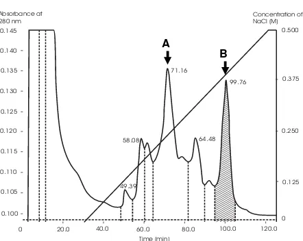
The elution profile of bound proteins on CM-Sepharose CL6B eluted with a
linear (0-500 mM) gradientof NaCl in 5 mM phosphate buffer (pH 6.5) is shown
in Figure 1(a) Two major peaks were observed, which were eluted on 0.240 –
0.255 M NaCl and 0.33 – 0.37 M NaCl, respectively.
0.135
Zullies Ikawati et al.
6
Screening of protein fraction with activity for cleavage of supercoiled DNA
All protein samples were screened for their ability to cleave supercoiled DNA as an indicator for the existence of RIP-like activity. However, only protein fractions of the bound proteins eluted on 0.33 – 0.37 M NaCl were found to show ability to cleave supercoiled DNA. The protein concentration was 2.88 µg/µL, and the ability to cleave supercoiled DNA was increased with increasing amount of protein used in the experiment (Fig. 2 lane B - E). The amount of protein fraction capable to cleave supercoiled DNA to be nick-circular and linear form was started from 11.52 µg (lane D).The capability of protein fraction to cleave supercoiled DNA was comparable to that of ricin, a well known RIP
(Fig. 2 lane F), despite less potent, supporting that the protein from M. jalapa L
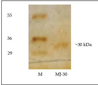
have RIP-like property. The active fractions are depicted with hatched areas marked in the peaks (Fig. 1a) in chromatogram. All the active fractions in bound protein were then pooled and analysed on SDS-PAGE and assayed for cytotoxicity against cell line. SDS-PAGE analysis revealed that the pooled protein fraction had a protein of molecular weight 30 kD (Fig. 1 b).
Fig. 1(b): Analysis by 12 % (w/v) SDS-PAGE of the active protein fraction. The protein fraction was observed on moleculer weight of 30 kDa, therefore, it was designated as MJ-30 (MJ for Mirabilis jalapa, and 30 for 30 kD). M = marker protein.
55
36
29
~30 kDa
7
Cytotoxicity against Tumour Cell Lines of a RibosomeBand 1
Band 2 Band 3
- + + + +
A B C D E F
A
Fig. 2: Effect of protein fractions of M. jalapa L (MJ-30) eluted on 0.33 – 0.37 M NaCl with increasing amount on the cleavage of DNA plasmid (pUC18). Lane A: pUC 18 control; lane B – E: pUC18 treated with 1, 2, 4, 8 µL, respectively, of protein with concentration of 2.88 µg/µL. Lane F: pUC18 treated with 2 µg of ricin. The ability of the MJ-30 to cleave supercoiled DNA was started from the amount of 11.52 µg (lane D). Band 1: supercoiled form of DNA; band 2: nick-circular form of DNA, band 3: linear form of DNA.
Cytotoxic activity
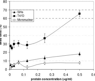
Protein fractions of bound protein with DNA cleaving activity demonstrated different levels of cytotoxic activity against T47D and SiHa cell lines, and very less toxic against human mononuclear cells (Fig. 3). After 24 hour of incubation,
LC50 of the MJ-30 against T47D, SiHa, and human mononuclear cells were found
to be 0.36 µg/mL, 5.6 µg/mL and 21.04 µg/mL, respectively. Percentage of
dead cells caused by the highest concentration of protein used (0.55 µg/mL) to
treat T47D, SiHa, and mononuclear cells were 65.39%, 18.14%, and 7.61 %, respectively.
The results of the present study demonstrated that the protein fractions
isolated from extract of the leaves of M. jalapa L contain protein with RIP-like
Zullies Ikawati et al.
8
from plants may be classified into type 1 or 2 according to their single- or
double-chainstructure (Barbieri, Batteli and Stirpe 1993). Besides the classical
type 1 and 2 RIPs, a 60-kDa RIP (called JIP60) has been identified in barley
(Hordeum vulgare) that consists of an amino-terminaldomain closely related to
the RIP enzymatic chain linked to anunrelated carboxyl-terminal domain with
unknown function, which may be classified as type 3 RIP (Reinbothe et al. 1994).
Our protein is most likely similar to type 1 RIP as it has molecular weight in the
range of 30 kD. Several isoforms of RIPs have been identified in the seeds of M.
jalapa, designated as Mirabilis antiviral protein (MAP) with molecular weight of 27.788 kD, MAP-2 (30.412 kD), MAP-3 (29.771 kD), and MAP-4 (29.339 kD)
(Bolognesi et al. 2002). We did not confirm yet whether our MJ-30 is similar to
MAP-2, however, it is likely that RIPs may be different in different tissue.
0
Fig. 3: Cytotoxic effect of MJ-30 on T47D and SiHa cells line compared to that on human peripheral mononuclear cells. Phosphate buffer pH 6.5 was used as negative control for protein fraction. Data are means of 4-6 experiments in triplicate.
9
Cytotoxicity against Tumour Cell Lines of a Ribosomefinding shows that MJ-30 relatively less cytotoxic for normal cells, and produces more cytotoxic activity against tumor cells despite in different levels.
The cytotoxicity of MJ-30 seems to be varied on the type of target cell line. The RIP showed markedly higher cytotoxicity on T47D cell line, but only slightly cytotoxic against SiHa cell lines. T47D cell line is originally derived from pleural effusion of 54 years old human female with breast carcinoma, while SiHa cells are derived from a surgically removed cervical carcinoma, which carry one or two copies of human papillomaviruse (HPV) 16 DNA as an
integrated form in chromosome (Friedl et al. 1970). The difference in cell type
may account for the difference cytotoxic response against anti cancer agents. In the case of RIP, variation of cytotoxicity against different cells was also
observed in another RIP, trichosantin (Chan et al. 2002). Chan et al. (2002)
employed three different cell types IC21, JAR and Vero cell lines, which were shown to be high, medium and low sensitivity to trichosantin. A good relationship was demonstrated between intracellular trichosantin concentration and toxicity. It seemed that variation of cytotoxicity of RIPs in different cells may be dependent upon the mechanisms affecting its internalization.
This phenomenon may also be applied generally for other RIP, including MJ-30. It has been proposed that type I RIPs enter cells through passive mechanisms such as fluid-phase pinocytosis. However, some observations, such as the difference in sensitivity of type I RIPs among different cell types, and the organ-specific toxicity of type I RIPs, indicate a specific mechanism for the entry
of these proteins into target cells (Cavarallo et al. 1995). A receptor responsible
for the binding and endocytosis of RIP is alpha 2-macroglobulin receptor (alpha 2MR). A study with saporin and trichosantin, a potent type I RIPs, indicated that general mechanism of complex interactions between RIPs and cellular
membranes is mediated by alpha 2-macroglobulin receptor (Cavarallo et al.
1995; Chan et al. 2000). The alpha 2-macroglobulin receptors are expressed in
cancer cell line, including breast cancer cell line, T47D and BT-20 cell line (Li et
al. 1998). Despite the lack of data concerning the expression of alpha
Zullies Ikawati et al.
10
Other possibilities that may explain the different cytotoxicity of the RIP toward different kind of cells is the difference in the downstream death pathways. Indeed, the inhibition of protein synthesis by cleavage of the N-glycosidic bond of a specific adenine of 28 S rRNA has been accepted as the mechanism by which plant ribosome-inactivating proteins (RIPs) cause cytotoxicity. However, beside inhibition of protein synthesis, RIPs have been
shown to induce apoptosis or programmed cell death (Olmo et al. 2001).
Apoptosis is not a general direct consequence of protein biosynthesis inhibition, as deduced from the comparative analysis of the effects of alpha-sarcin and
cycloheximide (Olmo et al. 2001). Olmo and colleagues reported that
cycloheximide does not induce apoptosis even at concentrations far beyond its
IC50, where protein biosynthesis is null. It indicates that there may be several
mechanisms of cytotoxicity induced by RIPs. Consequently, more mechanisms may contribute to more cytotoxic activities of RIPs against tumor cells. In SiHa cancer cells, for example, p53 are disrupted by human papillomaviruses (HPVs) E6 and E7, respectively (Hausen 1996), while T47D cells still express normal
level of p53 (Hurd et al. 1995), this may cause the SiHa cells become more
resistant to apoptosis than T47D cells. This may explain the different sensitivity between SiHa and T47D cells against RIPs.
The variation of sensitivity of two tumor cells employed in our study against RIPs is not fully understood yet. There are still some contradictory reports whether the cytotoxic effect of RIPs is dependent or independent of a surface receptor-mediated pathway. These hypothesis need further elucidation, however, these results may give insight to develop anticancer agent derived from RIPs for certain type of cancer.
CONCLUSION
The protein with ribosome inactivating protein (RIP)-like activity isolated from
the leaves of Mirabilis jalapa showed high cytotoxicity against T47D and SiHa
cell lines with different extent (LC50 : 0.36 µg/ml and 5.6 µg/ml, respectively),
11
Cytotoxicity against Tumour Cell Lines of a RibosomeACKNOWLEDGEMENT
This work was supported by grants from Quality of Undergraduate Education (QUE) Program Batch III Faculty of Pharmacy Gadjah Mada University. We are thank to Ms. Leny Wirawan, Helena Siboro, and Ni Kadek Darsini for technical assistance.
REFERENCES
BARBIERI, L., BATTELI, M.G. & STIRPE, F. (1993) Ribosome Inactivating Protein from Plants, Biochimica et Biophysica Acta, 1154:237—84.
BOLOGNESI, A., POLITO, L., LUBELLI, C., BARBIERI, L., PARENTE, A. & STIRPE, F. (2002) Ribosome-inactivating and Adenine Polynucleotide Glycosylase Activities in Mirabilis jalapa L Tissues, Journal of Biology Chemistry, 277: 13709—716.
CAVALLARO, U., NYKJAER, A., NIELSEN, M. & SORIA, M.R. (1995) Alpha 2-macroglobulin receptor mediates binding and cytotoxicity of plant ribosome-inactivating proteins, European Journal of Biochemistry, 232:165—71.
CHAN, W.L., SHAW, P.C., TAM, S.C., JACOBSEN, C., GLIEMANN, J. & NIELSEN, M.S. (2000) Trichosanthin interacts with and enters cells via LDL receptor family members, Biochemistry Biophysics Research Communication, 270:453—7.
CHAN, W.L., ZHENG, Y.T., HUANG, H. & TAM, S.C. (2002) Relationship between trichosanthin cytotoxicity and its intracellular concentration, Toxicology, 177:245—51.
FRIEDL, F., KIMURA, I., OSATO, T., ITO, Y. (1970) Studies on a new human cell line (SiHa) derived from carcinoma uterus. I. Its establishment and morphology, Proceedings of the Society for Experimental Biology and Medicine, 135:543-545
HAUSEN, H. (1996) Papillomavirus infections--a major cause of human cancers, Bioc.himica et Biophysics Acta, 1288:F55—78.
Zullies Ikawati et al.
12
IKAWATI, Z., SUDJADI, SISMINDARI, ELLY, W. & PUSPITASARI, D. (2003) Induction of apoptosis by protein fraction isolated from the leaves of Mirabilis jalapa L on HeLa and Raji cell-line, Oriental Pharmacy and Experimental Medicines, 3:1516.
KREITMAN, R. J. & PASTAN, I. (1998) Immunotoxins for targeted cancer therapy, Advance Drug Delivery Review, 31: 53-88, 1998.
LI, Y., WOOD, N., DONNELLY, P. & YELLOWLEES, D. (1998) Cell density and oestrogen both stimulate alpha 2-macroglobulin receptor expression in breast cancer cell T-47D, Anticancer Research, 18: 1197—202.
LING, J., LIU, W. & WANG, T.P. (1994) Cleavage of supercoiled double stranded DNA by several ribosome inactivating proteins in vitro, FEBS Letters, 345:143—6.
MOSMANN, T. (1983) Rapid colorimetric assay for cellular growth and survival: application and cytotoxicity assay, Journal of Immunology Methods, 65:55—63.
OLMO, N., TURNAY, J., BUITRAGO, G.H., SILANES, I.L., GAVILANES, J.G. & LIZARBE, M.A. (2001) Cytotoxic mechanism of the ribotoxin α-sarcin: induction of cell death via apoptosis, European Journal of Biochemistry, 268: 2113—2123.
OSBORN, R.W. & HARTLEY, M.R. (1990) Dual effects of the ricin A chain on protein synthesis in rabbit reticulocyte lysate. Inhibition of initiation and translocation, European Journal of Biochemistry, 193: 401 – 407.
REED, L.J. & MUENCH, H. (1938) Simple method of estimating fifty percent endpoint, American Journal of Hygiene, 27:493—497.
REINBOTHE, S., REINBOTHE, C., LEHMANN, J., BECKER, W., APEL, K. & PARTHIER, B. (1994) JIP60, a Methyl Jasmonate-Induced Ribosome-Inactivating Protein Involved in Plant Stress Reactions, PNAS, 91:7012—6.
SISMINDARI (NO FIRST NAME) & LORD, J.M. (2000) Ribosome-inactivating RNA N-glycosidase activity of Mirabilis jalapa L , Morinda citrifolia L, and Carica papaya L. Indonesian Journal Biotechnology, 12:342—5.


