Research Article
Confirmation on Status of Chaetocnema basalis(Coleoptera: Chrysomellidae) as A Vector of Stewart Wilt Disease
Konfirmasi Status Chaetocnema basalis(Coleoptera: Chrysomellidae) sebagai Vektor Penyakit Layu Stewart
Heri Widodo1)*, Arman Wijonarko2), Witjaksono2), & Suputa2)
1)Agricultural Quarantine Station of Tanjung Balai Karimun,
Jln. Jend. Sudirman Tanjung Balai Karimun, Kepulauan Riau 29661
2)Department of Crop Protection, Faculty of Agriculture, Universitas Gadjah Mada
Jln. Flora 1, Bulaksumur, Sleman, Yogyakarta 55281 *Coresponding author. E-mail: heri.widodo@ymail.com
ABSTRACT
Chaetocnema pulicariaand C. denticulataare recognized as vectors of Stewart wilt disease caused by Pantoea stewartii
on maize. These insects have not been reported yet in Indonesia, but Stewart wilt disease has been reported in Java and Sumatera Islands. Genus Chaetocnema which presented in Indonesia is C.basalis. It is not cleared whether C. basalis
is a vector for Stewart wilt disease like C. pulicariaand C. denticulata. This reseach was aimed to conduct the confirmation on status whether C. basalis have a role as vector of Stewart wilt disease on maize or not. C. basalis imago were collected from maize growing areas in Yogyakarta, and then starved for 24 h. Treatments were applied by placing imago of C. basalis on infected-P. stewartiiplants for 72 h. Five insects were then transferred to each plot of healthy plant (1 plot consisted of 5 plants) for 72 h. For control, imago of C. basalis were put on healthy plants for 72 h and five insects were then transferred to other healthy plant (1 plot consisted of 5 plants) for 72 h. Each treatment was repeated three times. On the fifteenth days after transmission, PCR assays were carried out on leaf samples and isolates of bacteria. All sampled leaves analysis showed that there were no Stewart wilt diseases transmission based on PCR assay and bacterial isolates. This concluded that C. basalis is not a vector for Stewart wilt disease on maize.
Keywords: Chaetocnema basalis, Stewart wilt, vector
INTISARI
Chaetocnema pulicariadan C. denticulatamerupakan serangga vektor penyakit layu stewart yang disebabkan oleh bakteri Pantoea stewartiipada tanaman jagung. Kedua serangga ini belum pernah dilaporkan keberadaannya di Indonesia tetapi penyakit layu stewart telah ditemukan di pulau Jawa dan pulau Sumatera. Serangga Genus Chaetocnema yang ada di Indonesia adalah Chaetocnema basalis. C. basalisbelum diketahui secara pasti sebagai vektor penyakit layu stewart seperti halnya C. pulicariadan C. denticulata. Penelitian ini bertujuan untuk melakukan konfirmasi status apakah
C. basalisberperan sebagai vektor penyakit layu stewart pada tanaman jagung atau tidak. Serangga uji berupa imago
C. basalisyang dikoleksi dari pertanaman jagung di Yogyakarta, lalu dilaparkan selama 24 jam. Pengujian perlakuan dilakukan dengan menempatkan imago C. basalispada tanaman terserang P. stewartiiselama 72 jam. Kemudian di-pindahkan pada tanaman sehat sejumlah 5 ekor per plot tanaman bersungkup (1 plot terdiri dari 5 tanaman) selama 72 jam. Perlakuan kontrol dilakukan dengan menempatkan imago C. basalispada tanaman sehat selama 72 jam, kemudian dipindahkan pada tanaman sehat yang lain sejumlah 5 ekor per plot tanaman bersungkup (1 plot terdiri dari 5 tanaman) selama 72 jam. Masing-masing perlakuan diulang sebanyak tiga kali. Pada hari ke-15 setelah penularan, dilakukan uji PCR daun tanaman sampel dan isolat bakteri. Hasil pengujian semua sample daun menunjukkan negatif sehingga dipastikan bahwa C. basalis bukan merupakan vektor penyakit layu stewart pada tanaman jagung.
Kata kunci: Chaetocnema basalis, layu stewart, vektor
INTRODUCTION
Chaetocnema is a grass flea beetlewhich belongs to family of Chrysomellidae and distributes almost throughout the world. It is approximately 437 of about
630 species had been identified (Konstantinovet al.,
2011). In Indonesia, Kalshoven (1981) reported the presence of genus Chaetocnema populations on paddy,
maize and grasses which were assumed as Chaetocnema
basalis.
Stewart wilt disease caused by bacterium Pantoea
stewartii has generated large problems for maize producing countries such as United States of America (USA), resulting yield loss up to 95%. Currently, this disease has been reportly distributed in all over the world such as: Austria, Bolivia, Brazil, Canada, Costa Rica, Guyana, Mexico, Peru, Puerto Rico, USA, China, India, Malaysia, Thailand, Vietnam, and Indonesia (Anonymous, 2016a). In Indonesia, it was considered as new disease on maize and had been reported distributing in Pasaman Barat, West
Sumatera (Rahma, 2013), Bogor (Rahma et al., 2014),
and Yogyakarta (Anonymous, 2015). Rahma (2013) detected the presence of this disease in maize producing center of West Sumatera with disease incidence ranging 1−15%.
Stewart wilt disease dispersed through seed and
vector (Pataky, 2004).Chaetocnema pulicaria and
C. denticulata were reported as vectors of this disease (Poos & Elliot, 1936). These two spesies have not been found yet in Indonesia. Genus Chaetocnema which was found associating with maize in Indonesia is C. basalis. This research was aimed to investigate
whether C. basalis that grouped into same genus
with C. pulicaria andC. denticulatais able to transmit
Stewart wilt disease on maize.
MATERIALS AND METHODS
Research was conducted in a green house, Faculty of Agriculture, Universitas Gadjah Mada, from Desember 2016 until Januari 2017. Imago of
C. basaliswere collected from maize growing areas in Sleman, Yogyakarta (110,4000 BT; 7,7099 LS). Bonanza variety of sweet corn was used as a test plant. The 3-days old bacterial isolate on yeast dextrose calcium carbonate (YDC) medium was then diluted into sterile aquadest to obtain bacterial suspension. Turbidity level of suspension was measured using spectrophotometer at wavelength of 600 nm and OD of 0.3 which was estimated containing bacterial cell
about 1.6×10⁹cell ml-1. The suspension was then injected
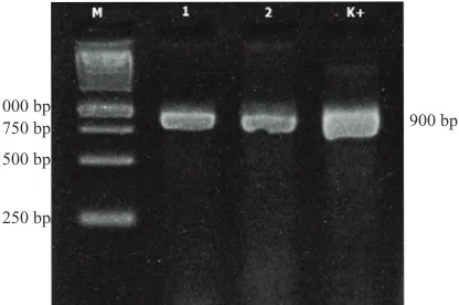
on 8-days old maize plant, and confirmed to be infected with Stewart wilt through molecular assay (Figure 1),
then 24 h-starvedC. basalis were put on infected
plant for 72 h. Five C.basaliswere then transferred
to plot of 8-days healthy plant (each plot consisted of 5 covered plants) for 72 h, furthermore considered as Treatment 1 (P1).
The collected and 24 h-starved C. basalis were
placed on healthy plant for 72 h. Five insects were then transferred to plot of 8-days plant (each plot consisted of 5 covered plants) for 72 h, here in after used as control (P0), each treatment was repeated three times (Figure 2).
On the fifteenth day after transmission, leaves of each plant were sampled and analysed using two methods, i.e. direct plant extraction and isolation of Nutrient Agar (NA) medium. DNA extraction was
performed using method developed by Goodwin et
al. (1994).
Figure 1. Molecular test of artificially inoculated of Pantoea stewartiito plant: 1=plant 1, 2=plant 2, M=1 kb DNA ladder, K+ = positive control
Figure 2. Flow chart of transmission assay of Chaetocnema basalisagainst Pantoea stewartiion maize
1000 bp 750 bp
500 bp
250 bp
Leaf samples were washed with running water, surface sterilized with 70% ethanol for 1 min, rinsed 3 times with sterile water, and dried on sterile paper towels. Symptomatic leaf samples were aseptically cut into 10 mm×10 mm pieces and put into NA medium, incubated over night to allow release of bacteria into the medium. The bacterial suspension was then trans-ferred and purified on YDC, the DNA of 3-days pured cultures were then extracted using CTAB
method of Goodwin et al.(1994). Extracted pellet was
checked to find out the presence of DNA by mixing the pellet with loading dye in 1:4 ratio, and then put into well of agarose 1% and electrophoreted (50 Volt for 40 min). Afterwards, DNA was amplified
according to Coplin et al. (2002), molecular assay
was carried out with Polymerase chain reaction
(PCR) using primer of HRP3c 5’-GCG GCA TAC CTA ACT CC-3’ and HRP1d 5’-CA CTC ATT CCG ACC AC-3’, in following mixed composition: 1 µl of DNA pellet, 1 µl of primer HRP3c, 1 µl of primer
HRP1d, 12.5 µl of go taq reaction, and 9.5 µl of dd
H2O. The protocol were as follows: predenaturation
of 94oC for 1 min, denaturation of 94oC for 15 s,
annealing of 55oC for 15 s, extension of 72oC for 30 s,
and final extension of 72oC for 5 minutes. Steps of
denaturation, annealing, and extension were cycled for 25 times.
PCR products were then put into well of 1.5% and electrophoreted (50 Volt for 45 min). Electrophoresis product visualized on UV transillluminator. Percentage
of disease incidence was analysed with t-test with
α level of 5% (p = 0.05).
RESULTS AND DISCUSSION
Transmission test showed that C. basaliscould
not transmit P. stewartii, pathogen of Stewart wilt.
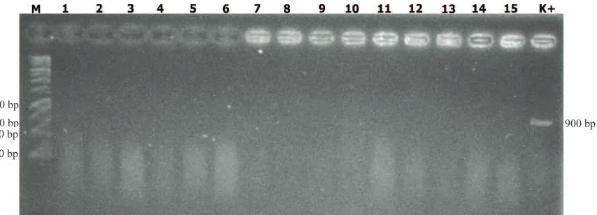
Molecular assay as shown on Figure 3, 4, and 5, in which all of samples and analyses methods showed negative results. Figure 3 on test control plants (P0),
Figure 3. Molecular test of extracted leaves directly on control (P0): 1=P011, 2=P012, 3=P013, 4=P014, 5=P015, 6=P021, 7=P022, 8=P023, 9=P024, 10=P025, 11=P031, 12=P022, 13=P023, 14=P024, 15=P025; M=1 kb DNA ladder, K+ = positive control
Figure 4. Molecular test of extracted leaves directly on treatments 1 (P1): 1=P111, 2=P112, 3=P113, 4=P114, 5=P115, 6=P121, 7=P122, 8=P123, 9=P124, 10=P125, 11=P131, 12=P122, 13=P123, 14=P124, 15=P125; M=1 kb DNA ladder, K+ = positive control
1000 bp
750 bp 500 bp
250 bp
900 bp 1000 bp
750 bp 500 bp
250 bp
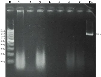
indicating negative results of all repetition and tested plant have been extracted and molecular analysed. Figure 4 showed that on treated plants (P1) also revealed negative results in all repetition and tested plant has been extracted and molecular analysed. Figure 5 showed that transmission test on all treatments and isolated plant samples, leaf samples exudating bacterial colony and then purified and extracted. Molecular assay expressed negative results on all sampled isolates.
Disease transmission through vector was a complicated interaction between insect, plant and
pathogen (Martini et al.,2015). Feeding manner of
insect, imunity reaction of insect and plant, and characteristics of pathogen were some considerating
factors. C. basalis was beetle with mouthparts type
of bitting and chewing, acquition of pathogenic bacteria occured through its mouthpart during feeding process. Transmission happened through saliva containing pathogen which would penetrate via lession occuring for feeding process, so that the pathogen could spread through xylem vessels and cell cavities (Orlovskis
et al.,2015). However, feeding process of C. basalis
did not generate the infection of Stewart wilt, the occurred lession was the enter for other pathogen.
The strange objects which entered into insect bodies could be pathogenic or symbiotic (Ammar
et al.,2014). They would establish defense systems
when foreign objects penetrated into their bodies (Chapman, 2013). The same reaction also occurred on plants when extraneous particles got into their tissues, i.e. utilizing induced resistance systems which were considered as a normal mechanism functioning in restriction of pathogen growth and spread on plant (Agrios, 2005).
P. stewartii associated with digestive tracts of insect vectors (foregut, midgut, hindgut, and malpigian tubule). Pathogen could be observed in midgut and
hindgut until 12 days after acquisition (Ammar et al.,
2014). Pathogen would be brought by blood circulation and dissolved in saliva. Such condition took place when the pathogen was persistent in vector.
P. stewartii used two type III of secretion systems (T3SS) to establish colony on host plant and its vector. Colony establishment on host plant was initiated by producing injectisomere or pili to transfer effector protein to host plant. T3SS was important factor for
pathogenicity of P. stewartiion maize plant. Secretion
system of second T3SS was the establishment of
Pantoea secretion island 2 (PSI-2) which was required by pathogen to be persistent in insect bodies.
Mutagenesis of PSI-2 psaN gene was very influential
in reducing the persistency of P. stewartii in gut of
vector and the ability of beetle in transmitting the
pathogen on host plant (Correa et al. 2012). The
incompetence of Chaetocnema basalisin transmitting
Figure 5. Molecular test of bacterial isolates from leaf isolation, 1=P115, 2=P122, 3=P124, 4=P131, 5=P132, 6=P133, 7=P135; M=100 bp DNA ladder, K+ = positive control
Stewart wilt was much affected by the inability of P. stewartiito survive in C. basalis.
Generally, interaction of pathogen and vector
was specific, in this case, C. pulicariawas the specific
vector for P. stewartii. The presence of other Chaetocnema
species in pathosystem of Stewart wilt was not
necessarily vector for dispersal of P. stewartii. The
existence of C. basalison maize was also restricted
as herbivores on those plants.
Stewart wilt dispersed through seed and vector, the seed dispersal intensity was less than 0.3%, so that insect vector was considered as main factor in
distribution of this disease. C. Pulicaria and C.
denticulatawere effectively recognized in dispersing of this disease, and the spread by other vector was assumed less effective (Pataky & Ikin, 2003).
In several areas reporting the incidence of Stewart wilt disease, pathogen could not survive and establish in outside of North America (Pataky & Ikin, 2003). Austria, Greece, Poland, Rumania, dan Russia documented the incidence of this disease, however the pathogen could not survive in those regions. China, Vietnam, Thailand, and Malaysia had also recorded the prevalence of this disease, but the pathogen could not survive as well (Anonymous, 2016b). Pathogen survives in insect bodies and host tissue, those factorsshould be simultaneously for pathogen to be able to survive and establish in certain
areas. Yet found of C. pulicaria and C. denticulata
can be considered in evaluation the status of Stewart wilt disease. Although the disease prevalence has been reported, pathogen can not survive and establish in Indonesia. So that it is required sustainable monitoring of this disease and its vector. Moreover, the improvement of supervision on pathways at entry points should be performed in order to ensure that Indonesia could be free from Stewart wilt disease.
ACKNOWLEDGEMENT
This article is part of author’s thesis. Author would like to express a great thank to Masanto and Bambang Trianom for the assistance in DNA extraction, as well as to Indonesia Agricultural Quarantine Agency as the donatur of scholarship.
LITERATURE CITED
Agrios, G.N. 2005. Plant Pathology. Fifth edition.
Academic Press, New York. 922 p.
Ammar, E.D, S.A. Hogenhout, V. Correa & M.G Redinbough. 2014. Immunofluorescence Localization and Ultrastructure of Stewart’s Wilt Disease Bacterium
Pantoea stewartiiin Maize Leaves and in its Flea
Beetle Vector Chaetocnema pulicaria(Coleoptera:
Chrysomelidae). Journal of Microscopy and Ultrastructure
2: 28–33.
Anonymous. 2015. Laporan Pemantauan Organisme
Pengganggu Tumbuhan Karantina 2015. Balai Karantina Pertanian Kelas II Yogyakarta, Yogyakarta. 155 p.
Anonymous. 2016a. http://www.plantwise.org/Knowledge Bank/PWMap.aspx? speciesID=31363&dsID= 21939 & loc=global, modified 05/02/17.
Anonymous. 2016b. Pantoea stewartii subsp. stewartii.
EPPO Bulletin 8: 30–35. https://www.eppo.int/ QUARANTINE/data_sheets/bacteria/ERWIST_ds. pdf., modified 04/09/16.
Chapman R.F. 2013. The Insect “Structure and
Function”. Cambridge University Press, Cambridge, UK. 929 p.
Coplin, D.L, & D.R. Majerczak. 2002. Identification of Pantoea stewartiisubsp.stewartiiby PCR and
Strain Differentiation by PFGE. Plant Disease86:
304–311.
Correa, V.R, E.D. Ammar, S.A. Hogenhout, & M.G.
Redinbough. 2012. The Bacterium Pantoea stewartii
Uses Two Different Type III Secretion Systems to
Colonize its Plant Host and Insect Vector. Applied
and Environmental Microbiology78: 6327–6336.
Goodwin, P.H. B.G. Xue, C.R. Kuske, & M.K. Sears. 1994. Amplification of Plasmid DNA to Detect Plant Pathogenic Mycoplasmalike Organisms.
Annals of Applied Biology124: 27–36.
Konstantinov, S.A, A. Baselga, V.V. Grebennikov,
& S.W. Lingafelter. 2011. Revision of the Paleartic
Chaetocnema Species (Coleoptera: Chrysomellidae: Geluricinae: Alticini). Pensoft Publisher, Moscow. 366 p.
Martini, X, M. Hoffman, M.R. Coy, L.L. Stelinski, & K.S. Pelzstelinski. 2015. Infection of an Insect Vector with a Bacterial Plant Pathogen Increases its
Propensity for Dispersal. PLoS ONE10: 1–16.
Orlovskis, Z, M.C. Canale, V. Thole, P. Pecher, J.R.S. Lopes, & S.A. Hogenhout. 2015. Insect-Borne Plant Pathogenic Bacteria: Getting a Ride
Goes beyond Physical Contact. Current Opinion in
Insect Science9: 16–23.
Pataky, J. & R. Ikin. 2003. Pest Risk Analysis: The
Pataky, J. K. 2004. Stewart’s Wilt of Corn. The Plant Health Instructor. http://www.apsnet.org/ edcenter/ intropp/lessons/prokaryotes/Pages/StewartWilt.aspx. modified 09/05/17.
Poos, F.W & C. Elliott. 1936. Certain Insect Vectors
of Aplanobacter Stewarti. Journal of Agricultural
Research52: 585−608.
Rahma, H. 2010. Penyebaran Penyakit Stewart oleh
Bakteri Pantoea stewartii sebagai Penyakit Baru pada Tanaman Jagung (Zea mays) Studi Kasus di Pasaman Barat.http://repository.unand. ac.id/4112/ 1/ARTIKEL_Haliatur.doc. Project Report. Universitas Andalas, Padang. 11 p. (Unpublished).
Rahma, H, M.S. Sinaga, M. Surahman, & Giyanto. 2014. First Report of Stewart’s Wilt of Maize
Caused by Pantoea stewartii subsp. Stewartii in
Bogor District, Indonesia. Journal of the International
Society for Southeast Asian Agricultural Sciences