Corresponding author: [email protected]
Comparing P-Selectin (CD62P) expression
in patients receiving non-leukodepleted
versus leukodepleted thrombocyte
concentrates
Teguh Triyono1*, Budi Mulyono1, Sutaryo2, Abdul Salam Sofro3
1Departement of Clinical Pathology, 2Departement of Pediatry, Faculty of Medicine, Universitas Gadjah Mada, Yogyakarta, 3Faculty of Medicine, Universitas YARSI, Jakarta, Indonesia
DDOI: http://dx.doi.org/10.19106/JMedSci004903201704
ABSTRACT
Thrombocyte concentrate (TC) transfusion is an important supportive therapy in patients with thrombocytopenia. The risks in platelet transfusions may be related to the content of TC including the contaminant leukocytes. The aim of this study was to assess the risk of increased level of P-Selectin (CD62P) expression of non-leukodepleted TC transfusions. This was a quasi-experimental study. Subjects were children patients aged 1-18 years who received a non-leukodepleted or a leukodepleted TC transfusions. Comparison of the proportion of increased expression of CD62P in both groups expressed as relative risk. The subjects consisted of 51 patients who received non-leukodepleted and 52 patients who received leukodepleted TC transfusions. The risk of increased expression of CD62P in patients receiving non-leukodepleted TC transfusions were 2.38 (95%CI:1.60-3.53) times higher than those who received leukodepleted TC. Non-leukodepleted have signiicant
higher risks of increased CD62P expression than leukodepleted TC transfusions.
ABSTRAK
Transfusi thrombocyte concentrate (TC) merupakan terapi pendukung yang penting pada pasien yang mengalami trombositopenia. Risiko yang terjadi dapat terkait dengan komponen yang terkandung dalam TC termasuk adanya lekosit kontaminan. Penelitian ini bertujuan untuk menilai risiko berupa kejadian peningkatan ekspresi P-selectin (CD62P) pada transfusi TC non-lekodeplesi. Penelitian ini merupakan penelitian eksperimental semu. Subjek penelitian adalah pasien anak usia 1-18 tahun yang mendapat transfusi TC non-lekodeplesi dan lekodeplesi. Perbandingan proporsi peningkatan ekspresi CD62P pada kedua kelompok dinyatakan dalam risiko relatif. Subjek terdiri dari 51 pasien yang mendapatkan transfusi TC non-lekodeplesi dan 52 pasien yang mendapatkan transfusi TC lekodeplesi. Risiko peningkatan ekspresi CD62P pada pasien yang mendapatkan transfusi TC non-lekodeplesi adalah 2,38 (95%CI:1,60-3,53) kali lebih tinggi dibandingkan yang mendapatkan transfusi TC lekodeplesi. Transfusi TC non-lekodeplesi memiliki risiko yang lebih tinggi bermakna dibandingkan TC lekodeplesi.
INTRODUCTION
Globally, total blood donations are approximately 107 million units annually,
which is almost ifty percents collected in
high-income countries. In Indonesia 4,644,863
units of blood products were processed in 2014, which were consisted of 16.18% whole blood and 83.82% components. From those components, 58% were packed red cells (PRC), 22% were fresh plasma, 13% were thrombocyte concentrate (TC), 6% were fresh frozen plasma (FFP), 0.18% were washed erythrocyte, 0.65% were cryoprecipitate, and 0.17% were apheresis platelet.1 At Dr. Sardjito General Hospital, Yogyakarta, approximately 36,000 units of blood components transfusion are registered annually. The increase of TC use has been noted from 29.52% in 2011 to 37.2% in 2015. Pediatry ward was the largest
TC user, and there has been a steady increased
of TC use from 51.20% in 2011 to 67.01% in
2015.2
Platelet transfusion is associated with post-transfusion reaction including allergic reaction and febrile non-hemolytic reaction.3 To reduce the risk of leukocyte-associated transfusion adverse event, blood component should be leukoreduced before usage.4 So far, there is little concern about leukocyte
contaminants within blood component
especially in developing countries. One of many methods described to eliminate thrombocyte transfusion reaction is by doing TC leukodepletion. Leukodepletion is a procedure to remove leukocyte contaminants
present within blood component, using a
speciic ilter.5
Nielsen et al.6 reported accumulation of chemical mediators as the result of leukocyte
intracellular granule release during blood component storage (PRC or TC) such as
could affect the function of platelets by releasing any chemical substances for example cathepsin G, reactive oxygen species, nitric
oxide,7,8 and myeloperoxidase (MPO).6
Myeloperoxidase is originally an enzyme stored inside the granule of polymorphonuclear cell (PMN), its detectable level in circulation relects the degranulation of PMN and PMN
cells that undergo necrosis during blood
component storage also produce it. This enzyme
is secreted along with other biochemical
substances like protease and peroxydase to surrounding extracellular environtment.9
MPO-induced platelet activation is marked by increasing expression of P-selectin (CD62P)
and Platelet Endothelial Cell Adhesion Molecule-1(PECAM-1).10-12 This study was aimed to compare the CD62P expression in
patients receiving non-leukodepleted versus leukodepleted thrombocyte concentrates.
MATERIALS AND METHODS
Subjects
Study subjects of this research were pediatric patients receiving transfusion of either non-leukodepleted or leukodepleted TC
during their care in Estella ward Dr. Sardjito
General Hospital, Yogyakarta. The subjects were selected in consecutive sampling basis
according to inclusion and exclusion criteria.
Randomization could not be performed properly because of a limitation in preparing leukodepleted TCs. Inclusion criteria of this
study were all pediatric patient age 1-18 years old diagnosed with malignancy and
indicated to receive platelet transfusion. The study subjects would be excluded from research if he had bleeding manifestation, DIC, hypersplenisms, fever, receiving
antipiretic or antithrombotic therapy or other
grup received leukodepleted TC transfusion. Protocol of this study has been approved by the Medical and Health Research Ethics
Committee of Faculty of Medicine, Univesitas Gadjah Mada, Yogyakarta. Leukodepleted
thrombocyte concentrates were
platelet-pooled (2-4 units) derived from whole blood
with CPDA1 processed using PRP method
and iltered to remove leukocyte contaminants before stored in agitator at 20 – 24 ᴼC for maximum 5 days. Non-leukodepleted
thrombocyte concentrates were produced using the same process without undergone
leukocyte iltration.
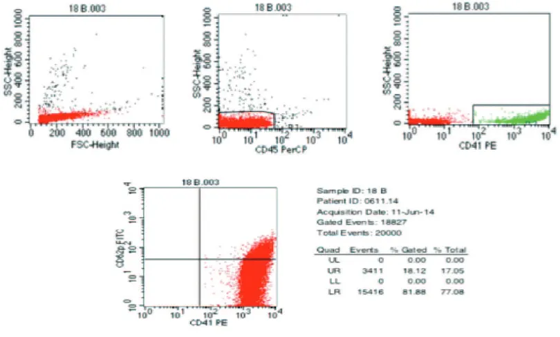
Measurement of CD62P expression using lowcytometry method
The method applied in this research
refered to previous method developed by
Kim and Lim,13 which minimalize platelet activation artiicially, eliminating the need of centrifugation for washing and ixation before
labelling and simultaneously label monoclonal antibody to the patient platelets. CD62P
expression was measured by lowcytometry
method using monoclonal antibody
anti-CD62P-FITC against whole blood sample without addition of agonist (ADP). The
measurement was conducted on blood sample
before and 1 hour after TC transfusion (pre vs. post-transfusion). For analytical purpose,
gates were made in predetermined platelet
area with addition of anti-CD41-PE (FIGURE
1). Data were presented as platelet percentage
that positively expressed CD62P.
Data analysis
Subject characteristics was presented
descriptively. The percentage of platelet (%)
that expressed CD62P was presented as mean
value. The comparison of increased CD62P
expression in both groups was presented
as relative risk. Statistical analysis was performed at signiicance value p < 0.05 with conidence interval of 95%.
RESULTS
Total subjects enrolled in this study
were 103 patients, divided into 2 groups; irst group consist of 51 patients receiving non-leukodepleted TC and another group of 52 patients receiving leukodepleted TC transfusion. Subject characteristics were summarized in TABLE 1. Overall, there were no statistically signiicant difference (p >0.05) between two groups in term of gender, age, body weight, height, body surface area (BSA) and body mass index (BMI).
TABLE 1. Characteristics of subjects
Characteristic Non-leukodepleted
(n=51)
Leukodepleted
(n=52) p
Gender
Male 32 (62.7%) 30 (57.7%) 0.600
Female 19 (37.3%) 22 (42.3%)
Age (mean ± SD year) 8.69 ± 4.83 9.93 ± 5.10 0.209
1-5 16 12
5-10 17 17
10-14 7 6
14-18 11 17
Weight (mean ± SD kg) 26.12 ± 15.84 26.67 ± 12.84 0.847
Height (mean ± SD m) 1.22 ± 0.28 1.28 ± 0.29 0.279
BSA (mean ± SD m2) 0.925 ± 0.379 0.975 ± 0.354 0.491
BMI (mean ± SD kg/m2) 16.048 ± 4.10 15.279 ± 2.64 0.259
In this study, CD62P expression on platelet
was measured from patient venous blood sample before and 1 hour after TC transfusions. Mean expression of CD62P before transfusion was not statistically signiicant difference between two groups i.e. 25.42 ± 19.52% in non-leukodepleted group and 27.16 ± 10.31% in leukodepleted group respectively (p > 0.05). On the contrary, there was statistically
TABLE 2. Mean expression of pre and post-transfusion CD62P
Group
Non-leukodepleted (n = 51)
%
Leukodepleted (n = 52)
%
p
Pre-transfusion 25.42±19.52 27.16±10.31 0.574
Post-transfusion 35.85±20.31 24.12±7.16 0.000
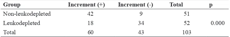
Relative risk of increased CD62P expression in patients receiving non-leukodepleted TC compared to non-leukodepleted group was presented in the TALE 3. It was
seen that the increased CD62P expression
was 2.38 times higher in the group receiving non-leukodepleted TC than leukodepleted TC transfusions.
TABLE 3. Relative risk of increased CD62P expression in non-leukodepleted group
Group Increment (+) Increment (-) Total p
Non-leukodepleted 42 9 51
Leukodepleted 18 34 52 0.000
Total 60 43 103
Relative risk = 2.38; (95%CI=1.60-3.53)
DISCUSSION
The increased expression of post-transfusion CD62P was greater in the group receiving non-leukodepleted TC than leukodepleted TC probably caused by addition effects of pretransfusion activated donor platelets or the effect of increased level of MPO in transfused TCs. Nomura et al.14 have conducted a research with transfusion of 100 TCs involved 28 hematologic and
non-hematologic malignancy patients, it was concluded that platelet CD62P expression
was diminished signiicantly after leukocyte iltration process or leukodepletion.
There were some markers to evaluate platelet activation such as β-Thyroglobulin (β-TG), Glycoprotein-1β (GPIB/CD42), platelet aggregation test or GPIIb/IIIa (CD41), soluble P–selectin, surface P-selectin (CD62P), and among them CD62P is the best marker.15-17 CD62P also known as α-granule platelet
membran protein-140 (GMP-140), expressed
on the surface membrane during platelet activation, but remain undetectable in resting
platelets. CD62P reported to be expressed
rapidly on the surface membrane when platelet is activated.18 CD62P is the most sensitive and speciic marker which relects activation of platelets. Among other activated thrombocyte surface marker, CD62P expression shows the most signiicant difference between activated
and resting platelet state.18,19
There was a correlation between
P-selectin expression and platelet survival after transfusion, stronger expression of P-Selectin on the platelet surface, associated with easier destruction of platelet after being transfused.18,19 After activation, platelets will
release proinlammatory chemokine, cytokine and adhesion molecule appear onto its surface, including CD62P and CD40 ligand (CD40L).
Interaction between platelet and neutrophyl
and it contribute toward host immunity and thrombosis process. Human neutrophil
α-defensins (HNPs) consist of 50% protein from azurophilic granules. It can be released from azurophilic granule into extracellular environtment after PMN cell activation and then undergo degranulation, leakage, cell death and lysis during inlammation.20
Clinical relevance of CD62P expression level to predict in vivo platelet behaviour after transfusion is still debatable and not fully understood. It is partly based on fact that dynamicity of platelet and loss of CD62P surface protein.21 There was evidence that P-selectin level increased during storage, whereas its activatability undergo reduction.22 Increased level of CD62P signiicantly unrelated to body mass index (BSA), carotid intima media thickness and inlammatory
mediator (CRP).17
CONCLUSION
In conclusion, non-leukodepleted have signiicant 2.38 times higher risk of increased post-transfusion CD62P expression than leukodepleted TC transfusions. 2014: Pelayanan darah di Indonesia. Jakarta: Kementrian Kesehatan Republik Indonesia,
2015.
2. UPTD RSUP Dr. Sardjito. Laporan pengeluaran darah dan rekapitulasi
3. Kaufman RM, Djulbegovic B, Gernsheimer
T, Kleinman S, Tinmouth AT, Capocelli KE,
et al. Platelet transfusion: A clinical practice guideline from the AABB. Ann Intern Med 2015; 162 (3): 205–13. http://dx.doi. org/10.7326/M14-1589.
4. Xie ZT, Chen C, Zhang SH, Yang HM, Tao
ZH. Effect of leukocyte iltration on the P-selectin expression of apheresis platelets. Genet Mol Res 2015; 14(2):5979-85. http:// dx.doi.org/10.4238/2015.June.1.15.
5. Sharma, RR., Marwaha, N. Leukoreduced
blood components: Advantages and strategies for its implementation in developing countries. Asian J Transfus Sci 2010; 4(1):3-8. http://
dx.doi.org/10.4103/0973-6247.59384
6. Nielsen HJ, Reimert CM, Pedersen AN,
Brunner N, Edvardsen L, Dybkler L, et al.
Time-dependent, spontaneous release of white
cell and platelet-derived bioactive substances
from stored human blood. Transfusion 1996;
36(11-12): 960-5.
h t t p : / / d x . d o i . o r g / 1 0 . 1 0 4 6 / j . 1 5 3 7 -2995.1996.36111297091738.x
7. Faraday N, Scharph RB, Doddo JM, Martinez EA, Rosenfeld BA, Dorman T. Leukocytes
can enhance platelet-mediated aggregation
and thromboxane release via interaction of P-selection glycoprotein ligand 1 with P-selectin. Anesthesiology 2001; 94: 145-51.
http://dx.doi.org/10.1097/00000542-200101000-00025
8. Faint RW. Platelet-neutrophil interactions:
their signiicance. Blood 1992; 6(2):83-91.
http://dx.doi.org/10.1016/0268-960X(92) 90010-N
9. McFaul SJ, Corley JB, Mester CW, Nath J. Packed blood cells stored in AS-5 become proinlammatory during storage. Transfusion
10. Kolarova H, Klinke A, Kremserova S, Adam M, Pekarova M, Baldus S, et al.
Myeloperoxidase induces the priming
of platelets. Free Radic Biol Med 2013; 61:357-69. http://dx.doi.org/10.1016/j. freeradbiomed.2013.04.014
11. Eiserich JP, Baldus S, Bernnan ML, Ma W,
Zhang C, Tousson A, et al. Myeloperoxidase
a leukocyte-derived vascular NO oxidase.
Science 2002; 296(5577):2391-4.
http://dx.doi.org/10.1126/science.1106830 12. Baldus S, Heitzer T, Eiserich JP, Lau D, Mollnau
H, Ortak M, et al. Myeloperoxidase enhances
nitric oxide catabolism during myocardial
ischemia and reperfusion. Free Radic Biol Med 2004; 37(6):902-11. http://dx.doi.
org/10.1016/j.freeradbiomed.2004.06.003 13. Kim SW, Lim YA. Establishment of reference
values for platelet activation markers by lowcytometry. Korean J Lab Med 2006;
26(5):323-8.
http://dx.doi.org/10.3343/kjlm.2006.26.5.323 14. Nomura S, Okamae F, Abe M, Hosokawa M,
Yamaoka M, Ohtani T, et al. Platelets expressing P-selectin and platelet-derived microparticles in stored platelet concentrates bind to PSGL-1 on iltrated leukocytes. Clin Appl Thromb Hemost 2000; 6(4):213-21. http://dx.doi. org/10.1177/107602960000600406.
15. Plaza EM, Cespedes P, Fernandez H, Sanchez-Guiu MI, Egea JM, Vicente V, et al. Quality assessment of buffy-coat-derived leukodepleted platelet concentrates in PAS-plasma, prepared by the Orbi Sac or TACSI automated system. Voxsanguinis 2013;
106(1):1-7.
16. Bashir S, Nightingale MJ, Cardigan,
R. Ensuring that blood transfusion sets
an effective dose of functional blood components. Transfus Med 2013; 23(4):226 -30. http://dx.doi.org/10.1111/tme.12045
17. Nagy BJ, Debreceni IB, Kappelmayer J. Flowcytometry in the clinical laboratory lowcytometric investigation of classical and alternative platelet activation. EJIFCC 2012;
23(4):1-11.
18. Vetlesen A, Holme PA, Lyberg T, Kjeldsen-Kragh J. Recovery, survival, and function of transfused platelets and detection of platelet engraftment after allogeneic stem cell transplantation. Transfusion 2012; 52(6):
1321-32.
http://dx.doi.org/10.1111/j.1537-2995. 2011.
03442.x.
19. Ritchie JL, Alexander HD, Rea IM. Flowcytometry analysis of platelet P-selectin
expression in whole blood-methodological
considerations. Clin Lab Haematol 2000; 22(6):359-63. http://dx.doi.org/10.1046/ j.1365-2257.2000.00339.x
20. Horn M, Bertling A, Brodde MF, Muller A, Roth J, Van Aken H, et al. Human neutrophil alpha-defensins induce formation of ibrinogen and thrombospondin-1 amyloid-like structures and activate platelets via glycoprotein IIb/IIIa. J Thromb Haemost 2012; 10(4):647-61. http://dx.doi. org/10.1111/j.1538-7836.2012.04640.x. 21. Levin E, Jenkin C, Culibrk B, Issa G, Maria
IC, Serrano K, et al. Development of a quality monitoring program for platelet components: a report of the irst four years’ experience at Canadian Blood Services. Transfusion 2012;
52(4):810-18.
h t t p : / / d x . d o i . o r g / 1 0 . 1111 / j . 1 5 3 7 -2995.2011.03402.x.