Indo. J. Chem., 2007, 7 (2), 214-217 214
SYNTHESIS OF UK-3A ANALOGUE AND ASSAY ON P 388 MURINE LEUKEMIA CELLS
Y Anita1,*, M Hanafi1, AMJ Putra1, S Arifin 2, Y Usuki3 , and H Iio3
1 LIPI-Research Centre for Chemistry, Kawasan Puspiptek –Tangerang 15314
2 Department of Chemistry, University of General Soedirman, Purwokerto, Indonesia
3Osaka City University, Department of Material Science, Osaka-Japan
Received 16 April 2007; Accepted 1 June 2007
ABSTRACT
UK-3A is secondary metabolite of Streptomyces sp.517-02 which has IC50 38 µg/mL against P388 Murine Leukemia cells. An analogue of UK-3A was synthesized from L-methyl serine as the starting material by amidation and esterification. An analogue UK-3A was analyzed and identified by TLC, FT-IR, LC-MS and NMR spectrometer. It was found to have IC50 15.4µg/mL against the same Leukemia cells. The overall yield was 87.10%.
Keywords: UK-3A, Streptomyces sp. 517-02, Anticancer, P388 Murine Leukeumia cell
INTRODUCTION
UK-3A compound had been isolated from the
mycelium of Strepyomyces sp. 517-02 [1]. The
compound was then acknowledged to have a high activity against mouse leukemia cells, P-388 (IC50 = 38
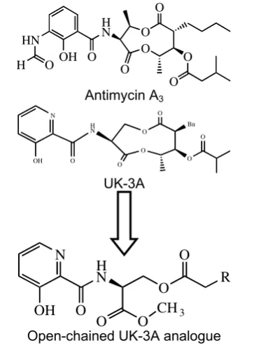
μg/mL) [2]. The structure of UK-3A is almost identical to the one of previously known antibiotics, Antimycin A3.
Both Antimycin A3 and UK-3A consist of 9-membered
dilactone rings linked via an amide bond to an aromatic acid moiety (Fig 1).
Antimycin A3 is a fit ligand of protein Bcl2, which is involved in the intrinsic apoptosis sequences of cancer cells [3]. Antimycin is already known to induce apoptosis of human Leukemia cells, HL-60 [4]. While Bcl2 is known to be over-expressed in 90% of colon cancer cells, 80% of B-cell lymphomas, and 70% of breast cancer cells, it is reasonable to expect Antimycin A3 to induce apoptosis of those cells as well. Thus, it is also reasonable to expect UK-3A (or its analogs) to have similar or higher anti-cancer activities.
* Corresponding author.
Email address : [email protected]
This research focused on the 9-membered dilactone ring itself, which was replaced by suitable mimetic structure, as shown in Fig 1, and report the result of preliminary studies on the preparation and cytotoxic assay of open-chained UK-3A analogue. Cancer is still a life-threatening disease in Indonesia nowadays [5], and it is our social responsibility to discover effective drugs for the disease.
EXPERIMENTAL SECTION
Column chromatography was carried out using Merck silica gel 60 GF254 and for TLC analysis, precoated silica gel plates (Merck Kiesel-gel 60 GF254, 0.25mm) were used. Visualization of TLC plates was performed using Ninhydrin spray reagents and UV lamp 254 nm. The identity and purity of the compounds were established by Spectrometer LC-MS HP 5972 series,
O
Open-chained UK-3A analogue Fig 1. Structures of AA, UK-3A and analogue
.
FT-IR Spectrophotometer Shimadzu 2010 A. 1H and 13
C-NMR spectra were recorded at 500 MHz on JEOL ECA.
Material
The Compounds were prepared from L- Serine methyl ester-HCl, 3-Hydroxypicolinic acid, and Octanoic acid as main materials pTSOH, DCC and DMAP were used as catalyst and activator. Pyridine and chloroform were used as solvent.
Procedure
The synthesis of open chained UK-3A analogue consists of two step reaction : amidation and esterification reaction, as shown in scheme 1.
Indo. J. Chem., 2007, 7 (2), 214-217 215
To a stirred solution of 3-hydroxypicolinic acid (0,5606 g; 4 mmol ) to afford the corresponding L-serine methyl-HCl (0,3120 g; 2 mmol ) in pyridine (10.0mL) was added DCC (0,5061 g; 2,2 mmol) and DMAP (0,048 g; 0,4 mmol) successively to afford the corresponding L-serine methyl-HCl (0,3120 g; 2 mmol ). After stirring for 24 h at 55 oC, the mixture was acidified with HCl 2 % and extracted with dichloromethane. The combined organic layers were washed with 1 % NaOH solution and
brine, and dried over MgSO4. The filtrate was
concentrated and purified by silica gel column chromatography (hexane-ethyl acetate) to give the condensation product.
The final step in this synthesis of UK-3A analogues is the coupling of amidation product with each carboxylic acid .
To stirred solution of amidation product (0,0919 g; 0,4) mmol and octanoic acid (0,095 mL; 0,6 mmol) in chloroform (5 mL) was added DCC (0,1246 g; 0,6 mmol) and DMAP (0,0132 g; 0,1 mmol). After stirring for 4 h at 25 oC, the mixture was extracted with dichloromethane. The combined organic layers were washed with 1 % NaOH solution and brine, and dried over MgSO4. The filtrate was concentrated and purified by silica gel column chromatography (hexane-ethyl acetate) to give the ester product.
Cytotoxicity assay [6-8]
P-388 cells were seeded into 96-well plates at an initial cell density of approximately 3x10⁴ cells cm-3. After 24 h of incubation for cell attachment and growth, varying concentrations of samples were added. The compounds added were first dissolved in DMSO at the required concentration. Subsequent six desirable concentrations of samples were prepared using PBS (phosphoric buffer solution, pH 7.30-7.65). Control wells received only DMSO. The assay was terminated after an 48 h incubation period by adding MTT reagent
[3-(4,5-dimethyl-thiazol-2-yl)-2,5-diphenyl tetrazolium bromide; also named as thiazol blue] and the incubation was continued for another 4h, in which the MTT-stop solution containing SDS (sodium dodecyl sulphate) was added and another 24 h of incubation was conducted. Optical density was read by using a microplate reader at 550 nm. IC50 values were taken from the plotted graph of percentage live cells compared to control (%), receiving only PBS and DMSO, versus the tested concentration of compounds (µM). The IC50 value is the concentration required for 50% growth inhibition. Each assay and analysis was run in triplicate and averaged.
RESULT AND DISCUSSION
UK-3A analogue was prepared as shown in Fig 2. Amide formation with 3-hydroxypicolinic acid proceeded smoothly to afford the corresponding L-serine methyl-ester-HCl in the presence of DCC/DMAP. The resulting amide formation was purified by SiO2 column chromatography that showed a positive reaction with ninhydrin reagent and obtained as white smooth crystals with mp. 90-91 oC in yield 61.20%. The identity of this compound was established by LC-MS : 240.2768 m/z [M+] is the molecular weight of amidation product. The presence of amide (CONH) and phenolic (OH) were identified by the FT-IR absorption at 3360 cm-1 and 3412 cm-1, respectively.
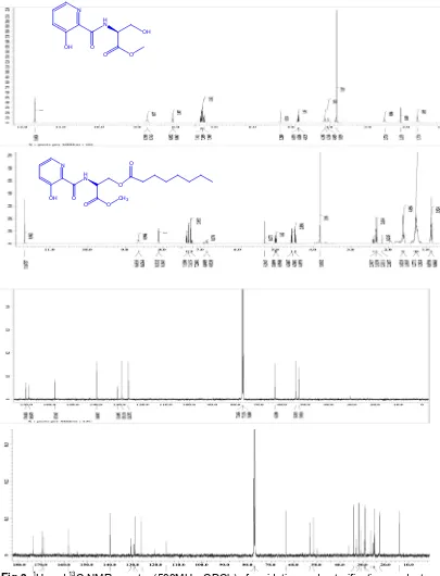
The product was confirmed as well by 1H and 13
C NMR spectra (500MHz, CDCl3), as shown in Fig 3. A signal 1H for the phenolic OH proton was seen at
δH11.68 (s). This downfield-shifted signal suggests the formation of an intramolecular hydrogen bond between the phenolic OH proton and the carbonil oxygen of an amide bond. The signal 1H and 13C for CONH were
3-Hydroxypicolinic acid amidation product
+
Fig 2. Synthesis of UK-3A analogue
Indo. J. Chem., 2007, 7 (2), 214-217 216
H N
OH
O O N
O OH
N
OH H N
O
O
O O
CH3
O
Fig 3. 1H and 13C NMR spectra (500MHz, CDCl3) of amidation and esterification product.
The final step in this synthesis of UK-3A analogue is the coupling of amidation product with carboxylic acid (octanoic acid).The purity and identity were conducted by SiO2 column chromatography that showed positive reaction with ninhydrin reagent and obtained product in 87.10%. The product was established by LC-MS and 371.87 m/z [M+] and FT-IR absorption at 1217 cm-1 (C-OOR).
The product was confirmed as well by NMR spectra (500MHz, CDCl3), as shown in Fig 3. A signal 1H for the phenolic OH proton was seen at δH 11.67, This downfield-shifted signal suggests the formation of an intramolecular hydrogen bond between the phenolic OH proton and the carbonil oxygen of an amide bond. The presence of octanoil aliphatic chain was determined signal of proton at δH 0.88-2.40. The signal of octanoil aliphatic chain was also determined
Indo. J. Chem., 2007, 7 (2), 214-217 217
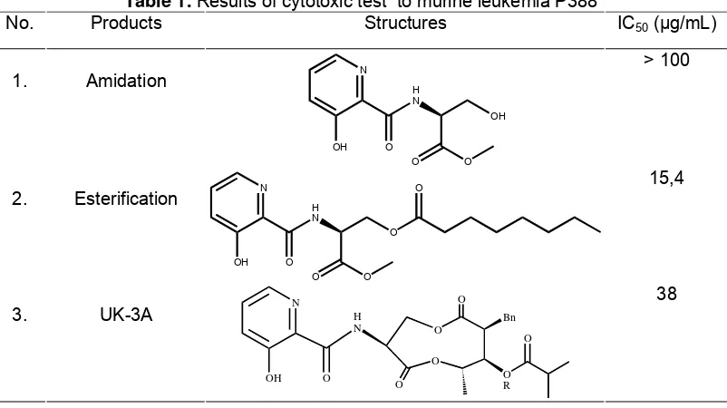
Table 1. Results of cytotoxic test to murine leukemia P388
No. Products Structures IC50 (μg/mL)
1. Amidation H
N
OH
O O
N
O OH
> 100
2. Esterification H
N
O
O O
N
O OH
O 15,4
3. UK-3A
O O
O
O
Bn
O R H
N N
O OH
O
38
δC 22.76–34.19 and for the ester (COO) at δC 168.94 (downfield-shifted signal).
The cytotoxicity of compound was evaluated against murine leukemia P-388 cells. The result indicated that the compound was cytotoxic in vitro, with inhibitory concentration (IC50) value of 15.4 µg/mL. The compound showed higher inhibitory activity than UK-3A (IC50 38 µg/mL) and the amidation product, as shown in Table 1. This fact suggested that the length of alkyl chain and the hydrophobicity for membrane permeability of the structure had some effect on anticancer potency.
CONCLUSION
The 9-membered dilactone ring in UK-3A and Antimycin A3 could be substituted for open-chained ester. However, anticancer activity depends on the structure or hydrophobicity of the open-chained ester, as cell membrane permeability is necessary.
ACKNOWLEDGEMENT
This research was supported by LIPI and the Japan Society for the Promotion of Sciences (JSPS), which are gratefully appreciated.
REFFERENCES
1. Ueki, M. and Taniguchi, M. 1997, J.Antibiotic. 50, 152.
2. Hanafi, 1995, Studies of Novel Antibiotic
Metabolites from Streptomyces sp. 517-02. Department of Chemistry, Faculty of Science, Osaka City University.
3. Liu X., 2003, Curr Med Chem Anti-Canc Agents, 3, 217.
4. King, E., 2002, Cytometry, 49, 106.
5. www.kompas.com/kompas-cetak/0404/07/ humaniora/954503.htm, 2004, November 1.
6. Sahidin, and Hakim, EH. 2006, Cytotoxic
Properties of Oligostilbenoids from the Tree Barks of Hopea Dryobalamides. 60c, 723.
7. Usuki, Y., Adachi, N., Fujita, K-I., Ichimura, A.., Iio,
H., and Taniguchi, M. 2006, Bioorg.
Med.Chem.Lett. 16, 3319.
8. Shimano, M., Kamei, N., Shibata, T., Inoguchi, K., Itoh, N., Ikari, T., and Senda, H. 1998, Tettrahedron. 54, 12745.