P-ISSN : 1978-225X; E-ISSN : 2502-5600 DOI: https://doi.org/10.21157/j.ked.hewan.v11i2.5400
DETECTION OF FIVE VIRUS INFECTIONS IN THE LAYER FARM WITH
RUNTING-STUNTING SYNDROME IN SUKABUMI AND TANGERANG
USING POLYMERASE CHAIN REACTION TECHNIQUE
Risza Hartawan1
* and Ni Luh Putu Indi Dharmayanti1
1Laboratory of Virology, Indonesian Research Center for Veterinary Science, Bogor, Indonesia
*Corresponding author: [email protected]
ABSTRACT
The objective of this study was to identify the five different viral infections including avian influenza, Newcastle disease, avian reovirus, avian
encephalomyelitis, and Marek’s disease in the runting-stunting syndrome outbreak in several commercial layer farms in Sukabumi and Tangerang in November 2014 using polymerase chain reaction technique. As results, this study identified mix infection of three viruses in the field samples,
including Newcastle disease, reovirus, and avian encephalomyelitis; however, it was negative for avian influenza and Marek’s disease viruses. Subsequently, the inoculation of several samples into embryonated chicken eggs confirmed the growth of these three viruses. As a consequence, disease control management should be conducted in the affected farms by implementing effective biosecurity and vaccination program. ____________________________________________________________________________________________________________________ Key words: layer, runting stunting syndrome, Sukabumi, Tangerang, viral infection
ABSTRAK
Penelitian ini bertujuan mengidentifikasi lima jenis infeksi virus yaitu avian influenza, Newcastle disease, avian reovirus, avian encephalomyelitis dan Marek pada kasus kekerdilan yang terjadi pada peternakan ayam petelur komersial di Sukabumi dan Tangera ng pada bulan November 2014 dengan menggunakan teknik polymerase chain reaction (PCR). Analisis pada sampel lapang menunjukkan adanya infeksi campuran virus Newcastle disease, reovirus dan avian encephalomyelitis tetapi negatif terhadap virus avian influenza dan Mare k. Selanjutnya, inokulasi sampel lapang pada telur ayam bertunas mengonfirmasi pertumbuhan ketiga jenis virus tersebut. Tiga jenis infeksi virus teridentifikasi pada kasus kekerdilan ayam yaitu virus Newcastle disease, avian reovirus, dan avian encephalomyelitis .
____________________________________________________________________________________________________________________ Kata kunci: ayam petelur, sindrom kekerdilan, Sukabumi, Tangerang, infeksi virus
INTRODUCTION
Runting-Stunting Syndrome (RSS) was firstly identified at the end of 1970 in broiler chicken characterized by slow growth, abnormal quill growth (helicopter wing syndrome), diarrhea, and the other abnormalities (Jones, 2008). These diseases lead to economic loss as a result of underweight chicken (below target), increase of interpretation rate, and feed inefficiency. The cause of RSS in chicken is still not known definitively. Some disease agents are suspected playing a role in resulting this stunting, while the other cause, such as stress, toxin, and environment condition are the disease predisposition factors (Goodwin et al., 1993).
Viral infection as the causative agent had been identified to cause RSS such as reovirus, toravirus, parovirus, enterovirus-like virus, adenovirus, calicivirus, and togavirus-like virus, however the certain etiology of this disease is still unclear due to the experimental studies of some viruses failed to manifest clinical symptoms consistently (Guy, 1998). Goodwin
et al. (1993), in Georgia, has identified the infection of avian nephritis virus, enterovirus, and reovirus in RSS. In Northern Germany, Otto et al. (2006) identified an infection of avian rotavirus in RSS. Kang et al. (2012) found infection of chicken astrovirus and avian nephritis virus in the case of chicken stunting. Zsak et al. (2013) revealed that chicken parvovirus infection experimentally might cause stunting symptom in young broiler chicken. The result of these studies indicated an essential role of viral infection in chicken stunting symptoms in various regions of the world.
However, chicken stunting syndrome in the field did not only attack broiler chicken, but also laying chicken. The stunting outbreak that struck commercial layer farms occurred in Sukabumi and Tangerang District in November, 2014. This study aimed to identify infection of some viruses in stunting case. The examination method used was reverse transcriptase polymerase chain reaction (RT-PCR) test for avian influenza (AI), Newcastle disease (ND), avian reovirus (REO), and avian enchepalomyelitis (AE) test for ribonucleid acid (RNA) virus and polymerase chain reaction (PCR) test for Marek’s disease (MD) test for deoxyribonucleid acid (DNA) virus. Identification of virus-related infection was carried out focusing on only five viruses which commonly struck the commercial laying chicken farms, namely AI, H5N1 subtype, ND, REO, AE, and MD. The selection of the five viruses also regarded the clinical symptoms such as stunting, neurological symptoms, and paralysis.
MATERIALS AND METHODS
Field Samples and Research Field
Risza Hartawan and Ni Luh Putu Indi Dharmayanti
66
Primer Set and Virus Control
The specific Primer set was used for PCR and RT-PCR examination on five types of viruses (AI, ND, REO, AE, and MD) as presented in Table 1. Positive control of viruses was taken from virus collections of BBLitvet Virology Laboratory and commercial source. Isolate of A/Ck/WestJava/PWT-WIJ/2006 (BBLivtet) and RIVS (BBLivtet) were used as control for AI and ND, while Reo 1133 and Calnek 1143 strain AE (Nobilis) isolates were used as control for REO and AE. As control for MD, MDV-1 CV1988 Rispens strain and FC128 strain HVT (Merial) isolates were used.
Isolation of Genetic Materials for RNA and DNA Virus Group
Isolation of RNA virus (AI, ND, REO, and AE) was performed using commercial kit QIAamp®
Viral Mini
Kit (Qiagen) while the isolation of DNA virus (MD) was performed by DNeasy Mini Kit (Qiagen). Results of RNA and DNA extraction were stored at -20 C for further analysis.
RT-PCR test for identification of AI, ND, REO, and AE virus
RT-PCR examination to identify AI, ND, REO, and AE virus was performed by SuperScript®
III One Step RT-PCR System with Platinum® Taq DNA Polymerase (Invitrogen). Each reaction with a total volume of 25 μl consist of 12.5 μl 2X reaction mix; 1 μl 5 mM MgSO4; 1
μl each of forward and reverse primer (20 μM); 1 μl
SuperScript® III RT/Platinum® taq mix; and 8.5 μl RNA template. RT-PCR process initiated a reverse transcriptase at temperature of 45 C for 45 minutes, followed by inactivation of enzyme at temperature of 92 C for 2
Table 1. Sequence of primer set used in this study
Virus type Gene target Primer code Sequence (5’-3’) PCR product Literature
AI H5 H5-155F ACACATGCYCARGACATACT
545 bp Lee et al. (2001) H5-699R CTYTGRTTYAGTGTTGATGT
ND F NCD3 GTCAACATATACACCTCATC 309 bp Stäuber et al. (1995)
NCD4 GGAGGATGTTGGAGCATTT
REO σA σA1 ATTACGCAGAGGCATTT
241 bp Ke et al. (2006)
σA2 CCCACTGCCGAATACA
AE VP2 MKAE1 CTTATGCTGGCCCTGATCGT
619 bp Xie et al. (2005)
MKAE2 TCCCAAATCCACAAACCTAGCC
MDV-1 meq meqF GAATCTTCCCTGCATTGTGTC
196 bp
Islam et al. (2006); Renz et al. (2006)
meqR ATCTGGCCCGAATACAAGGAA
GaHV3 DNApol DNApolF GTCTGCCCTCGTCTTAGC
283 bp DNApolR ACTCGCTTCCTCCAATTCG
HVT Sorf1 Sorf1F AAGCGCTTGTATGTGTAGG
350 bp
Sorf1R TATGGACGTCATGCAGTTGG
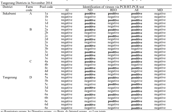
Table 2. Identification of viral infection on samples of chicken organs that experiencing stunting symptom in Sukabumi and
Tangerang Districts in November 2014 Dsitrict Farm
code Pool code AI Identification of viruses via PCR/RT-PCR test ND REO AE MD
Sukabumi A 1a negative positive positive positive negative
1b negative negative negative negative negative
1c negative positive positive positive negative
1d negative positive negative positive negative
1e negative positive negative positive negative
B 2a negative positive positive positive negative
2b negative negative negative positive negative
2c negative negative positive positive negative
2d negative positive negative positive negative
2e negative negative negative positive negative
3a negative positive negative positive negative
3b negative negative negative negative negative
3c negative positive positive positive negative
3d negative positive negative positive negative
3e negative positive negative positive negative
C 4a negative positive negative positive negative
4b negative negative negative negative negative
4c negative positive positive positive negative
4d negative positive negative negative negative
4e negative positive negative positive negative
Tangerang D 5a negative positive positive positive negative
5b negative negative negative negative negative
5c negative negative positive positive negative
5d negative positive negative positive negative
5e negative negative negative positive negative
6a negative positive positive positive negative
6b negative negative negative negative negative
6c negative negative positive positive negative
6d negative positive negative positive negative
6e negative negative negative positive negative
minutes. Gene amplification was performed for 40 cycles consisting of denaturation (92 C, 30 seconds), annealing (55 C, 90 seconds) and elongation (72 C, 2 minutes). A final extension was completed at 72 C for 10 minutes.
PCR Test for Identification of MD Virus
The PCR examination to identify three different serotype of MD virus was performed by HotStarTaq®
Plus Master Mix Kit (Qiagen). Each reaction with a total volume 20 μl consist of 10 μl 2X HotStarTaq Plus Master mix, 1 μl bovine serum albumine (10 mg/ml), 0.5
μl each of respective forward and reverse primer of meq, DNApol, and sorf1 gene, and 8.5 μl DNA template. The PCR process was initiated by initial heat activation at 95°C for 5 minutes and followed by 30 amplification cycles (denaturation at 92 C for 90 seconds, annealing at 60 C for 60 seconds, and elongation at 72 C for 60 seconds) with a final extension at 72 C for 10 minutes.
Visualization of PCR/RT-PCR Results
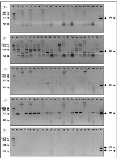
Visualization of PCR/RT-PCR results was done by electrophoresis with 100 volts for 30 minutes in 2% Agarose 1x TBE stained with ethidium bromide. Ladder DNA 100 bp (Invitrogen) was used as molecular marker. The profile of DNA was visualized using UV transilluminator apparatus and documented with Gel Doc machine (Vilber Lourmat).
Inoculation of Field Samples into Embryonated Eggs Isolation of virus was performed on field samples which positively identified viral infected by PCR or RT-PCR examination, through inoculation of the virus in specific negative antibody embryonated eggs (EE). Then the result of isolation was confirmed by respectivePCR and/or RT-PCR test to identify virus growth.
RESULTS AND DISCUSSION
Field Samples
The stunting syndrome occurred in laying pullets characterized by symptoms of slow weight-gaining, thinness, lethargy, drowsiness, dull quills where several chickens had paralysis and neurological symptom. A total of 30 pools of organ which grouped as respiratory organs, digestive organs, brain, internal organs, and leg joints were successfully collected from the field.
Identification of Infection of Viruses in Stunting Syndrome
The results of PCR and RT-PCR examination on five viruses from field samples in Sukabumi and Tangerang District was presented in Table 2 and Figure 1.The analysis of field samples on five virus infections showed the occurrence of mixed infection of three different types of virus: ND, REO, and AE virus. On the other hand, the analysis of all samples on H5 subtype AI and MD virus
Risza Hartawan and Ni Luh Putu Indi Dharmayanti
68
infection showed negative results. The rate of viral infection was dominated by AE virus (80%) then followed by ND (60%) and REO virus (33.33%). Most infections were a mixed infection among the three types of viruses, particularly ND and AE virus. The electrophoresis results in Figure 1 showed the difference of amplicon size product between field samples with positive control. The difference was probably caused by genetic difference between them, thus further confirmation by gene sequencing is needed.
In this study, avian reovirus was identified on a number of field samples. This reoviridae-family virus was indicated playing an important role in stunting chicken cases as well as viral arthritis paralysis (Van der Heide, 2000). The infection of avian reovirus had been spreading in some commercial laying chicken farms but most of the cases were non-pathogenic (Jones, 2008). In Indonesia, infection of avian reovirus was identified affecting some commercial farms especially in broiler chicken (Wahyuwardani et al., 2005a). Clinical study conducted by Van de Zande and Kuhn (2007) showed that ERS strain of reovirus not only causes stunting syndrome, but also infect the central nerve system resulting in tremor and torticollis symptoms. Bányai et al. (2011) reported that AVS-B strain reovirus was successfully isolated from
RSS case in Delaware, USA with symptoms of diarrhea, loss of appetite, and slow growth. Reovirus infection leads to immunosuppressive condition which resulted in more susceptible of the chicken to other diseases (Wahyuwardani et al., 2005b).
This study indicated the infection of ND virus on several field samples although no literature explains the correlation between this virus and the stunting syndrome. The paramyxoviridae-family virus caused disease with various degrees of clinical manifestation and severity, ranged from the subclinical infection, mild infection, respiratory and neurological disturbance, to death (Alexander and Jones, 2008). ND virus infection was probably a secondary infection due to immunosuppression by reovirus infection. However, it is possible that the infection was done by genotype VII ND virus, so that the existing vaccination program became ineffective (Dharmayanti et al., 2014).
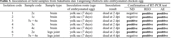
Beside REO and ND virus infections, this study also identified the presence of AE virus. There is no literature linking this virus which is from the picornaviridae family, with stunting syndrome. AE virus caused epidemic tremor syndrome in several types of poultry at a very young age (Tannock and Shafren, 1994). Welchman et al. (2009) identified the presence of AE virus infection in peasant Figure 2. Appearance of chicken embryo that inoculated with field samples from Sukabumi and Tangerang Districts in 2014
bird farms with neurological symptoms in the UK in 2007. Asasi et al. (2008) identified the AE virus infection in broiler farms in Shiraz, Iran with symptoms of ataxia, tremor, and stretching of the legs. The case of epidemic tremor in Indonesia was suspected occurred in Bogor in the 1970s (Maide et al., 1972). Problems that might arise by the presence of AE virus infection was the impact on egg production which lead to low surviving of chicken flock to achieve maximum egg production.
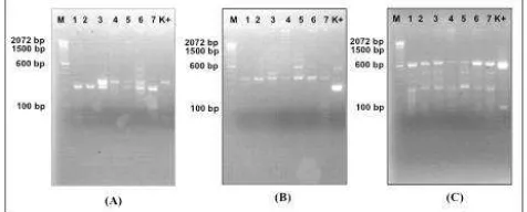
Inoculation of Field Samples into Embryonated Eggs The selected field samples of stunted laying chicken from Sukabumi and Tangerang District in 2014 that were tested for isolation stage were brain organs and legs joints due to the less risk of bacterial contamination. Virus isolation was performed by inoculation on EE with yolk sac route at 7 days. The inoculation of 7 field samples from Sukabumi and Tangerang District in 2014 was presented in Table 3.
In Table 3, the results of viral isolation into embryonated eggs showed that almost all field samples caused the death of embryos at 2 day post inoculation (dpi), except for the sample with the inoculation code seven, causing embryo death at 4 dpi. Figure 2 showed that embryo death at 2 dpi showed bleeding and destruction. Meanwhile, embryo death at 4 dpi showed no significant anatomic changes but remained severe hemorrhage. In Table 2 and Figure 3, the results of confirmation by RT-PCR test demonstrated that in all embryos inoculated with field samples showed the growth of REO and AE viruses. ND virus was also identified in some inoculated embryos. It is needed advance research to separate the three types of viral infections before proceed to the next step of biological characterization of each type of virus.
CONCLUSION
Three types of viral infections were identified in cases of chicken stunting in several commercial laying chicken farms in Sukabumi and Tangerang Districts in November 2014, namely Newcastle disease, avian reovirus, and avian encephalomyelitis.
REFERENCES
Alexander, D.J. and R.C. Jones. 2008. Paramyxoviridae. In Poultry Diseases. Mark, P., F.M. Paul, M.B. Janet, and J.A. Dennis (Eds.). 6th
end. WB Saunders, Edinburgh.
Asasi, K., A. Farzinpour, and A.K. Tafti. 2008. Clinico-pathological studies on avian encephalomyelitis in Shiraz, Iran. Turk. J. Vet. Anim. Sci. 32:229-231.
Bányai, K., E. Dandár, K.M. Dorsey, T. Mató, and V. Palya. 2011. The genomic constellation of a novel avian orthoreovirus strain associated with runting-stunting syndrome in broilers. Virus Genes. 42:82-89.
Dharmayanti, N.L.P.I., R. Hartawan, D.A. Hewajuli, and R. Indriani. 2014. Phylogenetic analysis of genotype VII of new castle disease virus in Indonesia. Afr. J. Microbiol. Res. 18:1368-1374. Goodwin, M.A., J.F. Davis, M.S. McNulty, J. Brown, and E.C. Player.
1993. Enteritis (so-called runting stunting syndrome) in Georgia broiler chicks. Avian Dis. 37:451-458.
Guy, J.S. 1998. Virus infections of the gastrointestinal tract of poultry. Poult. Sci. 77:1166-1175.
Islam, A., B.F. Cheetham, T.J. Mahony, P.L. Young, and S.W.
Walkden-Brown. 2006. Absolute quantitation of Marek’s disease virus and
herpesvirus of turkeys in chicken lymphocyte, feather tip and dust samples using real-time PCR. J. Virol. Methods. 132:127-134. Jones, R.C. 2008. Reovirus Infections. In Diseases of Poultry. Saif, Y.M.,
A.M. Fadly, J.R. Glisson, L.R. McDougald, L.K. Nolan, and D.E. Swayne (Eds.). 12th ed. Blackwell Publising, Iowa.
Kang, K.L., M. El-Gazzar, H.S. Sellers, F. Dorea, S.M. Williams, T. Kim, S. Collett, and E. Mundt. 2012. Investigation into the aetiology of runting and stunting syndrome in chickens. Avian Pathol. 41:41-50. Ke, G.M., H.L. Cheng, L.Y. Ke, W.T. Ji, J.L.C Chulu, M.H. Liao,
T.J. Chang, and H.J. Liu. 2006. Development of a quantitative light cycler real-time RT-PCR for detection of avian reovirus. J. Virol. Methods. 133:6-13.
Lee, M.S., P.C. Chang, J.H. Shien, M.C. Cheng, and H.K. Shieh. 2001. Identification and subtyping of avian influenza viruses by reverse transcription-PCR. J. Virol. Methods. 97:13-22. Maide, M.S., S. Hardjoutomo, and A. Makmur. 1972.
Encephalomyelitis infectiosa avium pada anak ayam di Indonesia. Bulletin LPPH. 3:8-23. 2006. Absolute quantification using real-time polymerase chain
reaction of Marek’s disease virus serotype 2 in field dust samples,
feather tips and spleens. J. Virol. Methods. 135:186-191. Stäuber, N., K. Brechtbühl, L. Bruckner, and M.A. Hofmann. 1995.
Detection of Newcastle disease virus in poultry vaccines using the polymerase chain reaction and direct sequencing amplified DNA. Vaccine. 13:360-364.
Tannock, G.A. and D.R. Shafren. 1994. Avian encephalomyelitis: A review. Avian Pathol. 23:603-620. patologi secara makroskopi dan mikroskopi pada ayam pedaging yang diinfeksi reovirus isolat lokal. JITV. 10:63-70. Wahyuwardani, S., H. Huminto, and L. Parede. 2005b. Efek
imunosupresif infeksi reovirus isolat lokal pada ayam pedaging. Prosiding Seminar Nasional “Teknologi Peternakan dan
Veteriner”. Bogor:1049-1055.
Welchman, D. de B., W.J. Cox, R.E. Gough, A.M. Wood, V.J. Smyth, D. Todd, and D. Spackman. 2009. Avian encephalomyelitis virus in reared pheasants: A case study. Avian Pathol. 38:251-256.
Xie, Z., M.I. Khan, T. Girshick, and Z. Xie. 2005. Reverse transcriptase-polymerase chain reaction to detect avian encephalomyelitis virus. Avian Dis. 49:227-230.
Zsak, L., R.M. Cha, and J.M. Day. 2013. Chicken parvovirus-Induced runting-stunting syndrome in young broilers. Avian Dis. 57:123-127.
Table 3. Inoculation of field samples from Sukabumi dan Tangerang Districts into embryonated chicken egg.