MECHANISM OF INFECTION Spodoptera litura Multiple Nucleopolyhedrosis Virus
(SpltMNPV) ON MIDGUT EPITHELIAL CELL ARMY WORM (Spodoptera litura)
OBSERVED BY TEM
Mahanani Tri Asri 1*), Siti Rasminah Ch. Sy2), Bambang Tri Rahardjo2) and Sutiman B. Sumitro3) 1) Department of Biology, Mathematics and Natural Sciences Faculty, State University of Surabaya
Jl. Ketintang no 3,4 Surabaya East Java Indonesia
2) Department of Plant Pests and Diseases Faculty of Agriculture University of Brawijaya Jl. Veteran Malang 65145 East Java Indonesia
3) Department of Biology. Faculty of Mathematics and Natural Sciences University of Brawijaya Jl. Veteran Malang 65145 East Java Indonesia
*) Coresponding author Phone: 031-8280009 E-mail: [email protected]
Received: June 12, 2012/ Accepted: 24 April 2013
ABSTRACT
Spodoptera litura is one of agricultural crop pests. They are resistant to chemical insecticides. One of alternate biological control is Spodoptera litura Multiple Nucleopolyhedrosis Virus (SpltMNPV) The research was conducted to determine how SpltMNPV infected midgut epithelial cells of S. litura larvae in vitro. The midgut monolayer epithelial cells were infected by SpltMNPV, then incubated for 6 and 24 hours. The mechanism of infection was observed by a transmission electron microscopy (TEM). The result of this observation showed that infecting midgut cell by SpltMNPV involved 5 phases, they are: 1) the attachment of SpltMNPV at the membrane of suitable host, 2) the penetration, formation of tunnels and release of protein envelope, 3) the biosynthesis of virus components in the cell nucleus, 4) the assembling of virus components, and 5) the releasing of MNPV/multiple nucleocapsid through budding.
Keywords: mechanisms of infection, Spodoptera litura multiple nucleopolyhedrosis virus, midgut army worm larvae cells
INTRODUCTION
Nuclear Polyhedrosis Virus (NPV) is one of the biological agents to control various insect pests. It can be used to control the larvae of Spodoptera litura. Nuclear Polyhedrosis Virus (NPV) is an obligate insect pathogen that is used to control insect populations. In general, NPV/Spodoptera litura Multiple Nucleopoly-hedrosis Virus (SpltMNPV) infects its host in
larvae phase through the digestive tract. Nuclear Polyhedrosis virus belongs to the genus of Baculovirus, Baculoviridae familia (Gothama et al., 1994). It is a DNA virus. It attacks the Spodoptera litura and it is better known as the NPV. This virus has a double stranded DNA wrapped in a protein coat (capsid). It is called nucleocapsid. Nucleo-capsids are infective to various insects and wrapped in sheaths/ envelopes called a virion. SpltMNPV has Multiple Nucleocapsid enveloped (MNE) (Gothama et al.,1994). Virions are generally crystalline and covered by matrix protein, multifacet called Polyhedral Inclusion Body (PIB). Hink (1982) has pioneered culture cabbage insects larvae cells (Trichoplusia ni TN-368) to produce Autographa californica Nuclear Polyhedrosis Virus (AcNPV).Midgley et al. (1998) have also succeeded in culturing the cell line (sf9) of Spodoptera fungiperda as the host of Baculovirus by monolayer and suspension cultures methods. Based on the research Asri et al. (2007), primary cells of the midgut larvae S. litura have successfully developed by monolayer method in Grace's medium enriched with Fetal Bovine Serum. The primary cells can be recultured to 27 generations.
Phase of SlNPV infection mechanism in host are: 1) ingesting virus particles by the host (0 h), 2) releasing virus particles into cytoplasm (4-8 hours), 3) undergoing first virus modification in the nucleus of infected cells (16 hours); 4) forming viroplasma (24 hours); 5) replicating of nucleocapsid (36 hours); 6) replicating poly-hedra (48 hours); 7) forming complete PIB (72 hours) (Falcon 1975 cit. Mangoendiharjo and Pollet,1991). In addition, based on Lua and Reid
Accredited SK No.: 81/DIKTI/Kep/2011
(2000). HaSNPV (Helicoverpa armigera Single Nucleopolyhedrosis virus) replication was observed in the virogenic stroma by appearance of nucleo-capsid at 16 h post infection (p.i), Polyhedron formation was detected by 24 h p.i, and the polyhedron envelope (PE) was completely formed by 72 h. p.i.
The research was aimed to observe the behavior of SpltMNPV through the completion of virus particles replication using TEM (transmission electron microscope).
MATERIALS AND METHODS
SpltMNPV In Vivo Propagation and Virus Purification
Larvae of S. litura were maintained using\ artificial feed until the third instar. Then they were infected by SpltMNPV using the feed contamination method. Infected larvae were maintained until their death. The typical death symptoms, that is, and easily broken when it is touched and releases liquid contains viruses. Viruses were isolated from the dead larvae using centrifugation at 3500 rpm for 15 minutes (Arifin, 2006).
SpltMNPV’s Infection on Midgut Cells of Larvae The SpltMNPV concentration in the culture was 7.6 x 10⁷ PIBs/ml. The volume of SpltMNPV infected to 4 ml media contained epithelial midgut cells was as much as 0.2 ml. Infection process of cell line was done after SpltMNPV had purified and its concentration was calculated using a haemocytometer. Polyhedra of SpltMNPV at first was broken using Na2HCO3 0.5 M before being infected in the cultured cells (in preliminary research). The infected cells were`incubated in 30oC for 6 and 24 hours.
Observation by TEM (transmission electron microscope)
Phases of the SpltMNPV infection mecha-nism was observed 2 times at 6 and 24 hours. Phase one and two of the infection mechanism were got within 6 hours incubation while phase three, four and five were got within 24-hour incubation.
The infection cycle of SpltMNPV in Spodop-tera litura epithelial midgut cells were divided in 5 phase, they are, 1) the attachment of SpltMNPV at the membrane of suitable host, 2) the the cell membrane of suitable host (Figure 1). In this phase was occurred the attachment of some MNPVs (multiple nucleopolyhedrosis viruses) on the host cell membrane. There was changes of compounds in the cell membrane so that the cell membrane changed to be brighter than previous condition (Phase 1A). Invagination was formed after Phase 1A (described in Phase 1B).
Figure 1. The first phase, the attachment of viruses in the host cell
A. The attachment of SpltMNPV at the membrane of suitable host. mnpv= multiple nucleopolyhedrosis virus, b= brightener (magnification x 50,000, bar = 100 nm ) B. The invagination of cell membrane. Inv = invagination (magnification x 30,000 bar = 200
nm)
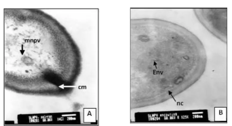
Figure 2. Phase 2, penetration and releasing nucleocapsid in the envelope to the cytoplasm A. Tunnel form by host cell membran. tn= tunnel membrane (Magnification x 40,000,
bar=200 nm)
B. The releasing of nucleocapsid from MNPV’s envelope. nc=nucleocapsid, Env=envelope (magnification x 25,000, bar = 200 nm).
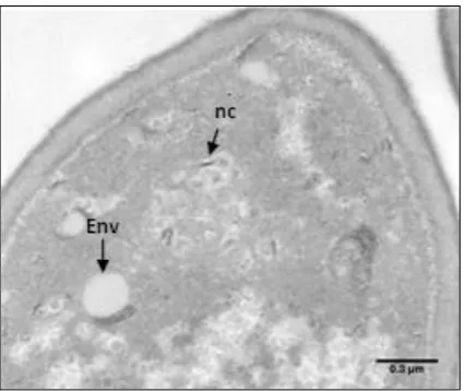
Figure 3. Phase 3, biosynthesis of the virus components (magnification x 5000, bar = 0.3 µm)
Remarks: nc: nucleocapsid, Env: envelope
Figure 4. Phase 4, the assembling of the virus components
A. The assembling of the nucleocapsid to multiple nucleocapsid such as mnc1, 2, 3 and 4 = multiple nucleocapsids (there were several arranged nucleocapsids i.e. four nucleocapsids arranged into in one cluster, and etc.) (magnification x 50,000, bar = 100 nm)
B. The mechanism of nucleocapsid entering the envelope. a. Nucleocapsid go to envelope. b.Envelope contains some nucleocapsid (magnification x 8000, bar = 1 µm)
Biosynthesis of the virus components was the third phase of SpltMNPV infection mechanism (Figure 3). This phase was the formation stage of virogenic stroma which contains genetic material and envelope. Figure 3 showed the nucleic acid
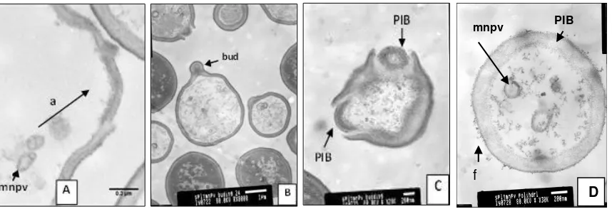
Figure 5. Phase 5, the releasing of SpltMNPV (Spodoptera litura Multiple Nucleopolyhedrosis Virus) A. Polyhedra was formed from host’s cell membrane (a) (magnification x 8000, bar = 1 µm). B. Budding of midgut cell membrane contained multiplenucleocapsid. Bud = budding
(magnification x 8000, bar = 1 µm).
C. Two SpltMNPV Polyhedra were released from the host cell (magnification x 28,000 bar = 200 nm) D. Polyhedra inclusion bodies of SpltMNPV after released from the host cell. f= filli of
polyhedra; PIB =Polyhedra inclusion bodies (magnification 30,000x, bar = 200 nm).
The assembling of the virus components was the fourth phase (Figure 4). In this phase occured a single rod-shaped nucleocapsid assembling into multiple nucleocapsid (1, 2, 3 and 4 line respectively) which have not been wrapped by the second protein envelope (Figure 4A). Single nucleocapsid entered into the envelope by curved structure to form multiple nucleopoly-hedrosis virus one by one (Figure 4B). The last phase of mechanism of infection SpltMNPV was releasing of SpltMNPV (Figure 5). In this phase occurs polyhedra formation from
host’s cell membrane, budding and releasing
polyhedra of SpltMNPV. Polyhedra which had filis released from the host cell wasn’t symmetric
DISCUSSION
The SpltMNPV infection mechanism phases were observed 2 times at 6 and 24 hours. Phase one and two of the infection mechanism were found in the 6 hours incubation, while phase three, four and five were found at 24-hour incubation. This fact is different from the previous theory that the phase of SlNPV infection mechanism in host cell (in vivo) have 72 hours (Falcon 1972 cit. Mangoendiharjo and Pollet, 1991). The infection cycle of SpltMNPV in Spodoptera litura epithelial midgut cell was divided in 5 phase.
The Attachment of Viruses in The Host Cell The attachment of viruses in the host cell is the first phase of SpltMNPV invation to host cell. (Figure 1). Some multiplenucleocapsid begin to recognize the host cell membrane by attaching to a specific place (receptor site) and release a particular compound that can change the cell membrane so that the cell membrane appears brighter (Figure 1A). Because of the conformation changing of the compounds. Such phase is continued in Figure 1B, some multiple-nucleocapside (MNPV) recognize the host cell membrane and attach to a specific place (receptor site) and release a compound that can cause particular changes in cell membrane so that the membran invaginates, brings the MNPV. According to Rohrmann (2008) at phase 1 multiplenucleocapsid attach larva midgut cells by interacting between vp91 (virus protein 91) and midgut cells. MNPV envelope has sensitive receptor sites so that it can bind proteinases and change the conformation of the compounds in the epithelial cell membrane. Therefore the cell membrane appears brighter (Figure 1A).
Penetration and Releasing Nucleocapsid in The Envelope to The Cytoplasm
Penetration and releasing nucleocapsid in the envelope to the cytoplasm is the second phase after the attachment of virus in the host cell. The second phase shows that MPNV has
mnpv PIB
f
already attached to the host and it causes the invagination to form a tunnel into the cell (Figure 2A). After the MNPV penetrates into the cell (Figure 2B), the envelope of MNPV will open and its contents are released, therefore the envelope becomes empty (Figure 2B). This fact is different from the previous theory. General viruses will penetrate and release the envelope to the cytoplasm by creating pores/holes in membran cell or by endocytosis and vesicles formation (Tortora et al.,1995; Rohrmann, 2008).
Biosynthesis of the Virus Components Biosynthesis of the virus components is the third phase of SpltMNPV after penetration and releasing envelope in cytoplasm. The third phase, shows the biosynthesis of the virus components. This phase is obtained from a virus infection in incubated cultured cells for 24 hours. The figure shows that virogenic stroma containing genetic materials and capsid protein (nucleocapsid) has been formed. The nucleo-capsid resembles rod-shaped threads scattered in the cell nucleus. The nucleus membrane of
Virogenic stroma is formed because some nucleocapsid can succeed in penetrating genetic material (DNA) into the cell nucleus. Nucleocapsid penetrates into the cell nucleus through the pores which are the size approximately 38 nm. Virogenic stroma formation is regulated by a virus protein called PP31 (Rohrmann, 2008).
The Assembling of the Virus Components The assembling of the virus components is the fourth phase after biosynthesizing the virus components. Figure 4 shows that the new nucleocapsids are formed in the incubation period of 24 hours. Beganing the nucleocapsid formation in the nucleus is separated (Figure 4A) then they nucleocapsids get closer each other and form a group of nucleocapsid which is called
multiplenucleocapsid. Figure 4A show that nucleocapsid in two line, 3 line and 4 line. The nucleocapsids have not been surrounded by envelope. The next mechanism can be observed in Figure 4B. It shows the mechanism of nucleocapsid entering the envelope. The longer rod-shaped nucleocapsid is curved to adjust the envelope shape and then enters the envelope. Furthermore nucleocapsids follow the previous step, that is, it is curved then enters the envelope. This mechanism enables the efficiency space can be achieved.The small envelope can be infiltrated by more than singgle nucleocapsid. This fact is different from the previous theory that nucleo-capsids have straight form (Adam, 1977 cit O’Reilly. et al., 1992). The nucleocapsids of SpltMNPV infecting larvae of S. litura epithelial midgut cells of have curved shape and they pile up each other in the envelope.
The releasing of SpltMNPV is the last phase after the assembling of virus components. In this phase the SpltMNPV are released from epithelial cells of larvae that is shown in Figure 5. Figure 5A shows that the cells begin to form a
budd’ midgut, and MNPV move to either nucleus
membrane or cell membrane to get polyhedral. Two MNPVs are going to the membrane of nucleus and membrane cell and then they buds (Figure 5B). Figure 5B shows that the cell membrane contains a budd of multiplenu-cleocapsid and whereas Figure 5C, Shows the cell membrane opens and releases SpltMNPV polyhedra. Figure 5C, shows that budding’s direction is not symmetrical and is not patterned. It is coused by viruses gathering as in a group in basal and lateral tip. The budding location will be away from the lumen but closer to the cell trachea as next cell target of infection (Rohrmann, 2008).
CONCLUSIONS
The infection mechanism of Spodoptera litura multiple nucleopolyhedrosis virus in midgut epithelial cells Spodoptera litura larvae were observed using a transmission electron micros-cope appear at 6-hour incubation.The six hour incubation has the following phases: 1) the attach-ment of MNPV (Multiple Nucleopolyhedrosis virus) on the host cell; 2) the penetration of MNPV into the cytoplasm. Beside of that, in the 24 hour incubation we can look the following phases: 3) the biosynthesis of virus components, 4) the assembling of virus components; and 5) the releasing of SpltMNPV in the form of polyhedral inclusion bodies.
ACKNOWLEDGEMENTS
This manuscript is a part of the first authors dissertation for the Doctoral Program funded by Post Graduate Scholarships at Agricultural Faculty University of Brawijaya Malang. Thank to the institution of Eijkman in Jakarta for the process of taking picture slides by TEM, and DP2M (Directorate General of Higher Education) as the funders of fundamental research for 2010 Grant Program.
REFERENCES
Arifin, M. 2006. Compatibility of SlNPV with HaNPV to control Armyworm and soybean caterpillar. Bogor. Indonesia. Jurnal Penelitian Tanaman Pangan. 25 (1). p.65-77. (in Indonesia)
Asri, M.T,.N. Ducha, and Dian P. 2007. An attempt to multyply SpltMNPV through in vitro As bioinsektisida using insects cell culture techniques. Research report of Competitive Grants. State University of Surabaya Surabaya. p.25-32 (in Indonesia)
Gothama, A.A.A., I.G.A.A. Indrayani, andS. Subiyakto. 1994. Prospects of using NPV to control fruit larvae of H. armigera and Armyworm of S. litura. Jurnal Pene-litian dan Pengembangan Pertanian 12(4): 106-110 (in Indonesia).
Hink. W.F. 1982. Production of Autographa california nuclear polyhedrosis virus in cells from large scale suspension cultures, in Microbial and Viral Pesti-cides.ed. E. Kurstaki Marcel Dekker. Ink. New York. p.493-506
Lua, H.L. and S.Reid. 2000. Morphogenesis virus of Helicoverpa armigera nucleopoly-hedrosis virus in Helicoverpa Zea Serum-free Suspension Culture. Journal of General Virology 81(10): 2531-2543 Mangoendiharjo, S. and A.Pollet. 1991. Insect
viruses and the possible application of nuclear polyhedrosis virus for control of Armyworm Spodoptera litura F.In Short article of Workshop on Integrated Pest Management of Soybean Plant. Depart-ment of Agriculture. Balittan. Malang. p.169-175.
Midgley,C.A., A.L. Craig, J.P. Hite and T.R. Thipp. 1998. Baculovirus expression and the study of the reglarvaion of The Tumor Supressor Protein. p 53 in Rapid, K.and R.I.Freshney (eds), DNA Transfer to Culture Cells. New York. Eiley-liss, p.27-54.
O’Reilly.D.R., L.K.Miller, and V.A.Luckow.1992.
Baculovirus expression vector. A. Laboratory manual.W.H. Freeman and Company. New York. p.6-11.
Rohrmann, G. 2008. Baculovirus molecular biology. Chapter 3.The baculovirus replication cycle: Effects on cells and insects. Department of Microbiology, Oregon State University, Corvallis. 33-43p.