The journal homepage www.jpacr.ub.ac.id ISSN : 2302 ‐ 4690
55
Study on iNOS Expression and Levels of Malondialdehyde
(MDA) in the White Rat (
Rattus norvegicus
) Exposed with
Aggregatibacter actinomycetemcomitans (Aa
)
Reviana Setin,1 Aulanni’am Aulani’am1* and Chanif Mahdi1
1
Department of Chemistry, Faculty of Mathematics and Natural Sciences, Brawijaya University, Jl. Veteran, Malang 65145, East Java, INDONESIA
Corresponding contact: [email protected]; Phone: +62341575838; Mobile: +6285246109179
Received 7 February 2013; Accepted 3 March 2013; Published online 1 May 2013
ABSTRACT
Aggregatibacter actinomycetemcomitant (Aa) is gram-negative bacteria composed of lipopolysaccharides (LPS) in the outer membrane. This bacteria is normally found in dental plaque and periodontal pockets causing parotid gland damage when it is present in excess amounts. LPS is an endotoxin and a complex glycolipid composed of polysaccharides (hydrophilic) and lipid A (hydrophobic) which can induces Reactive Oxygen Species (ROS), and increase the levels of malonaldialdehyde (MDA) and tissue damage. The aims of this research are to find the expression of iNOS parotid gland cell and to determine levels of MDA using thiobarbituric acid (TBA) spectrophotometric test at 533 nm. The results showed a significantly difference (P<0.05) of MDA levels in control rats (3.135 ± 0.0436 ppm) and Aa rats (6.307 ± 0.0473 ppm). iNOS expression is confirmed by the brown spots formed on the parotid gland, affording the enhancements of iNOS expression in Aa rats and control rats of 2.292 ± 0.038 and 0.166 ± 0.003, respectively (P<0.05).
Key word: Aggregatibacter actinomycetemcomitans, Reactive Oxygen Species, malondialdehyde, iNOS
INTRODUCTION
There are some microorganisms in human mouth which have beneficial functions, but there are also some which can be pathologenic. A report explained about 1011 bacteria per milligram of dental plaque can be found in teeth surface, although not all of these bacteria can harm, as they can live in the oral cavity1. In term of decreasing human immunity, the oral bacteria may turn to be pathogens, and spread in the body causing systemic infections. The progress on identifying of some oral bacteria revealed the relationship between the incidence of infections of the mouth, gums and teeth with various systemic diseases such as heart disease, diabetes and stroke.
Bacterium is one factor which able to cause periodontitis by inducing tissue damage through stimulation of inflammatory cells, and this able to damage tissue by releasing destructive enzymes, reactive oxygen species (ROS) and pro-apoptotic factors2. One of various kinds of microorganisms grows in the oral cavity as a source of pathogen is
Aggregatibacter actinomycetemcomitans (Aa). This bacteria are classified as pathogenic
The journal homepage www.jpacr.ub.ac.id ISSN : 2302 ‐ 4690
56
Aa bacteria found in dental plaque and periodontal pockets and in excess amounts can cause various diseases. Previous studies confirmed Aa was able to impair many types of host cells. Many evidences were reported this organism was able to invade endothelial cells and epithelial3. The Aa bacteria induced reactive oxygen species (ROS) in the oral cavity by producing free radicals to affect increasing levels of MDA, tissue damage and changing of protein profiles.
Production of ROS in cell its causes the oxidation of protein, DNA oxidation and changes in the constituent bases of DNA, which lead to triggering cell and tissue damages10. ROS also causes peroxidation of membrane lipid which one of its final products is MDA. One of the tissues found in the oral cavity possibly affected directly because of excessive ROS production by bacteria Aa in the parotid gland tissue. Tissue of parotid gland is the largest salivary gland located under the ear around the maxilla. Excess amount of ROS especially in can directly stimulate the tissue damage. The Aa bacteria in the body react with the blood, and affecting the whole system of the body. In this paper was reported the effect of Aa bacteria on the expression of iNOS and MDA level of parotid gland using white rats (Rattus norvegicus)5.
EXPERIMENTAL Treatment of Animals
A 10 males of white rats aged 3 months with weight of 150-180 grams and adapted for 12 days were used. Samples were divided into 2 groups, consisting of 5 rats for each group. Group 1 is the control and treated by injecting 200 μL of physiological NaCl (0.9%) whereas group 2 is the Aa rats and treated by injecting 200 μL of bacteria Aa with the dose of 108 CFU/mL. Injection is done twice on day 1 and day 7 in the upper gum located at the left gingiva. On the day 13, the white rat samples were surgered and subsequently their parotid gland and blood serum were harvested. All conditions and handling of animals in this study were conducted with protocols approved by the Brawijaya University Committee on Animal Use and Care (UB 106-UB-PEM).
Harvesting the parotid glands
After surgery on the day 13, parotid gland tissues of white rats are divided into two sections; ¼ part is treated with 10% paraformaldehyde (PFA) as a preparative sample while the remaining part is treated with a phosphate buffer saline-azide (PBS-azide) pH 7.4 for isolation of a protein tissue.
Protein isolation
The journal homepage www.jpacr.ub.ac.id ISSN : 2302 ‐ 4690
57
Preparation of Standard Curve of MDA
Each 100 μL of Kit MDA standard (density 0.997 mg/mL) with a series concentration of 0, 1, 2, 3, 4, 5, 6, 7 and 8 mg/mL is transferred into micro-tubes, and followed by addition 550 μL of distilled water. Then, into each tube was added 100 μL of 10% TCA, 250 μL of 1N HCl, and 100 μL of Na-thio 1%. These solutions are homogenized by vortex and incubated in a water bath at 100 ° C for 30 min, followed by cooling at room temperature. Measurements of each solution were conducted at the wavelength of 533 nm.
Measurement of MDA using Thio Barbituric Acid test (TBA)
Parotid gland is cut into small pieces, crushed using a mortar placed on an ice block and added with 1 mL of NaCl 0.9%. After homogenization, the mixture is transferred into micro tubes and centrifuged at 8000 rpm for 20 min. Then, 100 μL of the parotid gland supernatant is taken, and subsequently added with 550 μL of distilled water, 100 μL of 10% TCA, 250 μL of 1N HCl, and 100 μL of Na-thio1%. Every addition of reagent solution, the mixture is homogenized by vortex, followed by centrifugation at 500 rpm for 10 min. The supernatant was taken and then incubated in a water bath at 100 ° C for 30 min. After cooling the sample solution to room temperature, the absorbance is measured at 533 nm. The absorbance is then plotted on a standard curve to calculate sample concentrations.
Expression of iNOS Immunohistochemistry method
Staining maxillary parotid tissue preparations involves the binding of antigen-antibody (avidin-biotin). The addition of primary antigen-antibody (antigen-antibody NOS) conducted for 1 hour at room temperature, and washed in PBS pH 7.4 for 3 x 5 min. Further, secondary antibodies Anti-Rabbit IgG Biotin Labeled were added for 1 hour at room temperature, and washed in PBS pH 7.4 for 3 x 5 min. SA-HRP (Strep Avidin-Horseradish Peroxidase) were added for 30-60 minutes at room temperature and washed in PBS pH 7.4 for 3 x 5 min. Furthermore, chromogen DAB (3,3-diaminobenzidine tetra hydrochloride) were added for 10-20 minutes at room temperature and washed in distilled water for 3 x 5 min. Then counterstain was conducted with Hematoxylen for 5 minutes at room temperature, washed in distilled water for 3 x 5 min, followed by the mounting with entellan. The observation was preformed using a microscope with 400x magnification. Expression of iNOS Aa is noticeably from a brown color on the tissue preparation.
RESULTS AND DISCUSSION
Levels of MDA white rats parotid gland (Rattus norvegicus) were exposed to Aa
MDA is a major reactive aldehyde resulting from the lipid peroxidation reaction and usually used as a marker of oxidative stress. In a condition of high oxidative stress, MDA levels also increase significantly. Analysis of MDA level in rat parotid gland is be measured from each group of rats. MDA level in the parotid gland of rats exposed by Aa bacteria increases due to the LPS existing in the outer membrane of bacteria.
The journal homepage www.jpacr.ub.ac.id ISSN : 2302 ‐ 4690
58
specific receptors of macrophages to stimulate the production as well as release inflammatory cells7.
Table 1. MDA levels in parotid gland of white rats (n = 5) Treatment
group
Level of MDA (ppm) Figure 1. Reaction mechanism of formation of
LPS-LBP complex
Control Aa
3.135 ± 0.0436 6.307 ± 0.0473
The damage of the parotid gland tissue, which is caused by bacteria Aa exposure, would trigger ROS. ROS can deteriorate lipids and proteins as the major components of the tissue. Consequently, a lipid peroxidation reaction occurred and thus the levels of MDA in the parotid gland tissue increased. As shown in Table 1, the MDA level in Aa rats is significantly (P<0.05) rise in comparison to that of control rats.
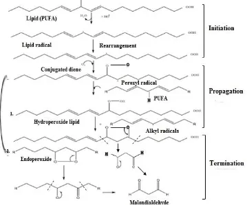
Figure 2. MDA formation through lipid peroxidation
The journal homepage www.jpacr.ub.ac.id ISSN : 2302 ‐ 4690
59
classified based on its radical activity, such as superoxide (O2•), hydrogen peroxide (H2O2) and hydroxyl radical (•OH). ROS serves to damage cells infected by a virus or bacteria. Highly reactive radicals can cause biochemical changes and damage to various components of living cells, such as proteins, lipids, carbohydrates and nucleic. The attachment of free radicals to cells membrane would lead to lipid peroxidation, which is indicated by the increase in MDA level. The latter compound is highly toxic to the cells since it is able to changes cell membrane fluidity and function. The mechanism of MDA formation is shown in Figure 2.
The C=C bonds of polyunsaturated fatty acid (PUFA) as the component of lipid in cells membrane is attached by ROS. The double bonds weaken C-H bonds in lipid, causing the reduction of H due to the attachment of OH•. As the result, the lipid radicals are formed. Moreover, the lipid radical is oxidized by O2 to form a peroxyl radical. Newly formed peroxyl radical can react with another PUFA by removing H, producing lipid hydro peroxides and other lipid radicals. This process is continuously propagated in a chain reaction. Lipid hydro peroxide is an unstable compound and it will produce MDA when fragmentation occurred. Additionally, peroxy radical can also undergo cyclization reaction to produce cyclic peroxide. This radical can be reduced to form hydro peroxide or subsequent cyclization reaction to form the bicyclic peroxide which then produces a molecule analogous to endoperoxide. The latter compound generates MDA formation9. So, the excessive numbers of free radicals increase the lipid peroxidation production followed by the increasing number of MDA.
Expression of iNOS white rats parotid gland (Rattus norvegicus) were exposed to Aa
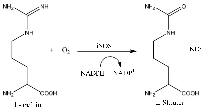
The expression of inducible nitric oxide synthase (iNOS) causes inflammation in many types of cell as a response to the foreign substances such as virus and bacteria. The catalysis of NO by iNOS normally involves the reaction of L-arginine to form L-sitrulin in the presence of NADP (Figure 3).
Figure 3. Catalysis reaction of NO• formation by iNOS
The journal homepage www.jpacr.ub.ac.id ISSN : 2302 ‐ 4690
60
antibody is bound by a marker for visualization, which can indicate the presence of the active ingredient in the tissue.
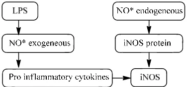
LPS could stimulate macrophage to produce and release inflammatory cells that causes pathophysiological effects such as infection up to tissue damage. Inflammatory processes in the cell will cause the migration of macrophages and produce cytokines8. Initially, LPS as an exogenous NO will activate macrophages. Then, the activated macrophages produce pro inflammatory cytokines and excessive inflammatory cells. Consequently, the increase in the effectiveness of the inflammatory enzyme iNOS is obtained. Moreover, iNOS could trigger the formation of free radicals endogenous NO* through the reaction catalyzed by NOS Figure 4. Additionally, the expression formed between antigen and antibody indicates the condition of the infected tissue pathology in the expression of iNOS. As shown in Table 2, it was found that there is a significantly (P<0.05) increase of iNOS expression in Aa rats in comparison to the rats control.
Table 2. Profile of the number of cells expression of iNOS in Aa rats and control rats
Treatment group
The average number of cells expressed
iNOS (%)
Control Aa
0.166 ± 0.003 2.292 ± 0.038 Figure 4. The mechanism of endogenous
NO* formation catalyzed by iNOS as a result of inflammation
A B
Figure 5. Parotid gland (Rattus norvegicus) by immunohistochemistry method 200x magnification (Blue arrow, A: Control rats; Red arrow, B : Aa rats).
The journal homepage www.jpacr.ub.ac.id ISSN : 2302 ‐ 4690
61
brown spots were detected as the result of high expression of iNOS. The high expression of iNOS in Aa rats indicates pathological conditions as a consequence of Aa bacteria exposure.
CONCLUSION
Based on the exposure to bacteria Aa known by relation between MDA and iNOS expression in the parotid gland. Levels of MDA for control and Aa rats by T-test respectively, 3.135 ± 0.0436 ppm and 6.307 ± 0.0473 ppm. Meanwhile the numbers of cells that express iNOS is 0.166 ± 0.003% for control rats and increase 2.292 ± 0.038 % for Aa rats.
ACKNOWLEDGEMENTS
The author would like to thanks for Dr. drg. Rini Devijanti Ridwan, M.Kes for providing all facilities access of research, and Dr. Sc Akhmad Sabarudin for reading this manuscript.
REFERENCES
1. Alikhani, M., Z. Alikhani, D.T. Graves, J. Dent. Res., 2004, 83(9), 671-676. 2. Atmar, R.L. and K.E. Mary, Clin. Microbiol. Rev., 2001, 14, 15-37.
3. Bank T.L, A. Dosen, R.F. Giese, E.M. Haase and S.T. Sojar, J. Nanosci. Nanotechnol., 2011, 11(10), 8450.
4. Cuschieri, J., B. Jens and V.M. Ronald., J. Leukocyte Biol.,2006, 80, 1289-1297.
5. Kusumawati, D., Bersahabat Dengan Hewan Coba, 2004, Universitas Gadjah Mada University Press, Yogyakarta.
6. Li X, K.M. Kolltveit, L. Tronstad and I. Olsen, Clin. Microbiol. Rev., 2000, 13(4), 547-558.
7. Schumann, R. R., S. R. Leong, G. W. Flaggs, P. W. Gray, S. D. Wright, J. C. Mathison, P. S. Tobias and R.J. Ulevitch, Science, 1990, 249, 1429–1431.
8. Tomlinson, J. L. and T.B. Anthony, J. Am. Vet. Med. Assoc. 2004, 224 (9), 1446-1452 9. Valko, M., W. Rhoades, C.J. Moncol, M.J. Izakovic and M. Mazur, Chem.Biol. Interact,
2006, 160 : 1-40.