Rekayasa Bakteri untuk Ternak dan Manusia: Pembuatan
Mutan
Escherichia coli
Penghasil Protein Rekombinan
Bacterial Engineering for Cattle and Human: Construction of
Escherichia coli Mutant for the Production of Recombinant
Proteins
Muhamad Ali1, Takako FUKUBA2, and Hideo NAKANO2
1Laboratory of Microbiology and Biotechnology, Faculty of Animal Sciences, University of Mataram,
Lombok 83125 Indonesia
2Laboratory of Molecular Biotechnology, Faculty of Bioagricultural Sciences Nagoya University,
Furo-cho, Chikusa-ku, Nagoya 464-01, Japan
KATA KUNCI KEYWORDS
ABSTRAK
ABSTRACT
mutan defisiensi-protease; transduksi phage P1; disrupsi kromosom
proteases-deficient mutant; P1 phage transduction; chromosomal disruption
Protein rekombinan seperti vaksin, antibodi, hormon, dan obat-obatan, semakin dibutuhkan oleh ternak dan manusia. Hambatan utama untuk menghasilkan protein rekombinan pada Escherichia coli sebagai inang yang digunakan paling luas adalah degradasi oleh enzim proteolitik. Hal ini disebabkan karena E. coli memiliki sejumlah enzim proteolitik yang tersebar di dalam sitoplasmanya. Untuk itu, lebih dari 90% degradasi protein terjadi di dalam sitoplasmanya. Pada penelitian ini, peneliti telah menghasilkan mutant E. coli BW25113 yang tidak memiliki gen penyandi enzim protease dengan menggunakan kombinasi metode pengerusakan kromosom dan metode transduksi phage P1. Pembuatan mutan tersebut dimulai dengan pengerusakan gen penyandi enzim protease pada kromosom bakteri dengan produk PCR yang memiliki bagian yang homolog dengan gen target. Mutan-mutan yang dihasilkan kemudian digunakan untuk menghasilkan mutan ganda dengan metode Transduksi phage P1. Analisis fenotif dan genotif menunjukkan bahwa kombinasi kedua metode tersebut sangat efektif untuk membuat lebih dari satu mutasi pada E. coli. Untuk itu, mutan E. coli yang telah diperoleh akan sangat bermanfaat untuk menghasilkan aneka protein rekombinan untuk ternak dan manusia.
These mutants were used to construct double mutants using P1 phage transduction. Phenotypic and genetic analysis showed that the combination of these methods were effective to construct more than one gene disruption in E. coli. Therefore, the obtained E. coli mutants would be absolutely useful for the generation of wide varieties of recombinant proteins for cattle and human.
In recent years, recombinant protein technology has become an important discipline spanning from scientific research to the pharmaceutical industry. It is considered so since recombinant proteins, such as vaccines, antibodies, hormone, or drugs, are increasingly needed for cattle and human health. The use of cowpox firstly as smallpox vaccine by Edward Jenner, followed by attenuated or killed virulent micro-organisms and recombinant proteins to fight disease has proven spectacularly successful. Appropriate administration of attenuated or killed Bacillus anthracis is very effective to prevent anthrax disease in farm animals. In addition, passive antibody therapies and immune sera could be used for treatment of certain infections in animal.
One of the challenges emerged in the biotechnology revolution to meet animals and humans demand is the development of techniques for the economical production of therapeutics recombinant proteins. Plant production system has been developed for the proteins production (Kersten et al., 2003; Valdes et al., 2003). Plants are potential
“biofarming factories” because they are capable of producing unlimited number and amount of recombinant proteins safely and inexpensively. However, some of the current hurdles include a long growth time, regula-tory elements uncertainties, and questions about the suitability of plant glycans for human therapeutics.
Among the most well documented and established systems used in various scales of recombinant protein production are the enterobacterium Escherichia coli. It is the most ubiquitous source of recombinant
protein as it is simple, cheap, and its technology is mature (Kristensen et al., 2005; Lombardi et al., 2005). Recombinant protein from bacteria, archaeabacteria, and eukaryotes are in many cases efficiently expressed and accumulated in E. coli
(Kristensen et al., 2005). In addition, high production levels of recombinant proteins are usually attainable when E. coli is used as the proteolytic enzymes distributed in its periplasm and cytoplasm. Previous studies demonstrated that endogenous proteases such as Lon, ClpP, DegP, and OmpT participated in rapid degradation of proteins
in vivo (Vasilyeva et al., 2000; Weichard et al., 2002; Jones, 2002; Ignatova et al., 2003; Okuno
et al., 2002). Therefore, more than 90% of the degradation occure in the cytoplasm.
One way to enhance the yield of recombinant proteins of interest in E. coli as a host is genetic manipulation. Ignatova et al., (2003) reported that the production of mature active penicillin amidase increased up to 10-fold when the protease-deficient strain E. coli
BL21 (DE3) was used as the host. Therefore, the use of protease-deficient strains as the host is a successful strategy to achieve higher productivity of a proteolysis-susceptible target protein.
Correspondence:
Genetic manipulation in bacteria genome can be achieved by a variety of techniques including error-prone PCR, DNA shuffling, saturation mutagenesis, and family shuffling (Fuji et al., 2004). Error-prone PCR introduces random mutations during PCR by reducing the fidelity of DNA polymerase. The fidelity of DNA polymerase can be reduced by adding manganese ions or by biasing the dNTP concentration. The combination of error-prone PCR and saturation mutagenesis constitutes an efficient way to explore the protein mutation properties. However, the creation of mutants by the above methods has been limited partly by point mutations that can introduce only a limited range of amino acid substitutions. This limitation narrows the sequence space of mutant proteins that can be created.
In this work, we generated several single protease-deficient mutants of E. coli
using one-step chromosomal disruption method (Datsenko et al., 2000). Parallel efforts were performed for the construction of double protease-deficient mutants using P1 Phage transduction method (Miller, 1972). The combination of one-step chromosomal disruption and P1 phage transduction methods was effective to construct more than one gene disruption in E. coli and would be widely useful for generating effective host of recombinant protein expression in other bacteria.
MATERIALS AND METHODS
Materials
Escherichia coli strains, plasmids, and phage used in this study are listed in Table 1. Luria-Bertani medium containing 1% of tryptone, 0.5% of yeast extract, and 1% of NaCl were used for cultivation. Super Optimal Broth (SOB) medium consisted of 2% tryptone, 0.5% yeast extract, 10 mM NaCl, with antibiotics at the following concentra-tions i.e. 50 g/ml kanamycin; 10 g/ml chloramphenicol; and 50 g/ml ampicilin.
Methods respectivelly. PCR products were generated using template plasmids (pKD3, pKD4, and pKD13) using primer with 60-nt extentions (Table 2). PCR mixture consisted of 10x Ex taq
PCR Verification
Phenotypic selection using selective media and PCR analysis were used to verify strain construction and recombinant forma-tion. Primers (20-mers) (Table 2) were used to verify kanamycin or chlorampehnicol-encoding gene replacement in protease-encoding gene site. PCR mixture contained 10x Ex Taq buffer, 0.2mM dNTP, 0.5 M each primer, 2.5 l of mutant colony dilution, and
Ex Taq DNA polymerase. The mixture was incubated at 95oC for 5 min, followed by 30 cycles of 30 s at 94oC, 30 s at 50oC, 2 min at
72oC and a final extension step of 7 min at 72oC. PCR products were run on 1% agarose gel along with a /Eco T141 marker.
Elimination of Antibiotic Resistance Gene
Antibiotic resistance mutants were transformed with pCP20. Since the plasmid is an ampicilin plus chloramphenicol resistance plasmid, the results of transformation can be selected using selective media containing the antibiotic. Heat shock (43oC) for inducing FLP synthesis was performed for antibiotic resistance gene elimination.
Table 1. Bacterial strains, plasmids and phage used in the experiment
Strain or phage Chromosomal Markers/Description Source or reference
E. coli BW25113 (araD-araB)567, lacZ4787(::rrnB-3),
lacIp-4000(lacIQ), -, rph-1, (rhaD-rhaB)568, hsdR514 Datsenko et al., 2000 Plasmid
pKD3, pKD4pKD13 Template for PCR product Datsenko et al., 2000
pKD46 The Red helper plasmid Datsenko et al. 2000
pCP20 FLP recombinase Datsenko et al. 2000
Phage
P1 phage Laboratory stock
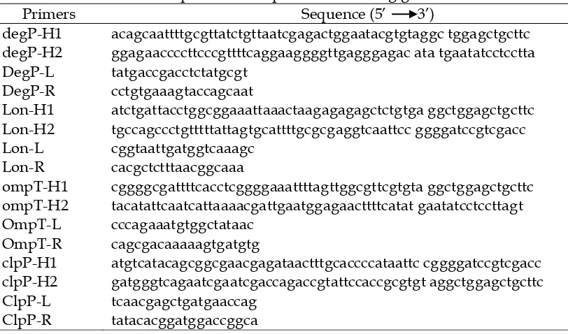
Table 2. List of primers used to verily kanamycein or chloramphonial encording gene replacement in protease encoding gene site
Primers Sequence (5’ 3’)
degP-H1 degP-H2 DegP-L DegP-R Lon-H1 Lon-H2 Lon-L Lon-R ompT-H1 ompT-H2 OmpT-L OmpT-R clpP-H1 clpP-H2 ClpP-L ClpP-R
acagcaattttgcgttatctgttaatcgagactggaatacgtgtaggc tggagctgcttc ggagaaccccttcccgttttcaggaaggggttgagggagac ata tgaatatcctcctta tatgaccgacctctatgcgt
cctgtgaaagtaccagcaat
atctgattacctggcggaaattaaactaagagagagctctgtga ggctggagctgcttc tgccagccctgtttttattagtgcattttgcgcgaggtcaattcc ggggatccgtcgacc cggtaattgatggtcaaagc
cacgctctttaacggcaaa
cggggcgattttcacctcggggaaattttagttggcgttcgtgta ggctggagctgcttc tacatattcaatcattaaaacgattgaatggagaacttttcatat gaatatcctccttagt cccagaaatgtggctataac
cagcgacaaaaagtgatgtg
atgtcatacagcggcgaacgagataactttgcaccccataattc cggggatccgtcgacc gatgggtcagaatcgaatcgaccagaccgtattccaccgcgtgt aggctggagctgcttc tcaacgagctgatgaaccag
RESULTS
PCR products, which consist of antibiotic resistance gene with homologous regions, were generated by PCR from plasmids template (pKD3, pKD4 and pKD13) using primers with 60-nt homologous extensions to the target. Each plasmid contains different antibiotic resistance genes that are flanked by directly repeated FRT site. pKD3 plasmid contain chloramphenicol resistance gene, pKD4 and pKD13 contain kanamycin resistance gene. The PCR products were purified, and digested with
Dpn I. Transformation of the PCR products which contain a homologous region were then performed into E. coli BW25113 using electroporation method. After plating on selective media and incubating at 30oC overnihgt, the growing colonies were then used as template for PCR confirmation.
The generated single mutants were Lon-deficient mutant ( lon), ClpP-deficient mutant ( clpP), DegP-deficient mutant ( degP), and OmpT-deficient mutant ( ompT). Electrophoresis result of PCR amplification of ClpP-deficient mutant ( clpP::Kmr) were shown in Figure 1 A. The band size of ClpP protease-deficient mutant (ΔclpP::Kmr) was higher than that of wild type (W1-W2) because of antibiotic resistance gene insertion between the homologous regions.
All of the single mutant that were still containing antibiotic resistance gene were subsequently used as donor cells for double mutant construction by means of P1 phage transduction. Receptor cells were prepared from these mutants following isolation of the antibiotic resistance gene. FRT sites placed in
the same chromosome will lead to a deletion or inversion of the antibiotic resistance segment by FLP recombinase produced by the strain with an easy curable FLP-expressing plasmid (pCP20) (Cherephanov et al., 1995). After excision of the antibiotic-resistance determinant, as shown in electrophoresis results of clpP (Figure 1A), the band size of the mutant was lower than that of wild type.
Double-protease deficient mutant produced in this study were DegP + OmpT-deficient mutant ( degP-ompT), DegP + Lon-deficient mutant ( degP-lon), Lon + OmpT-deficient mutant ( lon-ompT), and OmpT+ClpP-deficient mutant ( ompT-clpP). We also tried to construct triple mutant, but no colony was obtained.
Electrophoresis result of PCR amplification of OmpT and ClpP-deficient mutant were shown in Fig. 1 B. Replacement of protease gene by PCR products (antibiotic resistance segment) was increased the band size of these mutants (ΔompTclpP::Kmr) comparing to wild type. As occurred in single mutant, the band size become lower than wild type after elimination of the antibiotic resistance cassette (ΔompTclpP).
A B
Fig. 1. Electrophoresis results of PCR confirmation of E. coli BW25113 protease-deficient mutants. A = single mutants (ΔclpP::Km = ClpP-deficient mutants containing kanamycin-encoding gene; ΔclpP = ClpP-deficient mutants without kanamycin-encoding gene;W1, W2, W = wild type, 1-4 = single mutants). B = double mutants (ΔompTclpP::Km = OmpT and ClpP-deficient mutant containing kanamycin-encoding gene; ΔompTclpP = OmpT and ClpP-deficient mutant without kanamycin-encoding gene; WT = wild type, 1-6 = double mutants). M = λ DNA marker.
6 5
4 3 2
1 0
0.0 0.5 1.0 1.5
Time Course (h)
OD660
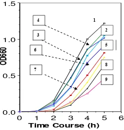
Fig. 2. Time course of E. coli mutant growth Cells were grown at 37 oC. 1 =
wild type/control, 2 = ompT, 3 = clpP, 4 = degP, 5 = clpP-ompT, 6 = lon, 7 =
degP-lon, 8 = lon-ompT, 9 = degP-ompT.
1
2 3
4
5 6
7
8
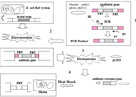
Heat Shock
Fig. 3. A schematic showing simple steps of gene disruption strategy for mutant construction. 1 = PCR products were generated from plasmid (pKD3, pKD4 and pKD13) using primer with 60-nt extentions, 2 = Electroporation of PCR products into E. coli Red system for gene target replacement, 3 = electroporation of FLP-expressing plasmid
(pCP20) into E. coli mutants for antibiotic resistance gene elimination, 4 = elimination of antibiotic resistance gene using heat-schock method. H1 and H2 refer to the homology regions, P1 and P2 refer to priming site. FRT = FLP recognition target.
DISCUSSION
Since E. coli posseses a number of proteolytic enzyme, many recombinant proteins are rapidly degraded when expressed in the bacteria. Lon is the primary protease degrading abnormally folded proteins in E. coli. Outer membrane protease, OmpT, can cleave many cytoplasmic protein after lysis cells. In the periplasm, DegP is protease which has a serine active site. In addition, ClpP can associate with ATPase subunit to form protease (Gottesman, 1996; Hengge and Bukau, 2003).
The procedure for the construction of protease-deficient mutants using one-step chromosomal disruption method are shown diagrammatically in Figure 3 The basic principle of the chromosomal disruption method is to replace a target gene with a selectable antibiotic resistance gene and homologous regions. Linear DNA containing antibiotic resistance gene flanked by homologous regions of bacterial chromosome is then transformed or electroporated into recombination-proficient E. coli BW25113 strain. Recombination between both ends of the linear DNA fragment and the bacterial
E. coli Red system
target gene
FRT FRT
PCR
PCR Product
Antibiotic gene
Electroporation
H1
P1
antibiotic gene
FRT FRT
Electroporation
FRT
pCP20
antibiotic resistence gene Plasmid (pKD3,
pKD4, pKD13)
H2 P2
1
2
3
4
chromosome results in gene replacement. The recombinations can be easily selected by the presence of the antibiotic resistance marker.
The antibiotic resistance gene was generated by PCR from plasmids template (pKD3, pKD4 and pKD13) using primers with 60-nt homologous extensions to the target. Each plasmid contains different antibiotic resistance genes that are flanked by directly repeated FRT site and homologous regions. pKD3 plasmid contain chloramphenicol resistance gene, pKD4 and pKD13 contain kanamycin resistance gene. The homologous regions were provided by primers with 60-nucleotides homologous extensions.
Digestion of PCR products using Dpn I was done to eliminate methylated (unamplified) template DNA. The linear DNA (PCR product) was then electroporated into transformants (E. coli BW25113) carrying the Red helper plasmid. In one-step chromosomal disruption method, target gene replacement with antibiotic resistance gene was conducted based on the Red system. Transformants, which carrying pKD46, have three genes (exo, and ) that facilitate and are necessary for recombination (Datsenko et al., 2000). The exo gene encodes the Red
protein, a 5’ to 3’ exonuclease that processively degrades the 5’-ended strand of a linear double-stranded DNA (dsDNA)
fragment to produce 3’-ended single-stranded DNA (ssDNA) overhangs. The gene encodes a pairing protein (Red ) that binds to
the 3’-ended ssDNA overhangs created by the Red protein and promotes renaturation of complementary strands, and is capable of mediating strand annealing and exchange reaction in vitro (Li and Wilkinson, 1998). The recombination function of Red and Red proteins are further assisted by the -encoded Gam protein, which inhibits the host RecBCD exonuclease V, an intracellular exonuclease that degrade the linear pieces of DNA in E.
coli, in order to linear DNA (PCR products) transformable.
All obtained mutants were verified by PCR colony wich tested for the presence of new locus- and junction-specific fragments. As shown in Figure 1, The presence of new locus (antibiotic resistance gene) was appeared by increasing size of band in single
and double mutants, ΔclpP::Kmr and
ΔompTclpP::Kmr.
The resistance gene was then eliminated by using a helper plasmid (pCP20) expressing the FLP recombinase, which act on the directly repeated FRT (FLP recognition target) sites flanking the resistance gene. FRT sites placed in the same chromosome will lead to a deletion or inversion of the antibiotic resistance segment by FLP recombinase that produced by the strain with an easy curable
FLP-expressing plasmid (pCP20)
(Cherephanov et al., 1995). After excision of the antibiotic-resistance determinant such a sequence would be left in the chromosome at the site of the initial cassette insertion. Elimination of the antibiotic resistance gene from these mutants reduce the size of band.
Several double mutants were also difficult to survive in a viable condition especially on solid growth media. These mutants show a variety of other phenotypic alteration which seem to be resulted from their decreased ability to degrade certain short-lived protein (Gottesman, 1996). We had attempted to make triple mutants, however all of such trials were unsuccessful, suggesting that deletion of double or more certain protease genes might give harmful effects on the growth and viability of E. coli
cells. This suggestion is based on the role of these proteases in the rapid turnover of short-lived regulatory protein for balanced growth of E. coli (Hengge and Bukau, 2003). Moreover, as reported previously, deletion of the above protease in E. coli leads to mucoidy and reduced strain fitness (Goff and four single and double protease-deficient mutants in E. coli using combination of one-step chromosomal disruption and P1 phage transduction methods. Using the methods, desired mutations can be made in any part of the DNA, independent of the presence of appropriate restriction enzyme sites. The combination of these methods allowed us to make more than one mutation, suggesting that the method is useful to create molecular diversity to other bacteria.
thank Hirotatsu SUZUKI (Laboratory of Molecular Biotechnology Nagoya University) for technical support during this research.
REFERENCES
Cherepanov PP and Wackernagel W 1995. Gene disruption in Escherichia coli: TCR and KmR cassettes
with the option of Flp-catalyzed excision of the antibiotic-resistance determinant. Gene, 158, 9-14. Datsenko KA and Wanner BL 2000. One-step
inactivation of chomosomal genes in Escherichia coli
K-12 using PCR products. Proc. Natl. Acad. Sci., USA, 97, 6640-6645.
Fujii R, Kitaoka M, and Hayash K 2004. One-step random mutagenesis by error-prone rolling circle amplification. Nucleic Acids Research, 32: e145. Goff SA and Goldberg AL 1987. An increased content of
Protease La, the lon gene product, increases protein degradation and blocks growth in Escherichia coli. J. Biol. Chem., 262, 4508-4515.
Gotesman S 1996. Proteases and their targets in E. coli. Annu. Rev. Genet. 30, 465-506.
Hengge R and Bukau B 2003. Proteolysis in prokaryotes; protein quality control and regulatory principles. Mol. Microbiol., 49; 1451-1462.
Ignatova Z, Mahsunah A, Giorgieva M, and Kasche V 2003. Improvement of postranslational bottlenecks in the production of penicilin amidase in recombinant
Escherichia coli strains. Appl. Environ. Microbiol., 69, 1237-1245.
Jiang XP, Oohira K, Iwasaki Y, Nakano H, Ichihara S, and Yamane T 2001. Reduction of protein degradation by use of protease-deficient mutants in cell-free protein synthesis system of Escherichia coli. J. Biosci. Bioeng., 93, 151-156.
Jones CH, Dexter P, Evans AK, Liu C, Hultgren SC, and Hruby DE 2002. Escherichia coli DegP protease cleaves between paired hydrophobic residues in a natural substrate: the PapA pilin. J. Bacteriol., 184, 5762-5771.
Kersten B, Feilner T, Kramer A, Wehrmeyer S, Possling A, Witt I, Zanor MI, Stracke R, Lueking A, Kreutzberger J 2003. Generation of Arabidiopsis protein chips for antibody and serum screening. Plant Mol. Bio., 52, 999-1010.
Kristensen J, Petersen HUS, Mortensen KK, Sorensen HP 2005. Generation of monoclonal antibodies for the assessment of protein purification by recombinant ribosomal coupling. Int. J. Biol. Macromolecules, 37, 212-217.
Lombardi A, Sperandei M, Cantale C, Giacomini P, Galeffi P 2005. Functional expression of a single-chain antibody specific for the HER2 human oncogene in a bacterial reducing environment. Protein Expr. Purif., 44., 10-15.
Miller JH 1972. Experiments in molecular genetics, p. 201-205. Cold Spring Harbor Laboratory Press, Cold Spring Harbor Laboratory, New York.
Okuno K, Yabuta M, Ohusye K, Ooi T, and Kinoshita S 2002. An analysis of target preferences of Escherichia coli outer-membrane endoproteases OmpT for use in theraupetic peptide production: efficient cleavage of substrates with basic amino acids at the P4 and P6 positions. Biotechnol. Appl. Biochem., 36, 77-84.
Surpuran CT, Scozzafa A, and Clare BW 2002. Bacterial
protease inhibitors. Med. Res. Rev. 22, 329-372. Weichart D, Querfurth N, Dreger M, and Aronis RH
2002. Global role for clpP-containing proteases in stationary-phase adaptation of E. coli. J. Bacteriol.,
185, 115-125.
Valdes R, Reyes B, Alvarez T, Garcia J, Montero JA, Figuroa A, Gomez L, Padilla S, Geada D, Abrahantes MC, Dorta L, Fernandez D, MendozaO, Ramirez N, Rodriguez M, Pujol M, Borroto C, Brito J 2003. Hepatitis B surface antigen immunopurification using a plant-derived specific antibody produced in large scale. Biochem. Biophys. Res. Commun., 310, 742-747.