Rapid and Simple Amplification of
Genomic DNA Sequences Flanking Transposon
ARIS TRI WAHYUDI
Department of Biology, Institut Pertanian Bogor, Darmaga Campus, Bogor 16680, Indonesia Phone/Fax: +62-251-622833, E-mail: [email protected]
A rapid and simple method to amplify genomic DNA sequences flanking mini-Tn5 transposon insertion was developed. This technique can be used to determine the location and orientation of the transposon insertion within genomic DNA of the bacteria. Based on the mini-Tn5Km1 transposon sequence, PCR primers can be designed to specifically amplify the DNA sequences flanking mini-Tn5 transposon by inverse polymerase chain reaction (inverse PCR) directly, upstream and downstream of the transposon insertion. The method involves: (i) digestion with a restriction enzyme that does not cut mini-Tn5Km1 sequence; (ii) self-ligation under conditions favoring the production of monomeric circles; and (iii) inverse PCR reaction using primers designed from mini-Tn5Km1 sequence to amplify the DNA sequences flanking mini-Tn5Km1 transposon insertion. Feasibility and reliability of this method were demonstrated with mini-Tn5Km1 mutants of the microaerobic magnetic bacterium Magnetospirillum magneticum AMB-1 which are defective in magnetosomes synthesis. The inverse PCR products amplified from these mutant genomes showed the correct fragments as determined through Southern hybridization and DNA sequence analysis.
Key words: inverse PCR, Transposon Mini-Tn5Km1, Southern Hybridization, DNA sequencing _____________________________________________
Polymerase chain reaction (PCR) is a technique for the in vitro amplification of specific region of DNA without conventional cloning procedures. However, limitation of this method is it amplifies only the region of DNA between two convergent primers. Any method for the in vitro amplification of the DNA sequences flanking a known sequence of DNA would have many useful applications in genetics such as amplification and identification of the sequence flanking a transposable element (transposon) (Ochman et al. 1988).
A mini-Tn5 transposon has been constructed that simplifies substantially the generation of insertion mutants of variety of gram negative bacteria (DeLorenzo et al. 1990). This transposon has become a popular tool to study molecular genetics of gram negative bacteria because it stably inserts into the genome (Hererro et al. 1990). Thus, this is an important genetic tool to localize the gene of interest to be isolated. Isolation of the DNA sequences or gene interrupted by the transposon is usually achieved by cloning the DNA containing the transposon, by the ligation-mediated polymerase chain reaction (LMPCR) (Prod’hom et al. 1998) or by the inverse polymerase chain reaction (inverse PCR) (Rich and Willis 1990). The inverse PCR has been used to amplify DNA sequences flanking Tn5 transposon insertion such as genomes of Hypomicrobium facilis (Feseveldt et al. 1997), E. coli (Ochman et al. 1988), Pseudomonas abietaniphila (Martin and Mohn 1999), and Pseudomonas fluorescens (Mirleu et al. 2000).
In this study, in order to amplify the genomic DNA sequences flanking mini-Tn5 transposon, an inverse PCR technique was developed based on the mini-Tn5Km1 sequence to design the primers directed outward from the transposon. To demonstrate this method, non-magnetic mutants of Magnetospirillum magneticum AMB-1 were generated by mini-Tn5Km1 transposon mutagenesis (Wahyudi et al. 2001) that were unable to synthesize magnetosomes, membrane-bound magnetic particles of
magnetite (Fe3O4) synthesized in the cell. DNA sequences flanking the transposon were directly isolated by inverse PCR. The inverse PCR products obtained were confirmed by southern hybridization and DNA sequence analysis.
MATERIALS AND METHODS
Bacterial Strains, Plasmids, and Media. Escherichia coli
cells were grown in Luria broth (LB) medium (tryptone 10.0 g l-1, NaCl 5 g l-1, yeast extract 5.0 g l-1) at 37 oC. Antibiotics were added when required with the following concentration (µg ml-1): kanamycin, 25; ampicillin 50. Magnetospirillum magneticum AMB-1 (Matsunaga et al. 1991) culture was grown at 25 oC in magnetic spirillum growth medium (MSGM) (Blakemore et al. 1979), and non-magnetic mutants of AMB-1 were grown in MSGM supplemented with kanamycin 5 µg ml-1 and incubated microaerobically. Plasmid pGEMT-Easy (~3 kb) was used as a cloning vector of inverse PCR products.
Mini-Tn5 Transposon Mutagenesis. Fourteen non-magnetic mutants of M. magneticum AMB-1 defective in magnetosome synthesis, were obtained from transconjugation between AMB-1 with E. coli S17-1 (λ pir) harboring pUTmini-Tn5Km1 (DeLorenzo et al. 1990; Hererro et al. 1990). Transconjugants were selected on MSGM agar plate supplemented with kanamycin 5 µg ml-1. Kanamycin resistant colonies were subsequently cultured in a liquid medium of MSGM containing kanamycin 5 µg ml-1 for further analysis.
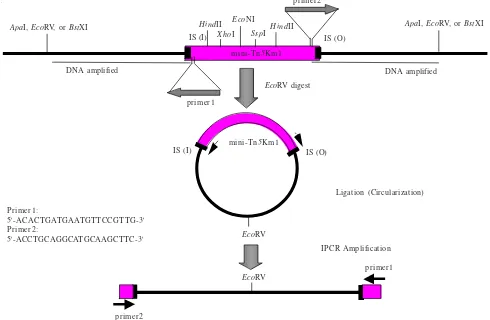
Figure 1 Strategy for amplification of DNA sequence flanking mini-Tn5 transposon by inverse PCR. Genomic DNA of non-magnetic mutant containing inserted transposon was digested with ApaI, BstXI, or EcoRV. IS: Insertion Sequence of the mini-transposon, 19 base pairs (inner and outer).
filter was carried out overnight using 10 x salt sodium citrate
(SSC) solution (3 M NaCl, 0.3 M sodium citrate, pH 7.0) as the transfer solution, and the DNA was fixed onto membrane by UV cross-linking. Blotted membrane was hybridized overnight at 42 oC with 1.8 kb of mini-Tn5Km1 transposon containing kanamycin resistance gene as a probe labeled with digoxigenin (Boehringer Manheim, Germany). Membrane was washed two times in 2 X wash solution (2 x SSC, 0.1%
sodium dodecyl sulfate (SDS) for 5 minutes at room temperature, and then two times in 0.1 x wash solution (0.1 x
SSC, 0.1% SDS) for 15 minutes at 68 oC. Detection of hybridization was conducted using X-ray film.
Preparation of DNA Templates for Inverse PCR. The strategy for inverse PCR is shown in Figure 1. DNA templates for inverse PCR were prepared from approximately 1 µg of genomic DNA from non-magnetic mutants digested with
EcoRV, BstXI, or ApaI (these restriction enzymes do not digest mini-Tn5Km1). Digested DNA was purified by phenol/ chloroform extraction followed by chloroform/isoamyl alcohol treatment and the DNA was precipitated with 100% ethanol and washed two times with 70% ethanol. DNA pellets were diluted in 5 µl of sterile milli Q and ligated with the same volume of DNA ligation kit version 2 solution I (Takara, Tokyo, Japan). The ligation reaction was incubated overnight at 15 oC and the circularized DNA was precipitated with 100% ethanol, washed with 70% ethanol, and the DNA pellets were diluted in 10 µl milli Q.
Conditions for Inverse PCR. The circularized DNA sequences flanking mini-Tn5 transposon (approximately 500
ng), was amplified using Gene Amp PCR 2400 (Perkin Elmer, USA) in a total volume of 50 µl containing 2.5 mM dNTP mixture, GC buffer II, LA Taq DNA polymerase (Takara, Japan), and 10 picomol of each primer designed from mini-Tn5Km1 near insertion sequence (IS). To amplify both upstream and downstream of the DNA sequences flanking mini-Tn5Km1 were used primer 1: 5’-ACACTGATGA ATGTTC-CGTTG-3’ and primer 2: 5-‘ACCTGCAGGCATGCA AGCTTC-3’ (Figure 1). A Gene Amp PCR System 2400 (Perkin Elmer, USA) was used to amplify DNA by: denaturation at 95 oC for 2 minutes; primer annealing at 60 oC for 1 minute; primer extension at 72 oC for 1 minute for 30 cycles and 10 minutes at 72 oC for the last cycle.
DNA Sequencing. The inverse PCR products were purified using gene clean III kit (Bio 101) and subsequently cloned in pGEM-T Easy (Promega, USA) and transformed into E. coli DH5α. The recombinant plasmids were extracted from E. coli and used as templates for cycle sequencing by PCR machine 9400. DNA sequencing was carried out using the dye terminator cycle sequencing kit (Applied Biosystems, USA) and M13 universal and M13 reverse primer were used as cycle sequencing primers. Sequencing of the DNA was performed using an automatic DNA sequencer ABI PRISM 377 (Perkin Elmer, USA).
RESULTS
Mini-Tn5 transposon mutagenesis generated eight non-magnetic mutants of M. magneticum AMB-1 defective in
ApaI, EcoRV, or BstXI
DNA amplified
HindII HindII XhoI
IS (I)
EcoNI
SspI
mini-Tn5Km1
primer2
IS (O)
primer1
EcoRV digest
DNA amplified
ApaI, EcoRV, or BstXI
mini-Tn5Km1
IS (O) IS (I)
Ligation (Circularization)
IPCR Amplification
primer1 EcoRV
EcoRV
primer2 Primer1:
5'-ACACTGATGAATGTTCCGTTG-3' Primer2:
5'-ACCTGCAGGCATGCAAGCTTC-3'
magnetosome synthesis. Frequency of transconjugation was about 2.7 x 10⁻⁷ cells per recipients. All the DNA fragments
of M. magneticum AMB-1 genome flanking mini-Tn5Km1 transposon could be amplified by inverse PCR (Figure 2). Analysis of eight genomic DNA of non-magnetic mutants by Southern hybridization, using kanamysin resistance gene (Km1) as a probe, revealed the presence of a single mini-Tn5
transposon insertion in the mutant’s genomic DNA, as shown in Figure 3. Sub-cloning of inverse PCR products using pGEM-T Easy as a cloning vector, and their sequence analysis revealed the presence of all insertion sequences (IS) coming from the mini Tn5Km1 transposon (19 bp inner and 19 bp outer), as well as 9 bases of the target sites of the transposon insertion were also identified (Table 1).
Southern hybridization analysis of EcoRI digestion of many non-magnetic mutant genomes indicated that mini-Tn5Km1inserts in the genomic fragment of approximately 12 kb in size (data not shown). This makes it difficult to clone the fragments containing the transposon into a plasmid vector, such as pUC18, pUC19, or pRK415 (data not shown). Therefore, the inverse PCR technique was developed to rapidly amplify the genomic DNA sequences of M. magneticum AMB-1 flanking the mini-Tn5Km1transposon. Primers oriented outward from the transposon permit directly amplification of those fragments, upstream, and downstream of the transposon. Flanking DNA sequence of M. magneticum AMB-1 up to 4.2 kb can be amplified by inverse PCR with EcoRV digestion.
DISCUSSION
In this study, the DNA fragments from restriction enzyme digestion were diluted and ligated under conditions that favor the formation of monomeric circles (Collin and Weissman 1984). The resulting ligation products are used as substrates for enzymatic amplification by PCR using primers near the end of the core sequence (mini-Tn5Km1) oriented outward from the known sequence region. The product resulting from amplification is a linear double- stranded DNA molecule including fragments situated both 5’ and 3’ to the known sequence region. The junction between the original upstream and downstream regions, can be identified as the restriction site of the restriction enzyme, in this case ApaI,
BstXI, or EcoRV, that was used to produce the linear fragments prior to self ligation.
Southern hybridization analysis of EcoRI or PstI digested from several non-magnetic mutant genomes indicated that mini-Tn5 inserted into the genomic DNA as a fragment of > 12 kb in size (data not shown). Such size renders difficulty in cloning a genomic fragment containing mini-Tn5 transposon into plasmids pUC19, pUC18, or pRK415 (data not shown). Therefore, we developed this simple technique to amplify the genomic DNA sequences flanking mini-Tn5 transposons. Primers oriented outward from the transposon permit directly amplification of those fragments, upstream and downstream of the transposon insertion.
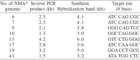
The expected size of the inverse PCR products generated by inverse PCR can be calculated based on the hybridization size minus the transposon mini-Tn5Km1 size (1.8 kb). Digestion of non-magnetic mutant 10 (NMA10) genome
using ApaI yielded a 1.3 kb of inverse PCR product (3.1-1.8 kb). Digestion of NMA 6 and 7 genomes with EcoRV, and NMA 9 genome with BstXI yielded 2.3; 2.3; and 2.1 kb, respectively (4.1-1.8; 4.1-1.8; 3.9-1.8 kb; respectively). On the other hand, NMA 15, 17, 19, and 41 genomes which were digested with EcoRV yielded 4.2; 2.8; 3.2; and 1.5 kb inverse PCR products, respectively (6.0-1.8; 4.8-1.8; 5.0-1.8; 3.2-1.8; respectively) (Figure 2 and 3; Table 1). To confirm consistence of the expected of the correct inverse PCR products generated in this study, DNA sequencing of the
(kb)
Figure 2 Agarose gel electrophoresis of 8 inverse PCR products amplified from non-magnetic mutant genomes by inverse PCR. M: Marker 1 kb DNA ladder. Numbers above of the figure indicate number of non-magnetic mutant genomes.
Figure 3 Southern hybridization analysis of non-magnetic mutant genomes digested with EcoRV, except no 9 and 10 were digested with BstXI and ApaI, respectively. M: Marker 1 kb DNA ladder. P: plasmid pUTmini-Tn5Km1 digested with EcoRI and PstI. Kanamycin resistance gene (Km1, 1.8 kb) isolated from pUTmini-Tn5Km1 was used as a probe.
Table 1 Size of DNA fragments flanking mini-Tn5Km1 transposon amplified from non-magnetic mutant genomes by inverse PCR, and confirmation by Southern hybridization. Nine bases of target sites of the transposon insertion were identified
inverse PCR products was performed. The inverse PCR products were cloned into pGEM-T plasmid vector and sequenced. Sequence analysis indicated that all of the inverse PCR products contained a 19 bp of insertion sequences coming from mini-Tn5Km1 transposon i.e. CTGTCTCTTGAT CAGATCT (inner) and ACTTGTGTATA AGAGTCAG (outer) (DeLorenzo et al. 1990). The presence of these insertion sequences of inverse PCR products indicated that the products of inverse PCR were correctly amplified because primers designed outward from the transposon passedthe 19 bases insertion sequences of the transposon. Thus, these results suggest that inverse PCR based on this method can be used to rapidly amplify both upstream and downstream of the genomic DNA fragment flanking mini-Tn5 transposon insertion of the bacterial genomes interrupted by this transposon.
The inverse PCR method permits the rapid amplification of regions of unknown sequence flanking specified segments of DNA. Since only regions of limited size can be enzymatically amplified by PCR, and since primers should be synthesized from known sequences, the inverse PCR approach is not amenable to proceeding very long distances in the genome. However, the technique described here is a simple and rapid method as an alternative to amplify and isolate genomic DNA sequences flanking mini-Tn5
transposon insertion. Furthermore, the inverse PCR products amplified by this method can be used to quickly obtain DNA sequence information on the region of the mini-Tn5
transposon insertion without sub-cloning of mutant genomic DNA containing transposon. In addition, we have successfully used these inverse PCR products as probes to isolate cosmid recombinant clones from a genomic library of wild-type M. magneticum strain AMB-1 (Wahyudi 2005). This method has also been successfully applied to amplify genomic DNA fragments flanking mini-Tn5Km1 transposon from acid-Al sensitive mutants of Bradyrhizobium japonicum
resulted by transposon mutagenesis (data not shown).
ACKNOWLEDGEMENT
Part of this work was conducted at Tokyo University of Agriculture and Technology, Japan, at the Department of
Biotechnology (Matsunaga-Takeyama Laboratory). Therefore, I grateful to Tadashi Matsunaga and Haruko Takeyama for the laboratory facilities and their valuable support.
REFERENCES
Blakemore RP, Maratea D, Wolf RS. 1979. Isolation and pure culture of a fresh water magnetic spirilum in defined growth medium. J Bacteriol 140:720-729.
Collin FS, Weissman SM. 1984. Directional cloning of DNA fragments at a large Distance from an initial probe: a circularization method. Proc Natl Acad Sci USA 81:6812-6816.
DeLorenzo V, Herrero M, Martinko JM, Parker J. 1990. Tn5 transposon derivatives for insertion mutagenesis, promotor probing and chromosomal insertion of cloned DNA in gram-negative eubacteria. J Bacteriol 172:6568-6572.
Feseveldt A, Poetsch M, Gliesche CG. 1997. Development of a species specific gene probe for Hypomicrobium facilis with the inverse PCR. Appl Environ Microbiol 63:335-337.
Hererro M, DeLorenzo V, Timmis KN. 1990. Transposon vectors containing non Antibiotic resistance selection markers for cloning and stable chromosomal insertion of foreign genes in gram negative bacteria. J Bacteriol 172:6557-6567.
Martin VJJ, Mohn WW. 1999. An alternative inverse PCR (IPCR) method to amplify DNA sequences flanking Tn5 transposon insertions. J Microbiol Met 35:163-166.
Matsunaga T, Sakaguchi T, Todokoro F. 1991. Magnetite formation by a magnetic Bacterium capable of growing aerobically. Appl Microbiol Biotechnol 35:651-655.
Mirleu P et al. 2000. Fitness in soil and rhizosphere of Pseudomonas fluorescens C7R12 compared with C7R12 mutant affected in pyoverdine synthesis and uptake. FEMS Microbiol Ecol 34:34-44.
Ochman H, Gerber AS, Hartl DL. 1988. Genetic application of an inverse polymerase Chain reaction. Genetics 120:621-623. Prod’hom G et al. 1998. A reliable amplification technique for the
characterization of genomic DNA sequences flanking insertion sequences. FEMS Microbiol Lett 158:75-81.
Rich JJ, Willis DK. 1990. A single oligonucleotide can be used to rapidly isolate DNA sequences flanking a transposon Tn5 insertion by the polymerase chain reaction. Nucleic Acids Res 18:6673-6676.
Wahyudi AT. 2005. [Construction of genomic library of Magnetospirillum magneticum AMB-1 and screening of genes involved in magnetosome synthesis]. [In Indonesian]. J Mikrobiol Indones 10:91-95.
Wahyudi AT, Takeyama H, Matsunaga T. 2001. Isolation of Magnetospirillum magneticum AMB-1 mutants defective in bacterial magnetic particles synthesis by transposon mutagenesis. Appl Biochem Biotechnol 91:147-154.