Leptospirosis
at Dr.
Cipto Mangunkusumo
and Persahabatan
Hospitat
Review
of
104
cases
Iskandar
Zulkarnain
Abstrak
Telah dilakukan suatu
studi
restropeldifunnk melihat
gambaran epidemiologi, klinis dan pengobatanpada
104 penderila leptospirosis yang dirawat di Rumah Sakit Cipto Mangunhuumo dan Persahabatan sejak Januari Tggj hingga besember 1b96. Darihasil
-studi didapatkan bahwa 76,9% penderita adalah laki-laki dan 43,79ô adalah wanita dengan umur
rita-rata
36,9 tahun (SD +l3)
dan 43,7 tahun (SD+
13,8), sedangkan angka kejadian meningkat pada bulanJanuai
hinggaApril.
Gejata klinis yang sertng timbul adalah mual dengan atau tanpa muntah, dandiilati
dengan keluhan demam, nyeriotàt,
sufusi koijungtiva,htntig,
sakit kepala, hepatomegali, batuk, perdarahan,rigor
dan splenomegali: sedangkan kelainan laboratoriimyor[
,"iing
timbul adalah meningkatnyalaiu
endap darahdiikuti
dengan meningkatnya amilase/urea darah, trombositopenia, teulasitosii, meningkatnya kreatinin/
bilirubin total/
lipase, proteinuri, meningkatnya SGOT/
SGPT dan anemia. Hasfluii
MAT menunjut:pan terdlapatnya leptospira tunggal (97,56% L.bataviae), leptospira ganda (680Â L.hardjo dan bataviae), leptospira irtpel (5094 L.iiterohaemoottogi"o, iavanica dan celledoni) pada 40,40%, 23% dan l,9oÀ penderita. Tidak lerdapat perbedaan bermakna antara penderira yangletaphidup dengan yang mati bila dihubungkan dengan jenis kelamin, umur, kadar hemoglobin, lekosit, trombosit,
irea
darah-, lcrâafinin,bilirubin total, jenis antibiotika, kecuali dengan uji MAT; demikian juga antara hasil
uji
MAT dengan lama demam sebelum masukrumah sakit dan lama perawatan. Tidak terdapat perbedaan hasil pengobatan baik dengan
pinisilin
maupun antibiotiça lain, termasuk sefalosporin generasi ketiga. Disimpulkan bahwa mual dengan atau tanpa muntah, demàm dan nyeri otot merupakan gejala klinis yang sering muncul, sedangkan meningkatnya laju endap darah merupakan lemuan laboratoium yang sering didapat.Tigkakematian lebih sering didapat pada kasus dengan hasil uji
MÀT
negatiJ. Penisilin masih tetap efektif untuk pingobat-an.Abstract
A
restrospeclive study of leptospirosis cases at Cipto Mangunkusumo and Persahabalan hospilal from January 1993 to December 1996 was done to determine the epidemiological, clinical and therapeutic characteristics.Fron t7l
patienrs, 76,9ok were male and 43,796 were female. The average age was 36,9 years (SD+ I3)
and 43,7 years (SD+
t3,S). The incidences were increased betweenver, muscle pain, respectively. The mylase and blood SGOT/SGPT, and 56%
L.
bataviae), double leptospira (68%L.
hardjo and bataviae),triple leptospira
(50%
L.
icterohaemonhagica,javanica,
and celledoni)
detectedii
lb.cN,'
23.1%and I.9oÀ
patients, respeclively.There wereno
signiJicant dillerences between sumive anddied
caseswith
sex, age, hemoglobin,. leukoiyte andt,
except MAT test and also no signiJicant di.fference and length of stay of leptospirosis casesin
hospital. included cephalosporin 3rd generation (p=0.35).lk
erythrocyte sedimentation rates was the mosr commonrtr"rt,oo'lfj',i,irltrt
!,#:t::,rî:Ïf;;!;,':::r:';::';î!;: !:'":#;'i,i[
negative MAT test. Procaine penicillin is still e/fectivefor
leptospirons.Keywords : Leptospirosis, epidemiological, clinical, therapeutic characrerisrics
D)teant
Depalzezl
a/
ine Universityof
Indonesia, Jakarta, Indones iaLeptospirosis
is
an
acute
generalized
infectious
disease
caused
DfgenDf
The causative
ag
ainterroga
species
of sp
multiple
272
Zulkarnainserovars
and
18 serogroups have beenidentified for
L
interrogans.l'2'3'4's
In
human
it
is a
zoonosis
thatinfected
from
animal
source. People
working
in
amilieu that
associatedwith
rats
or
infected livestock
with
water
are
especially
prone
to
infection.6'7Serologic
surveysof workers
athigh
risk confirm
thatsubclinical infection
is
common.
Less
than
l0% of
symptomatic
infection
result
in
severe,icteric
illness.None
of
the
presenting features
of
leptospirosis
arespecific,
because
clinical
findings ranging
from
asymptomatic infection
to
renal
failure
and
death.'Where leptospirosis
is
common, prediction may
bemade
on
clinical
suspicion
and
epidemiological
grounds.Leptospires
are sensitiveto
most antibiotics,
and antibiotics therapy should be given as early
aspossible
in
leptospirosis.r'8'e'r0The
aim of this
studywas
to
determine the epidemiological,
clinical
and therapeutic characteristicsof leptospirosis'
METHODS
This
study
is
a
retrospective
study
using
data
from
medical records
of
all
leptospirosis
casesadmitted
atCipto
Mangunkusumo
and
Persahabatan Hospital
between
January
1993
to December 1996.
Theinclusion
criteria
for
subjects
of
this
study
werepatients
diagnosed asleptospirosis that confirmed
by
clinical
features
and
microscopic
agglutination
(MAT).
Data
of
medical
recordswere investigated to
identiff
their
age, sex, occupation,
clinical
features,peripheral
blood
count, urinalysis, blood urea
andcreatinine,
bilirubin, SGOT,
SGPT, amylase,
lipase,result
of
serologic
test
(MAT),
treatment,
length
of
stay athospital
andmortality.
Statistical
analysis wasdone using
chi-square,
unpaired student t-test
andANOVA.
RESULT
Sixty-three (60.6%) patients
from
Cipto Mangunkusumo generalhospital and
forty-one
(39.4%) patients
from
Persahabatan
Hospital were included
in
this
study.The
characteristic
of
104 patients
who followed
the study were: 76.9% weremale
and43.70Â were female, the mean age was: 36.9 years(SD+13),
and 43.7 years(SDtl3.8)
respectively.
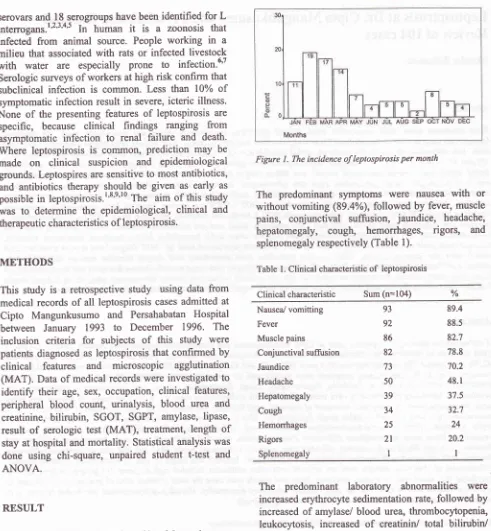
The
incidences
of
lepto-spirosis
cases
were
increased
between January
toApril
(Figure
l).
Med J Indones
[image:2.595.80.571.76.607.2]Months
Figure
l.
The incidence of leptospirosis per monthThe
predominant symptoms
were
nausea
with
or
without vomiting
(89.4%),
followed by
fever,
musclepains,
conjunctival
suffi.rsion,
jaundice,
headache,hepatomegaly,
cough,
hemorrhages,
rigors,
and splenomegalyrespectively (Table
l).
Table
l.
Clinical characteristicof
leptospirosisClinical characteristic Sum (n=104) %
Nausea/ vomitting 89.4
Fever
92
88.5Muscle
pains
86
82.7Conjunctival
suffusion
82
78.8Jaundice
73
70.2Headache
50
48.193
Hepatomegaly Cough
Hemorrhages
Rigors Splenomegaly
39
37.534
32.725
242l
20.2ll
The
predominant
laboratory
abnormalities
were increasederythrocyte
sedimentation rate,followed
by
increasedof
amylase/
blood
urea, thrombocytopenia,
leukocytosis,
increased
of
creatinin/
total
bilirubin/
lipase, proteinuria,
increased
of
SGOT/SGPT,
and anemia (Tabel 2).Serologic tests
by MAT
revealed single
leptospira(97.56%
L. bataviae), double leptospira
(68%
L.
hardjo
and
bataviae),
triple
leptospira
(50%
L.
icterohaemorrhagica,
javanica,
andcelledoni)
detectedTable 2. Laboratory abnormality
of
leptospirosisLaboratory abnormality Number
laboratory
Numberlaboratoryexamination
abnormalityPercentage
(%)
Increased erythrocyte sedimentation rate
(F>15, M>10)
Increased of amylase (>190 U/l)
Increased of blood urea (>120 mg/dl) Thrombocytopenia (< I 50,000 K/ul)
Leukocytosis (> I 0,000 IUul)
Increased of creatinine (>1.5 mgldl)
Increased of total bilirubin
(>l
mg/dl)lncreased of lipase (>120 U/l)
Proteinuria (protein urine>+)
Increased of SGOT (>37
Ull)
Increased of SGPT(>4lU/l)
Decreased of Hemoglobin (<10gÉl")37 36 34 97
9l
9l
87 75 32 59 50 39 t7 97.3 94.4 94.2 88.3 87.5 84.5 84.3 84.2 6s.6 60.2 47 16.3 36 103 103 104 103 89 38 90 83 83 104Table 3. Serologic test (MAT)
of
leptospirosisSerologic test (MAT)
Percentage(%)
Percentageserovars (%)Negative Single leptospira
Double leptospira
triple leptospira 34.6 40.4 23.1 bataviae hardjo hardjo+bataviae
icterohaemorrhagiae +bataviae
australisfbataviae
bataviae+tarassovi
celedoni +rachmati javanica+bataviae pomonatbataviae
icterohaemorrhagiaeljavanica+ celedoni
i cterohaemorrhagiae+batavi ae*tarassovi
97.56 2.44 68 t2 4 4 4 4 4 50 50 1.9
Table 4. Association between survive and died cases with variable factors
Variables
survlve
Died
DGender
Age Hb Leucocyte Thrombocyte Blood urea
Creatinin Bilirubin total
MAT test
Male
70Female
2237.3(SDll3.l)
lr.5(sDrl.6)
16220.6(SD16000) I 86456.5(SDr 144358.8)
r90.9(sDrl06.3)
4.e(sDr3.2))
8.5(SDrl
l.l)
Negative Single leptospira Double leptospira Triple leptospira Penicillin
Ampicillin
Chloramphenicol Tetracycline
Ampicillin+Chloramphenicol Cephalosporin 3rd generation
Male
l0
Female
247.8(SDr13.4)
I 1.8(SD1l.5) 2167s(SDrl 1298.3)
96416.7(SDr99t97 .2)
180.6(sDr77.7) 5.1(sDr3.2) 3.9(SD17.4)
Negative
l0
Single leptospira I Double
leptospira
I Tripleleptospira
076
Penicillin6
AmpicillinI
Chloramphenicol2
Tetracycline2
Ampicillin+Chloramphenicol4
Cephalosporin 3rd generation0.44*
0.97** 0.86+*
0.06++ 0.24++ 0.21 *+ 0.99+* 0.10** 0.002*
0.35 * 26 4t 23 2 Antibiotics 9 I 0 0 0 I 0
Ciprofloxacin
0
Ciprofloxacin274
ZulkarnainThere were
no significant
difference
betweensurvive
and died
caseswith
sex,
age,hemoglobin, leukocyte
and
thrombocyte
count,
blood urea, creatinin,
bilirubin
total, antibiotics
treatment, except MAT test.Table 5. Association between MAT test result with variable factors
Variables MAT test result
negative
singleleptospira
Med J Indones
Negative
MAT test was
found more frequently in
died cases, it- seems thatvery
low
antibody
in
severeill
patient.r3
In
our
study
we
found
single (40.4%),
double (23.1%)
and
triple
leprospira
(19%)
andnegative leptospira (34.6%)
basedon
MAT
test, therewere
no
correlation
with
duration
of
fever before
admitted
(p=0.75) and length
of
stay hospitalization
(p:0.82).
Effectiveness
of Penicillin
therapywere
notdiffer
with
other antibiotics included cephalosporin
3rd generation(p:0.35).
CONCLUSION
This
investigation concluded
that
nause
with or
without vomitting, fever and
muscle
pain were
themost common clinical
presentation
of
leptospirosis. Increased oferythrocyte sedimentation
rateswere
themost common laboratory abnormality.
Mortality
of
leptospirosis
is
more higher
in
caseswith
negativeMAT
test.
Procaine
penicillin
is still
effective for
leptospirosis.REFERENCES
L
WattG.
Leptospirosis.In:
StricklandGT,
ed. Hunter's Tropical Medicine. Philadelphia: WTI Saunders,l99l;
3t7-23.2.
Johnson RC. Leptospirosis.In:
WarrenKS,
Mahmoud AAF, eds. Tropical and geographical medicine;2nd edition. New York : Mc GrawHill,
1990; 889-93.3.
Sanford JP. Leptospirosis.In:
Braunwald E, IsselbacherKJ,
Peterdoerf RG, eds. Harisson principleof
Internal Medicine; l3th edition. Newyork
: Mc GiawHill,
1990; 740-3.4.
Syam AF, PohanHT,
ZulkarnainL
patogenesis dan Diagnosis Leptospirosis.Maj Kedokt Indon.
1997; 47:636-639.
5.
FaineS.
Leptospirosis.In:
HoeprichpD,
JordanMC,
,Ronald AR, eds. Infectious Diseases:
A
Treatiseof
Infectious Processes. philadelphia:JB
Lippincot
Co, 1994:619-24.6.
Sasaki DM, Pang L, Minette HP et al. Active surveillance and risk factorsfor
leptospirosisin
Hawaii. Am J Trop. Med. Hyg 1993;48: 35-43.7.
Fontaine A, PeslerbeX,
Gainere Jp. Occupationalhazard,of
unnoticed leptospirosisin
water ways maintenancestaff. Eur J Epidemiol 1992; B:228-32.
8.
Faine S. Clinical leptospirosis in humans. Leptospira andLeptospirosis.Florida: CRC Press Inc, 1994; 199-227.
9.
Marien F, Perolat P. Public health importance of humanleptospirosis
in
the South Pasific:
A five
year study in New Caledonia.Am
J Trop Med Hyg 1996;55 :174-178.10.
Marotto PF, Marotto MS, SantosDL,
SauzaNL,
SeguroA. Outcome of leptospirosis in children. Am J Trop Med Hyg1997;56:307-310.
pr
double
tripleleptospira leDtosDira
Duration 5.97
6.97
7.2
5
0.75fever
(SD+2.6)
(SDt3.9) (SDr3.5)
(SD+2.8) Lengthof
10.53
17.47
14.4115
0n
* ANOVA
There
were
no
significant difference
between
MAT
test
result
with
duration
of
fever before
admitted
tohospital, and
length
of
stay
of
leptospirosis
casesjn
hospital.DISCUSSION
Out
of
104 casesin
tÈis study male is more
frequentthan female
to
have leptosprosis, since male
patientsmore
likely work
outside and have occupational
risk
to
be infected than female
patients.l'eThe
incidencesof Ieptospirosis
were
increased between January
toApril
when flood
waters are
high. The
predominant
symptoms
in this
study were,
nauseawith or without
vomiting, followed by
fever, muscle pains,conjunctival
suffirsion,
juul^dlp".
This finding
is
not differ
with
other
authors.l'8'rrThe laboratory-
abnormality
of
this
study were
increased
of
erythrocyte
(9j.3%),
increased of amylase(94.4%),
increasedof blood
urea(94.2%),
thrombocytopenia
(88.3%),
leukocytosis
(87
.5%),
increased ofcreatinine (84.5%),
increasedof
total bilirubin (84.3%),
increased
of
lipase
(84.2%),proteinuria (65.6%),
increased
of
SGOT
(60.2%),
increased
of
SGPT
(47%),
decreasedof
hemoglobin
(163%).
These
results have
a
bit
different with
laboratory.^
abnormality
demonstrated
by
other authors,l'8'12 especially increased of amylase and lipasethat
indicated pancreatitis.
Although much
attentionand literatures are
concerned
with
renal and liver
complication, however we
alsofound
that pancreatitisfrequently
occuredin leptospirosis.
Heath CW, Alexander
AD,
GaltonMM.
Leptospirosis in the United States. Analysis of 438 Cases inMAN,
1949-1961. N Eng J Med 1965;'273:915-22.Martone, Kaufmann. Leptospirosis
in
humansin
theUnited States 1974-1978. J Infect Dis 1979; 140: 1020-22.
Turner LH. Leptospirosis
II.
Serology. Trans R Soc Trop Med Hyg 1968; 62:880-99.13.
ll