TELKOMNIKA, Vol.10, No. 3, September 2012, pp. 537~544 ISSN: 1693-6930
accredited by DGHE (DIKTI), Decree No: 51/Dikti/Kep/2010 537
Retinal Image Preprocessing: Background and Noise
Segmentation
Ibaa Jamal1, M. Usman Akram*2, Anam Tariq3 1,2,3
Bahria University, National University of sciences & Technology, Islamabad, Pakistan e-mail: [email protected], [email protected]*2, [email protected]
Abstrak
Citra medis merupakan area riset yang sangat populer sekarang ini yang melibatkan diagnosa berbantuan komputer untuk banyak penyakit, dengan menggunakan citra digital sebagai masukan. Citra retina digital digunakan untuk penapisan dan diagnosa penyakit retina mata akibat diabetes. Suatu sistem otomatis untuk diagnosis penyakit retina akibat diabetes seharusnya menonjolkan semua tanda penyakit yang ada dalam citra dan untuk meningkatkan akurasi sistem, kualitas citra retina harus ditingkatkan. Makalah ini mempresentasikan suatu metode peningkatan kualitas citra retina masukan dan metode ini merupakan langkah pemrosesan awal dalam diagnosa otomatis penyakit retina akibat diabetes. Pemrosesan awal terdiri dari estimasi latar belakang dan penghilangan derau dari citra retina dengan mengaplikasikan segmentasi kasar dan halus. Pengujian yang ekstensif dilakukan untuk validasi teknik pemrosesan awal yang diajukan menggunakan basis data citra baku permukaan belakang mata (fundus).
Kata kunci: pemrosesan awal, penyakit retina akibat diabetes, segmentasi latar belakang, segmentasi derau
Abstract
Medical imaging is very popular research area these days and includes computer aided diagnosis of different diseases by taking digital images as input. Digital retinal images are used for the screening and diagnosis of diabetic retinopathy, an eye disease. An automated system for the diagnosis of diabetic retinopathy should highlight all signs of disease present in the image and in order to improve the accuracy of the system, the retinal image quality must be improved. In this article, we present a method to improve the quality of input retinal image and we consider this method as a preprocessing step in automated diagnosis of diabetic retinopathy. The preprocessing consists of background estimation and noise removal from retinal image by applying coarse and fine segmentation. We perform extensive results to check the validity of proposed preprocessing technique using standard fundus image database.
Keywords: background segmentation, diabetic retinopathy, noise segmentation, preprocessing
1. Introduction
One out of ten is affected by diabetes, which result in vision loss, stroke and heart failure. Group of eye problems that people with diabetes may face is known as diabetic eye disease. People affected by diabetic are more facing eye problems like contracts and glaucoma and the main reason for vision loss is the disease affecting on retina[1]. The complications of diabetes affect the vascular structure of human retina and cause the leakage of blood on surface of retina which leads to blindness and it is known as diabetic retinopathy [1]. Diabetic retinopathy is a progressive disease as there are no such signs of disease at its early stages but as the time passes the disease turns into severe [2]. The common symptoms of diabetic retinopathy are blurred vision and even loss of vision if not treated in time [2].
highly illuminated while the noise increases closer to the edge of the retina [17]. So, Illumination equalization and noise removal are required to enhance the image quality.
In the beginning, before the detection of abnormalities and features in retinal image we must remove the noise and background from retinal image which will increase the quality of the image. This is done in preprocessing step and without this step the automated system will give poor result for feature extraction and abnormality detection.
The aim of preprocessing is to increase the quality of an image by reducing the amount of noise appearing in the image and highlighting features that are used in image segmentation. Two typical techniques used in preprocessing are filtering and contrast enhancing. Standard contrast stretching techniques have been applied by [4], [13] and [23] for segmentation and noise reduction. In [14], [15] and [16] the local contrast enhancement method is used for equalizing uneven illumination in the intensity channel of retinal images. A large mean filter, large median filter and collectively are used for retinal image have used intensity channel values to detect the dark regions from retinal image. Thresholding is also an important and widely used technique in image segmentation [19], because thresholding is effective and simple to implement. In thresholding, pixels within a defined range are selected as belonging to the foreground whereas gray-levels outside the range are rejected to the background [19].
In this paper, we present the retinal image preprocessing technique that detects the background using local mean and variance and removes noise using HSI color space. The proposed preprocessing method consists of two steps i.e. coarse and fine segmentation. In the first step, it does coarse segmentation that creates binary masks for background estimation and noise removal. In the second step, it does fine segmentation that combines background segmented mask and noise segmented mask and applies morphological operations to remove single pixel noise and edge pixels.
This paper is organized in four sections. Section 2 presents the step by step techniques required for color retinal image segmentation. Experimental results are discussed in section 3 followed by conclusion in section 4.
2. System Overview
Computer assisted diagnosis for various diseases is very common now a days and medical imaging is playing a vital role in such computer assisted diagnosis. We present an automated preprocessing system to improve the quality of retinal images so that the accuracy of automated screening of diabetic retinopathy can be improved. The proposed preprocessing method is used to improve the quality of image by extracting background and removing the noisy area from the retinal image. In automatic diagnosis of diabetic retinopathy, the processing of the surrounding background and noisy areas in retinal image is not necessary and consumes more processing time in all stages. Cutting or cropping out the region that contains the retinal image feature minimizes the number of operations on the retinal image. Moreover the noisy pixels may cause false features and reduce the accuracy of the automated classification so it is necessary to remove the noisy pixels. Figure 1 shows the flow diagram of our preprocessing technique. It shows the step by step outputs of proposed preprocessing technique.
2.1 Coarse Segmentation
Coarse segmentation creates mask for background and noise estimation using mean and variance method and HSI (Hue, Saturation, Intensity) color model respectively.
TELKOMNIKA
Figure 1.
2.1.1 Background Estimation A color retinal imag background. This dark backg feature extraction and lesion d is important to remove the bac We present a local me creates a binary background s greater than threshold value, belongs to background.
The algorithm for the b Step 1: Divide the acquired re Step 2: Compute the local me
Step 3: Use local mean value std(I) using equation 2
Step 4: Select threshold value Step 5: for each pixel, illumination is usually adequa located near the edge of the r abnormality detection. That is abnormalities.
In our technique, w area and it is applied on reti steps i.e. feature extraction convert RGB (Red, Green, Blu the way a human experience space [19]. The RGB retinal im
ISSN: 1693-6930
1. Flow Diagram of Retinal Image Preprocessing
ion
age consists of a (semi) circular region of int kground is initially never really black. It is not ne n detection algorithms on this area and it consume
ackground from input retinal image.
mean and variance based method [19] for backgro d segmentation mask by applying a threshold on std e, the block is considered as original retinal image
e background extraction mask is as below: retinal image into non-overlapping blocks
ean value M(I) using equation 1
ue computed in step 2 to compute the local standar n 2
lue empirically
>Threshold?
ixel in original retinal image area pixels ixel in background area pixels
Mask
al image is normally due to noise pixels and pixe exist in regions where illumination has been in quate in the center of the image, poor image qu e retinal image. Regions with poor image quality m
t is why they should be detected and removed be
we create binary noise segmentation mask which i etinal image to ensure not to process the noisy a n and abnormality detection. In this segmentatio Blue) retinal image into HSI color space because fi ces colors and secondly noise can be easily remo l image is converted into HSI color space using equ
539 ge area otherwise it
(1) may cause errors in before detection of
The algorithm for noise remov Step 1: Divide the input retina Step 2: Use histogram equaliz Step 3: Use a 3x3 median filte Step 4: Convert the equalized Step 5: Calculate N (noise fac Step 6: Select a threshold valu Step 7: for each pixel,
Calculate N(I)<Thres if true, add pixel in no
if false, add pixel in end_for Figure 2 shows the images. These segmentation removed in fine segmentation.
Figure 2. Coarse segmentatio
2.2 Fine Segmentation
Background and n
contain single pixel noise and from segmentation masks. In dilation, morphological erosio noise from binary masks [19]. operations [19]. Background s order to remove the black pix removes all black single p segmentation mask. Noise seg (Figure 2). In order to remov opening followed by erosion. single pixel noise and it giv segmentation masks and fine
oval mask is as below:
nal image I(i,j) into non-overlapping blocks with size lization to enhance the contrast
ilter to reduce the noise in background of image. ed and filtered RGB retinal image into HSI color spa factor) which is a ratio of Hue and Intensity
alue empirically.
reshold?
normal retinal image area pixels in noisy area pixels
e coarse background and noise segmentation m on masks contain single pixel and edge pixel no on.
tion. a) Original retinal image; b) Background segm Noise segmentation mask
noise segmentation masks that are formed by coa nd edge pixels. Fine segmentation is done to rem In fine segmentation, morphological operations i ion and morphological opening are applied to rem 9]. We have used 5 x 5 square structuring element d segmentation mask contains black single pixel no
pixel noise, square structuring element is used for pixel noise and edge pixels and it gives a segmentation mask contains white single pixel noise
ove the white pixel noise, square structuring ele n. Opening removes all edge pixels and erosion r gives a fine noise segmentation mask. Figure
e segmentation masks.
(5)
(6)
ize w x w.
space.
masks for retinal noise which will be
mentation mask; c)
TELKOMNIKA
Figure 3. Fine segmentati Segme
2.3 Final Segmentation Mask Final segmentation m and fine noise segmentation removed by filtering the comb segmentation mask is then a final segmentation masks for r
Figure 4. (a): Original Color from database; (b): Fine Segmentation Mask; (c): Segmentation Mask; (d): Fina
Mask
3. Experimental Results In medical image pr systems are very important diaretdb0 [12], diaretdb1 [20], method. Diaretdb0 and Diar resolution of 1500 X 1152 pi DRIVE and STARE database objective and subjective crit segmented retinal images and
Statistical results of o table 1 and table 2. Table 1 s noise segmentation mask and segmentation, fine segmen segmentation after applying m accurate segmented retinal im evaluate the proposed system experts and these manually la 3 shows the accuracy res segmentation masks.
ISSN: 1693-6930
ation. a) Original Color Retinal Images from databas entation Masks; c) Fine Segmentation Mask.
ask
mask is prepaid by combining fine background se ion mask. For more fine segmentation mask, s mbined mask by a medium size median filter [19] applied on retinal image for its segmentation. Fig
r retinal image segmentation.
lor Retinal Image e Background c): Fine Noise inal Segmentation
Figure 5 Subjective Validity: ( Retinal Images from datab Segmentation Masks; (c): F
Mask.
processing, the validity and evaluation of autom nt so we have used four standard retinal imag
0], DRIVE [21] and STARE [22] to extensively che iaretDB1 databases contain 130 and 89 retina pixels and of different qualities in terms of noise ses consist of 40 and 20 images respectively. We riteria for evaluation of proposed method. Figur nd it depicts the subjective validity of proposed met f our segmentation technique for all 279 images ar separately shows the accuracy of background seg and final segmentation mask. Table 2 shows the
entation after applying morphological opera g median filter. These tables show the number a l images and poorly segmented retinal images. In tem, manually labeled preprocessing masks are cr
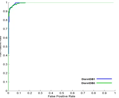
Table shows the background segm masks and final segmented re validity of our technique and s noisy areas. Figure 7 shows t by proposed algorithm. In ord order to check the validity o characteristics curves using segmented masks with it. F databases.
Figure 6. Preprocessing resu Color Retinal Images from dat Background Segmentation M Noise Segmentation Mask Segmentation Masks; (e): Fin
Images.
ble 2. Coarse and fine segmentation results
rately
ccuracy results for proposed preprocessing method
Database Accuracy
DRIVE 0.9982
STARE 0.9861
Diaretdb 0.9569
Average 0.9804
ifferent illumination and noise values are shown in gmentation masks, noise segmentation masks, fi retinal image for each color retinal image. These re d show that our technique gives good results for b s the manually created preprocessing masks and th rder to find out the accuracy, pixel by pixel compa of proposed system, we further calculate the re
TELKOMNIKA presented a method for the p diabetic retinopathy. The prop variance based method and divided our algorithm into two is prepaid by combining the improving the quality of segm mask to remove small regions images for their segmentation images taken from the standa ROC curves to validate the pro
References
[1] E J Susman, W J Tsiaras, K 3234.
[2] Effective Health Care - Com ulcers. Royal Society of Med [3] A Osareh, M Mirmehdi, B
Exudates in Digital Colour Im [4] S C Lee, E T Lee, R M Kin
early retinal lesions of diabe Arch. Clin. Exp. Ophtalmol. [5] C Sinthanayothin, J F Boyc
fovea and retinal blood vess 83(8): 231-238.
[6] M Foracchia, E Grisan, A Ru model of vessel structure. IEE
[7] N K M N Subhasis Chaudhu retinal images using twodim 8(3): 263-269.
[8] T Spencer, R P Phillips, microaneurysms in fluoresce [9] A J Frame, P E Undill, M J C computer based classificat fluorescein angiograms. Com
ISSN: 1693-6930
ing characteristic curves for proposed method using diaretDB1 databases
retinal images is that the quality of acquired imag en illuminated, blurred and noisy regions. It is very al images for reliable detection of abnormalities.
preprocessing of retinal image prior to screening oposed method consisted of background estimation d noise modeling and removal using HSI color m o phases i.e. coarse and fine segmentation. Final he background mask and noise segmentation m
gmentation mask, median filter are applied on fi ns. Final segmented masks are then applied on p tion. The results are confirmed by visual inspecti dard diabetic retinopathy databases. We further us proposed method.
, K A Soper. Diagnosis of diabetic eye disease. JAMA. 19
omplications of diabetes: Screening for retinopathy and M
edicine Press. 1999; 5(4): ISSN 0965-0288.
B Thomas and R Markham. Automated Identification r Images. British Journal of Ophthalmology. 2003; 87(10 Kingsley, Y Wang, D Russell, R Klein, A Wanr. Compari
betic retinopathy between a computer system and huma l. 2001; 119(4): 509-515.
yce, H L Cook, T. H.Williamson. Automated localizatio essels from digital color fundus images. British Journal o
A Ruggeri. Detection of optic disc in retinal images by mea
IEEE transactions on medical imaging. 2004; 23(10): 11 dhuri, Shankar Chatterjee, Michael goldbaum. Detection dimensional matched filters. IEEE transactions on med
, P F Sharp, J V Forrester. Automated detection an scein angiograms. Graefes Arch. Clin. Exp. Ophtalmol. 1
J Cree, J A Olson, K C McHardy, P F Sharp, J F Forrest ation methods applied to the detection of microaneur
omput. Biol. Med. 1998; 28(3): 225-238.
543
ing diaretDB0 and
ages is usually not ery vital to enhance . In this paper, we ng and diagnosis of ion using mean and model. We further al segmented mask mask. For further final segmentation poor quality retinal ction of segmented used accuracy and
1982; 247(
23),3231-d Management of foot
n of Diabetic Retinal ): 1220-1223. arison of diagnosis of man experts. Graefes
tion of the optic disc,
al of Opthalmol. 1999;
eans of a geometrical 1189-1195.
on of blood vessels in
edical imaging. 1989;
diabetic retinopathy in digital retinal images: a tool for diabetic retinopathy screening. Diabetes UK
Diabetic Medicine. 2003; 21(1): 84-90.
[15] C Sinthanayothin, V Kongbunkiat, S P Ruenchanachain, A Singlavanija. Automated screening system for diabetic retinopathy. Proc. of the 3rd International Symposium on Image and Signal Processing and Analysis, 2003: 915-920.
[16] A Osareh, M Mirmehdi, B Thomas, R Markham. Classification and localisation of diabetic-related eye disease. Proc. 7th European Conference on Computer Vision. Springer LNCS. 2002; 2353: 502-516,. [17] N P Ward, S Tomlinson, C J.Taylor. Image analysis of fundus photographs. Ophthalmology. 1989;
96(1): 80-86.
[18] H Wang, W Hsu, KG Goh, M L Lee. An effective approach to detect lesions in color retinal images.
Proc. IEEE Conference on Computer Vision and Pattern Recognition (CVPR). 2000: 181-187. [19] R C Gonzalez and R E Woods. Digital Image Processing, 2nd ed. Prentice Hall. 2002.
[20] Kauppi, T Kalesnykiene, V Kamarainen, J K Lensu, L Sorri, I Raninen A, Voutilainen R, Uusitalo, H Kälviäinen, H Pietilä, J DIARETDB1. Diabetic retinopathy database and evaluation protocol. Technical report. 2007.
[21] Hoover A, Goldbaum M. Locating the optic nerve in a retinal image using the fuzzy convergence of the blood vessels. Transaction on Medical Imaging. 2003; 22(8): 951–958.
[22] Staal, J Abramoff, M D Niemeijer, M Viergever M A , van Ginneken B. Ridge-based vessel segmentation in color images of the retina. IEEE Transaction on Medical Imaging. 2004; 23(4): 501– 509.