Complex calcium oscillations and the role of mitochondria
and cytosolic proteins
Marko Marhl
a,*, Thomas Haberichter
b, Milan Brumen
a,c, Reinhart Heinrich
baDepartment of Physics,Faculty of Education,Uni
6ersity of Maribor,Korosˇka cesta160,SI-2000Maribor,Slo6enia bInstitute of Biology,Theoretical Biophysics,Humboldt Uni
6ersity Berlin,In6alidenstr.42,D-10115Berlin,Germany cJosef Stefan Institute,Jamo
6a39,SI-1000Ljubljana,Slo6enia
Received 14 March 2000; received in revised form 31 May 2000; accepted 2 June 2000
Abstract
Intracellular calcium oscillations, which are oscillatory changes of cytosolic calcium concentration in response to agonist stimulation, are experimentally well observed in various living cells. Simple calcium oscillations represent the most common pattern and many mathematical models have been published to describe this type of oscillation. On the other hand, relatively few theoretical studies have been proposed to give an explanation of complex intracellular calcium oscillations, such as bursting and chaos. In this paper, we develop a new possible mechanism for complex calcium oscillations based on the interplay between three calcium stores in the cell: the endoplasmic reticulum (ER), mitochondria and cytosolic proteins. The majority (:80%) of calcium released from the ER is first very quickly
sequestered by mitochondria. Afterwards, a much slower release of calcium from the mitochondria serves as the calcium supply for the intermediate calcium exchanges between the ER and the cytosolic proteins causing bursting calcium oscillations. Depending on the permeability of the ER channels and on the kinetic properties of calcium binding to the cytosolic proteins, different patterns of complex calcium oscillations appear. With our model, we are able to explain simple calcium oscillations, bursting and chaos. Chaos is also observed for calcium oscillations in the bursting mode. © 2000 Elsevier Science Ireland Ltd. All rights reserved.
Keywords:Complex calcium oscillations; Bursting; Chaos; Mitochondria
www.elsevier.com/locate/biosystems
1. Introduction
Oscillations of cytosolic calcium concentration, known as calcium oscillations, play a vital role in
providing the intracellular signalling. Many cellu-lar processes, like cell secretion or egg fertilisation for instance, are controlled by the oscillatory regime of the cytosolic calcium concentration. Since the 1980s, when self-sustained calcium oscil-lations were found experimentally (Cuthbertson and Cobbold, 1985; Woods et al., 1986) many further experimental works have been published
* Corresponding author. Tel.: +386-62-2293643; fax: + 386-62-218180.
E-mail address:[email protected] (M. Marhl).
(for review, see Goldbeter (1996) and Berridge et al. (1999)). Additionally, many theoretical studies have been carried out to explain the mechanism of calcium oscillations as well as the phenomenon of calcium waves (Meyer and Stryer, 1988; Gold-beter et al., 1990; Somogyi and Stucki, 1991; Dupont and Goldbeter, 1993; Li and Rinzel, 1994; Jafri and Gillo, 1994; Jafri and Keizer, 1997; Marhl et al., 1997, 1998a,b; Ho¨fer 1999; Falcke et al., 1999; for review, see Goldbeter (1996)). Concerning the mechanism of calcium oscillations, it is widely agreed that the endoplas-mic reticulum (ER) represents the main calcium store in the cell. Calcium release from the ER plays the predominant role in generating sus-tained calcium oscillations. In some mathematical models, other intracellular calcium stores, such as cytosolic proteins, are included (Jafri et al., 1992; Jafri and Gillo, 1994; Marhl et al., 1997, 1998a,b). Recently, it has become even clearer that mito-chondria also play an important role in intracellu-lar calcium signalling and do not serve only as the metabolic powerhouses of the cell (Jouaville et al., 1995; Hehl et al., 1996; Babcock et al., 1997; Golovina and Blaustein, 1997; Babcock and Hille, 1998; Jouaville et al., 1998; Ricken et al., 1998; Simpson and Russell, 1998a,b; Simpson et al., 1998; Rizzuto et al., 1998; Verkhratcky and Pe-tersen, 1998; Drummond and Tuft, 1999). How-ever, except in previous theoretical work by the authors Marhl et al. (1998a) and in some other studies (Meyer and Stryer, 1988; Magnus and Keizer, 1997; Selivanov et al., 1998), mitochon-dria are usually not included into the modelling of calcium oscillations.
In the last decade, during the period of inten-sive mathematical modelling of calcium oscilla-tions, it is actually only simple calcium oscillations which have been studied, i.e. self-sus-tained calcium oscillations with repetitive spikes of equal amplitude. However, experimental results very often show more complex forms of self-sus-tained calcium oscillations as well (for review, see Borghans et al. (1997)). The most common pat-tern of such complex oscillations represents a periodic succession of silent and active phases. The silent phase is relatively quiescent, while dur-ing the active phase the system exhibits rapid
oscillations. This form of complex oscillations is known as bursting (cf. Vries, 1998) and the case of electrical bursting, i.e. bursting of the electric potential oscillations in excitable cells, is studied much more (e.g. Chay, 1996, 1997; for review, see Vries (1998)). The model presented by Chay (1996) is of special importance because it repre-sents a link between the electrical bursting and calcium bursting in excitable cells. This model was the first to be radically different from previous models of bursting excitable cells, whose mecha-nism is based on the ion channels in the plasma membrane and on the dynamics of intracellular Ca2+
concentration. This model presented a pos-sible explanation for calcium bursting oscillations in excitable cells. For non-excitable cells, Shen and Larter (1995) gave an early theoretical study of complex calcium oscillations. However, this field is relatively new and the mechanism of com-plex calcium oscillations, as well as its biological importance for intracellular calcium signalling, remains unexplained in many details.
Some possible mechanisms explaining complex calcium oscillations in non-excitable cells have been proposed by Borghans et al. (1997) and further mathematically analysed in Houart et al. (1999). The first main characteristic observed in these models is the idea that bursting could be a consequence of changes in IP3 production.
Be-cause IP3 has a direct influence on the opening
probability of the IP3-sensitive Ca
2+ channels in
the ER membrane, this approach is, of course, one of the most straightforward ways to explain the phenomenon of bursting in the case of intra-cellular calcium oscillations. Shen and Larter (1995) used a similar approach which also charac-terises the work of Olsen et al. (1999) and, re-cently, more detailed studies by Kummer et al. (paper in preparation, personal communication). The other mechanism explaining the complex cal-cium oscillations in Borghans et al. (1997) is also related to the activity of the Ca2+-release
chan-nels in the ER. Here, the cytosolic calcium con-centration is proposed to be the main regulator of both activation and inhibition of the Ca2+-release
a further mechanism with two intracellular cal-cium stores, one sensitive and one insensitive to IP3, is considered. As there is experimental
evi-dence that the ER acts partly as IP3-sensitive and
partly as IP3-insensitive store (Golovina and
Blaustein, 1997; Simpson et al., 1998), it should be noted that the two pools could be seen as two different parts of the ER and because of the specific Ca2+ kinetics, cannot be easily identified
with other intracellular calcium stores.
In the present paper, we give a new possible explanation of complex intracellular calcium oscil-lations based on calcium exchange between differ-ent calcium stores in the cell. In addition to the ER, representing the main Ca2+ store in the cell,
we also take into account the Ca2+ sequestration
in mitochondria and the Ca2+ binding to
cytoso-lic proteins. The importance of mitochondria for the modulation of simple calcium oscillations has been analysed in our previous paper (Marhl et al., 1998a) and the role of cytosolic Ca2+ binding
proteins for the frequency and amplitude of sim-ple calcium oscillations has been discussed in (Marhl et al., 1997, 1998b; Schuster et al., 1998). Here, the importance of mitochondria and the cytosolic Ca2+ binding proteins for the complex
calcium oscillations is presented. We show that the complex intracellular calcium oscillations can be explained by the specific Ca2+ kinetics of all
three intracellular Ca2+ stores included in the
model: the ER, mitochondria and the cytosolic Ca2+
binding proteins. We get simple calcium
oscillations, bursting and chaos. Chaos is also observed for calcium oscillations of the bursting type.
2. Mathematical model
The model system is schematically presented in Fig. 1. The main characteristics of the system are three different calcium stores: the ER, mitochon-dria and calcium binding proteins in the cytosol. We focus on calcium exchange between the cyto-sol and the three calcium stores, neglecting any exchange of calcium between the cytosol and the extracellular space. Considering the ER, three dif-ferent calcium fluxes are included in the model: the ATP-dependent calcium uptake from the cyto-sol into the ER (Jpump), the Ca
2+ efflux from the
ER through channels following the calcium-in-duced calcium release (CICR) mechanism (Jch)
and an additional Ca2+ leak flux from the ER
into the cytosol (Jleak). For the exchange of Ca2+
between the mitochondria and the cytosol we take into account the following Ca2+ fluxes: active
Ca2+ uptake by mitochondrial uniporters (J in),
calcium release through Na+/Ca2+ exchangers
combined with a flux through the mitochondrial permeability transition pores (PTPs) in a very low-conductance state, and a very small non-spe-cific leak flux (Jout).
In comparison to our previous model (Marhl et al., 1998a), some simplifications have been made,
whereas the calcium transport across the mito-chondrial membrane is described in some more details. The purpose here is to model a basic possible mechanism for complex calcium oscilla-tions. Therefore, in the present model the electric potential difference across the ER membrane is not taken into account and, consequently, the exchange of monovalent cations and monovalent anions between the ER and the cytosol is of no interest here. Furthermore, in the cytosol only one class of calcium binding proteins is included, the so-called buffering proteins. However, in addition to the mitochondrial Na+
/Ca2+
exchangers stud-ied previously (Marhl et al., 1998a) a new aspect of the Ca2+ release through the PTPs following
the mitochondrial calcium-induced calcium re-lease (mCICR) mechanism (Ichas et al., 1994a,b; Ichas et al., 1997; Jouaville et al., 1998) is implic-itly considered inJout.
In the present model, there are five variables: free cytosolic calcium concentration (Cacyt), free
calcium concentration in the ER (CaER), free
cal-cium concentration in mitochondria (Cam), the
concentration of free Ca2+ binding sites on the
cytosolic proteins (Pr) and the concentration of bounded Ca2+ binding sites on the cytosolic
proteins (CaPr). The number of model variables reduces to three independent variables applying the conservation relations for the total cellular calcium Catot,
Catot=Cacyt+
and for the total concentration of bound and unbound proteins Prtot,
Prtot=Pr+CaPr. (2)
Here rER and rm represent the volume ratio
between the ER and the cytosol and between the mitochondria and the cytosol, respectively. As-suming very fast unsaturated buffering of Ca2+
in the ER and mitochondrial compartments, we use constant factorsbER andbmfor relating the
con-centrations of free calcium in the ER and the mitochondria to the respective total concentra-tions (Li et al., 1995; Smith et al., 1996).
The time dependence of the free cytosolic cal-cium concentration,Cacyt, is determined by Ca2+
fluxes across the ER membrane, by Ca2+
ex-change with mitochondria and by the Ca2+
bind-ing to cytosolic proteins. Thus, it is described by the following equation:
dCacyt
dt =Jch+Jleak−Jpump+Jout−Jin+k−CaPr
−k+CacytPr, (3)
where k− and k+ denote the off and on rate
constants, respectively, of the Ca2+ binding.
The equation for the free calcium concentration in the ER, CaER, is linked with the fluxes across
the ER membrane as follows:
dCaER
dt = bER rER
(Jpump−Jch−Jleak). (4)
The equation for the free calcium concentration in mitochondria, Cam, reads:
dCam
dt = bm rm
(Jin−Jout). (5)
As in our previous publications (Marhl et al., 1997, 1998a,b), the ATPase-mediated Ca2+ flux, Jpump, into the ER lumen is taken as a linear
function:
Jpump=kpumpCacyt, (6)
where kpumpis the rate constant of the ATP-ases.
The other two Ca2+ fluxes across the ER
mem-brane, the channel flux Jch and the leak fluxJleak,
are also described as in our previous publications (Marhl et al., 1997, 1998a,b), but simplified by neglecting their dependency on the ER transmem-brane potential. Only the concentration gradient across the ER membrane is taken into account. The equations for both fluxes are given by:
Jch=kch
where kch represents the maximal permeability of
the Ca2+ channels in the ER membrane,
K1
rep-resents the half-saturation for calcium and kleakis
the rate constant for Ca2+ leak flux through the
ER membrane.
mitochon-dria through a specific uniporter (cf. Hehl et al., 1996; Babcock et al., 1997; Applegate et al., 1997) at free cytosolic calcium levels of more than :0.5 – 1.0 mM (cf. Jouaville et al., 1995; Bernardi and Petronilli, 1996; Hehl et al., 1996; Herrington et al., 1996; Hoth et al., 1997; Ricken et al., 1998). Therefore, for the mito-chondrial Ca2+ uptake by uniporters, J
in, a
step-like kinetics is considered (Marhl et al., 1998a).
where kinrepresents the maximal permeability of
the uniporters in the mitochondrial membrane and K2 represents the half-saturation for
cal-cium. As in our previous publication (Marhl et al., 1998a), in factor kin the constant value of
the mitochondrial transmembrane potential,
Dc(m), is implicitly included. The potential
dif-ference Dc(m) is usually strongly changed only
by fast release of calcium through the PTP. Note that under normal physiological conditions just a slow release of calcium from the mito-chondria takes place (Bernardi and Petronilli, 1996; Eriksson et al., 1999; Svichar et al., 1999). In the publication (Marhl et al., 1998a) we ex-plicitly speak about the Na+
/Ca2+ and H+ /
Ca2+ exchangers as providers of this slow
calcium release.
For the sake of generality, in addition to the Na+/Ca2+ and H+/Ca2+ exchangers also PTPs
are considered here. Their role is often corre-lated with the mitochondrial CICR (mCICR) as a possible candidate for the calcium spike mod-ulation (Ichas et al., 1994a,b, 1997; Jouaville et al. 1998). It seems to be of special importance in the study of complex calcium oscillations. Therefore, we take into account a mCICR-like kinetic of the PTPs, whereby we propose that the PTPs can only be transiently opened in a very low-conductance state and so have minimal consequence for changing the transmembrane potential Dc(m) (Babcock and Hille, 1998;
Simp-son and Russell, 1998a). Such modelling seems to be reasonable for biological systems with mi-tochondrial populations, as in our case. There is much experimental evidence that mitochondria
are clustered in the neighbourhood of the ER Ca-channels sites (Drummond and Fay, 1996; Golovina and Blaustein, 1997; Spacek and Har-ris, 1997; Applegate et al., 1997; Rizzuto et al., 1998). In contrast, it is experimentally shown that large openings of the PTP with consequent fast electric potential depolarisations are rather specific only for individual mitochondria (Hu¨ser et al., 1998) or generally in some extreme physi-ological conditions, as in the case of apoptosis as a type of cell death (Simpson and Russell, 1998a).
Combining the calcium release through Na+ /
Ca2+
exchangers, the flux through PTPs in a very low-conductance state comparable to that of the Na+
/Ca2+ exchangers, and a very small
non-specific leak flux, the mitochondrial Ca2+
efflux, Jout, can be expressed as:
Jout=
koutThe constant kout is the maximal rate for
cal-cium flux through Na+/Ca2+ exchangers and
PTPs in a very low-conductance state. The rate constant km stands for the non-specific leak flux.
Note that the mitochondrial Ca2+ release is
ex-perimentally observed to be of ten to 100 times slower then the uniporter uptake (Bassani et al., 1998; Falcke et al., 1999) and that under normal physiological conditions, no explicit evidence for the PTPs opening exists at all (Eriksson et al., 1999; Svichar et al. 1999). Therefore, the expres-sion for Jout implicitly includes both the kinetics
of the Na+/Ca2+ exchangers and the kinetics of
the PTPs in low-conductance state with only one rate constant determining the maximal cal-cium efflux from the mitochondria. The constant
K3 represents the half-saturation for calcium.
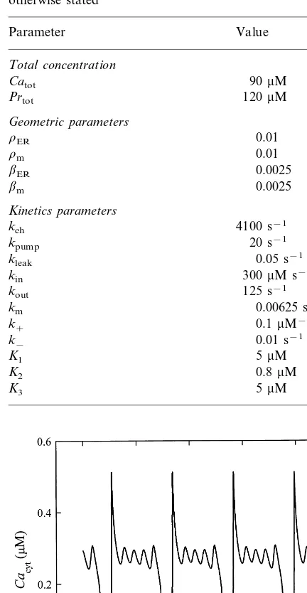
Table 1
Model parameters for which all results are calculated unless otherwise stated
Value Parameter
Total concentration
90mM
Catot
120mM
Prtot
Geometric parameters
0.01 rER
0.01 rm
bER 0.0025
0.0025 bm
Kinetics parameters
kch 4100 s−
1
20 s−1
kpump
0.05 s−1
kleak
kin 300mM s−1
125 s−1
kout
0.00625 s−1
km
k+ 0.1mM−1s−1
0.01 s−1
k−
5mM
K1
K2 0.8mM
5mM
K3
simple calcium oscillations to bursting and chaos. Here, we focus on complex calcium oscillations and therefore simple calcium oscillations are not shown. Parameter values, giving the simplest case of bursting are referred to as reference case and are listed in Table 1. It should be noted that all results are calculated for the parameter values given in Table 1 unless otherwise stated.
The results of numerical integration for the reference case are shown in Fig. 2. We plot the cytosolic calcium concentration (Cacyt) versus
time (t) as a characteristic result, which enables a direct comparison with usual experimental mea-surements. Fig. 2 shows a typical example of bursting of intracellular calcium oscillations. The superposition of high frequency oscillations to the basic calcium spikes is well expressed.
A closer look on this reference case allows analysis of the mechanism providing the calcium bursting oscillations. Fig. 3a shows time courses of model variables for one cycle, i.e. between two basic spikes. For a better explanation of the mechanism of bursting we divide the whole cycle into three different phases. In Phase I, the Ca2+
release from the ER is the dominating process. This leads to the rapid increase of cytosolic and mitochondrial calcium. Taking into account the Ca2+ buffering in the mitochondrial matrix (see
factor bmin Eqs. (1) and (5)), we can realise that
the largest fraction of the released calcium (: 80%) is sequestered in the mitochondria, which is in good agreement with recent experimental ob-servations (Babcock and Hille, 1998). At the end of calcium sequestration in mitochondria, Phase I is finished and a slow release of calcium from the mitochondria begins (Phase II). In agreement with experimental observations, calcium release from mitochondria is much slower than its uptake (Bernardi and Petronilli, 1996; Hehl et al., 1996; Bassani et al., 1998; Babcock and Hille, 1998; Falcke et al., 1999). The importance of this slow Ca2+ release is in Ca2+ transferring from
mito-chondria to the cytosolic proteins. This is a long-term process accompanied by much faster Ca2+
exchange between the ER and the cytosolic proteins causing small calcium spikes of bursting oscillations. Therefore, the kinetic properties of the ER channel as well as Ca2+ binding dynamics Fig. 2. Calcium bursting oscillations for the reference case (see
Table 1 for parameter values).
3. Results
of proteins are of crucial importance for the gen-eration of these high-frequency calcium oscilla-tions between the main spikes characterising the bursting oscillations. So, for example, slight changes of binding parameter kon significantly
influence the number of bursts, whereas a more drastic parameter changing results as simple cal-cium oscillations. In this case, the Ca2+
is trans-ferred rather directly from mitochondria to the ER (not shown here). At the end of the bursting phase (Phase II), the silent phase (Phase III) begins, during which the dissociation of the CaPr complex is the major process. The dissociation rate determines the duration of the silent phase.
In analysing the mechanism of calcium bursting
oscillations, a picture of the limit cycle is also very Fig. 4. Two-folded limit cycle of Ca2+ bursting oscillations.
Parameter values differing from those listed in Table 1,kch=
4000 s−1.
Fig. 3. Analysis of bursting oscillations for the reference case. (a) Time courses ofCacyt,CaER,CamandCaPrfor one cycle.
The concentration of unbounded Ca2+ binding sites (Pr) is
simply related withCaPr(Eq. (2)) and not shown explicitly; (b) Limit cycle in 3D-phase space ofCacyt,CaERandCam(in
mM).
helpful. The limit cycle for the reference case is shown in 3D-phase diagram of Fig. 3b. Note, that due to conservation relations (Eqs. (1) and (2)) our system is actually a 3D one. In particular, Fig. 3b gives good insight into the origin of superposed high frequency oscillations. These os-cillations can be recognised by the vertical spiral, which is the consequence of a slow decline of mitochondrial calcium accompanied by the fast cytosolic exchange of calcium between the ER and the cytosolic proteins.
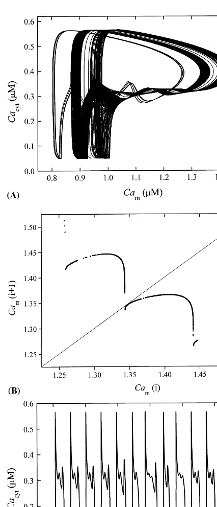
For the above discussion, we have chosen a set of model parameters that leads to simple limit cycle oscillations. In other regions of the channel permeability, kch, more complex behaviour is
ob-served. Fig. 4 shows an example of a 2-fold limit cycle in phase planeCacytversusCam. In this case,
we have bursting oscillations with two different patterns exchanging periodically in time.
Although a more detailed analysis of the dy-namic behaviour of the system in different regions of the parameter space is left for a sequel paper, it should be mentioned that period doubling, shown in Fig. 4, represents a typical transition to chaos. To demonstrate an example of chaotic behaviour of our system, we show the phase plane plot Cacyt
versus Cam for kch=2950 s−1 in Fig. 5a. The
succes-sive maxima of Cam are plotted against their
predecessors. If these maxima are plotted for an infinitely long time series, a continuous curve is obtained. Two important characteristics of the chaotic behaviour in our model should be pointed out. First, note that this is an example of chaotic bursting and second, the amplitudes of main cal-cium spikes remain nearly constant all the time. This is convincingly demonstrated in the time course of cytosolic calcium depicted in Fig. 5c.
4. Discussion
In the paper a possible mechanism explaining complex calcium oscillations is presented, which gives a new complementary aspect to the existing theoretical studies of complex intracellular cal-cium oscillations (Shen and Larter, 1995; Bor-ghans et al., 1997; Houart et al., 1999; Olsen et al., 1999). We show that complex calcium oscilla-tions arise as a consequence of Ca2+ exchanging
between the cytosol and three different intracellu-lar calcium stores: the ER, mitochondria and cytosolic proteins. For Ca2+ exchange between
the cytosol and a particular calcium store, simple plausible rates are used. We take into account the CICR from the ER, the ATP-ase active transport into the ER, the uniporter Ca2+ uptake by
mito-chondria, slow release from the mitochondria and Ca2+ binding to the cytosolic proteins. With the
model, we get simple calcium oscillations, burst-ing and chaos. Moreover, the chaotic behaviour of calcium bursting oscillations is obtained. All model results are in agreement with known exper-imental observations. The cytosolic calcium tran-sients have a typical spike-like form, reasonable frequencies and amplitudes. We emphasise that the amplitudes of main calcium spikes remain nearly constant in the whole range of the oscilla-tory regime. It is valid for all types of calcium oscillations, i.e. for simple calcium oscillations, for regular bursting, as well as for chaotic burst-ing calcium oscillations. As explained in our pre-vious publication (Marhl et al., 1998a), the constancy of amplitudes of calcium oscillations is a consequence of the specific mitochondrial kinetics.
Fig. 5. Chaotic behaviour of Ca2+ bursting oscillations: (a)
projection of trajectory in phase planeCacyt versusCam; (b)
return map for Cam (inmM); and (c) time course ofCacyt.
Parameter values differing from those listed in Table 1,kch=
The functioning of the intracellular calcium stores considered in the model agrees with known experimental observations. The fast calcium re-lease from the ER, characterised by the CICR kinetics, is confirmed by many experiments and used in almost all mathematical models for cal-cium oscillations (cf. Goldbeter, 1996; Heinrich and Schuster, 1996). Calcium binding to cytosolic proteins was also well studied experimentally. A short review of these works and their importance for calcium oscillations is given in our earlier publications (Schuster et al., 1998; Marhl et al., 1998b). The inclusion of mitochondria into the modelling of calcium oscillations is also motivated by many experimental works showing the impor-tance of mitochondria in cell signalling (Jouaville et al., 1995; Hehl et al., 1996; Babcock et al., 1997; Golovina and Blaustein, 1997; Babcock and Hille, 1998; Jouaville et al., 1998; Ricken et al. 1998; Simpson and Russell 1998a,b; Simpson et al., 1998; Rizzuto et al., 1998; Drummond and Tuft, 1999). It becomes clear that mitochondria actively sequester the Ca2+ released from the ER
(Hehl et al., 1996; Babcock et al., 1997; Applegate et al., 1997; Simpson and Russell, 1998a; Rizzuto et al., 1998; Drummond and Tuft, 1999). Some recent experiments also predict more accurate val-ues, namely that :80% of calcium released from the ER is cleared first into the mitochondria (Babcock and Hille, 1998). Our results confirm this value with surprisingly high accuracy.
In the model we show that, in addition to the fast Ca2+ uptake by mitochondria, a very slow
Ca2+ release back to the cytosol is of crucial
importance for understanding the mechanism of complex calcium oscillations. This calcium kinet-ics is in agreement with known experimental evi-dences showing that under normal physiological conditions Ca2+ efflux from the mitochondria is
much slower than Ca2+ uptake by uniporters
(Hehl et al., 1996; Bernardi and Petronilli, 1996; Babcock et al., 1997; Bassani et al., 1998; Verkhratcky and Petersen, 1998; Falcke et al., 1999). In our previous study (Marhl et al., 1998a), this calcium release from mitochondria was mod-elled explicitly by taking into account only the Na+/Ca2+ exchangers and no PTPs. Recent
ex-perimental results show that at physiological
con-ditions of normal cellular functioning the Ca2+
release occur via the Na+
/Ca2+ exchangers rather
than via the PTPs (Simpson and Russell, 1998b; Svichar et al., 1999; Eriksson et al., 1999).
However, since Ichas and collaborators have shown that PTPs posses the mCICR kinetics (Ichas et al., 1994a,b, 1997; Jouaville et al., 1998), which could serve as a calcium spike modulator, in this study of complex calcium oscillations we include also a mCICR-like kinetic of the PTPs in the model, whereas we consider just transient openings of the PTPs in a very low-conductance state. This is in agreement with the evidence that for PTPs several subconductance states are pro-posed (Simpson and Russell, 1998a; Eriksson et al., 1999). Although little experimental evidence exists about their regulation (Eriksson et al., 1999), it is well believed that at normal physiolog-ical conditions the PTPs should not be opened with large conductivities (Simpson and Russell, 1998a,b). Note that huge openings of the PTPs do not cause only the release of large quantities of matrix calcium but also activate a release of cell death factors from the mitochondrial matrix. There are several examples of different mitochon-drial matrix proteins or some intermembrane mi-tochondrial proteins such as cytochrome c, to be released from mitochondria via the PTPs and they appear to act as a direct activator of apoptosis (Simpson and Russell, 1998a). Larger openings of PTPs with consequent fast electrical potential de-polarisations are rather specific for individual mi-tochondria experiments and less specific in the case of mitochondrial populations, such as our case in the neighbourhood of the ER Ca-channels sites (Hu¨ser et al., 1998).
Model predictions show that at the very low conductivity of PTPs considered in the model, the specific mCICR-like kinetics of calcium efflux from the mitochondria seems to be of no crucial importance for complex calcium oscillations. Thus, we get complex calcium oscillations for a simple linear type of Ca2+ efflux from the
mito-chondria as well (not shown here, but the reader can check this by exchanging the expressionCacyt
2
ex-periments for excitable as well as for non-ex-citable cells, showing that PTPs do not partici-pate in the normal physiological regulation of calcium signals (Eriksson et al., 1999; Svichar et al., 1999). Additionally, in accordance to some experimental observations showing that PTPs may not be present in all mitochondria (Simp-son and Russell, 1998a), our model could even be seen as a yet more general one, giving an explanation of complex calcium oscillations in a larger variety of different cell types.
In future studies of the basic mechanism providing complex calcium oscillations, it will be important to focus on more detailed description of the intracellular Ca2+ stores considered,
rather than on attempts to include other Ca2+
stores. On the basis of known experimental re-sults, an inclusion of further Ca2+ stores seems
to be of no special importance for explaining the basic mechanism providing complex calcium oscillations. For example, it has recently been shown that changes in nucleus Ca2+
concentra-tion are very small in comparison to the other intracellular calcium stores (Xiong and Ruben, 1998). Additionally, the Ca2+ concentration in
nucleus changes simply concomitantly with the transient elevations of the cytosolic calcium. Therefore, from the point of view of mathemati-cal modelling, the nucleus, in responding to mathemati- cal-cium signals, plays a role similar to that played by signalling proteins (Marhl et al., 1998a,b), which have been excluded from the present model.
References
Applegate, T.L., Karjalainen, A., Bygrave, F.L., 1997. Rapid Ca2+ influx induced by the action of
dibutylhy-droquinone and glucagon in the perfused rat liver. Biochem. J. 323, 463 – 467.
Babcock, D.F., Hille, B., 1998. Mitochondrial oversight of cellular Ca2+ signaling. Curr. Opin. Neurobiol. 8, 398 –
404.
Babcock, D.F., Herrington, J., Goodwin, P.C., Park, Y.B., Hille, B., 1997. Mitochondrial participation in the intra-cellular Ca2+ network. J. Cell Biol. 136, 833 – 844.
Bassani, R.A., Fagian, M.M., Bassani, J.W.M., Vercesi, A.E., 1998. Changes in calcium uptake rate by rat car-diac mitochondria during postnatal development. J. Mol. Cell. Cardiol. 30, 2013 – 2023.
Bernardi, P., Petronilli, V., 1996. The permeability transition pore as a mitochondrial calcium release channel: A criti-cal appraisal. J. Bioen. Biomemb. 28, 131 – 137.
Berridge, M., Lipp, P., Bootman, M., 1999. Calcium sig-nalling. Curr. Biol. 9, R157 – R159.
Borghans, J.A.M., Dupont, G., Goldbeter, A., 1997. Com-plex intracellular calcium oscillations. A theoretical ex-ploration of possible mechanisms. Biophys. Chem. 66, 25 – 41.
Chay, T.R., 1996. Electrical bursting and luminal calcium oscillation in excitable cell models. Biol. Cybern. 75, 419 – 431.
Chay, T.R., 1997. Effects of extracellular calcium on electri-cal bursting and intracellular and luminal electri-calcium oscilla-tions in insulin secreting pancreatic b-cells. Biophys. J. 73, 1673 – 1688.
Cuthbertson, K.S.R., Cobbold, P.H., 1985. Phorbol ester and sperm activate mouse oocytes by inducing sustained oscillations in cell Ca2+. Nature 316, 541 – 542.
Drummond, R.M., Fay, F.S., 1996. Mitochondria contribute to Ca2+ removal in smooth muscle cells. Pflu¨gers Arch.
Eur. J. Physiol. 431, 473 – 482.
Drummond, R.M., Tuft, R.A., 1999. Release of Ca2+ from
the sarcoplasmic reticulum increases mitochondrial [Ca2+
] in rat pulmonary artery smooth muscle cells. J. Physiol. 516, 139 – 147.
Dupont, G., Goldbeter, A., 1993. One-pool model for Ca2+
oscillations involving Ca2+ and inositol
1,4,5-trisphos-phate as co-agonist for Ca2+ release. Cell Calc. 14, 311 –
322.
Eriksson, O., Pollesello, P., Geimonen, E., 1999. Regulation of total mitochondrial Ca2+in perfused liver is
indepen-dent of the permeability transition pore. Am. J. Physiol. 276, C1297 – C1302 Cell. Physiol. 45.
Falcke, M., Hudson, J.L., Camacho, P., Lechleiter, J.D., 1999. Impact of mitochondrial Ca2+ cycling on pattern
formation and stability. Biophys. J. 77, 37 – 44.
Goldbeter, A., 1996. Biochemical Oscillations and Cellular Rhythms. Cambridge University Press, Cambridge, UK. Goldbeter, A., Dupont, G., Berridge, M.J., 1990. Minimal
model for signal-induced Ca2+ oscillations and for their
frequency encoding through protein phosphorylation. Proc. Natl. Acad. Sci. USA 87, 1461 – 1465.
Golovina, V.A., Blaustein, M.P., 1997. Spatially and func-tionally distinct Ca2+ stores in sarcoplasmic and
endo-plasmic reticulum. Science 275, 1643 – 1648.
Hehl, S., Golard, A., Hille, B., 1996. Involvement of mito-chondria in intracellular calcium sequestration by rat go-nadotropes. Cell Calc. 20, 515 – 524.
Heinrich, R., Schuster, S., 1996. The Regulation of Cellular Systems. Chapman Hall, New York.
Herrington, J., Park, Y.B., Babcock, D.F., Hille, B., 1996. Dominant role of mitochondria in clearance of large Ca2+loads from rat adrenal chromatin cells. Neuron 16,
Ho¨fer, T., 1999. Model of intracellular calcium oscillations in hepatocytes: Synchronisation of heterogeneous cells. Bio-phys. J. 77, 1244 – 1256.
Hoth, M., Fanger, C.M., Lewis, R.S., 1997. Mitochondrial regulation of store-operated calcium signalling in T lymphocytes. J. Cell Biol. 137, 633 – 648.
Houart, G., Dupont, G., Goldbeter, A., 1999. Bursting, chaos and birhythmicity originating from self-modulation of the inositol 1,4,5-trisphosphate signal in a model for intracellu-lar Ca2+oscillations. Bull. Math. Biol. 61, 507 – 530.
Hu¨ser, J., Rechenmacher, C.E., Blatter, L.A., 1998. Imaging the permeability pore transition in single mitochondria. Biophy. J. 74, 2129 – 2137.
Ichas, F., Jouaville, L.S., Sidash, S.S., Mazat, J.-P., Hol-muhamedov, E.L., 1994a. Mitochondrial calcium spiking: a transduction mechanism based on calcium-induced per-meability transition involved in cell calcium signalling. FEBS Letts. 348, 211 – 215.
Ichas, F., Jouaville, L.S., Sidash, S.S., Mazat, J.-P., Hol-muhamedov, E.L., 1994b. Mitochondrial calcium spiking: the physiological face of permeability transition? In: Gnaiger, E., et al. (Eds.), Modern Trends in BioThermoKi-netics 3. Insbruck University Press, Insbruck.
Ichas, F., Jouaville, L.S., Mazat, J.-P., 1997. Mitochondria are excitable organelles capable of generating and conveying electrical and calcium signals. Cell 89, 1145 – 1153. Jafri, M.S., Gillo, B., 1994. A membrane potential model with
counterions for cytosolic calcium oscillations. Cell Calc. 16, 9 – 19.
Jafri, M.S., Keizer, J., 1997. Agonist-induced calcium waves in oscillatory cells: A biological example of Burgers’ equa-tions. Bull. Math. Biol. 59, 1125 – 1144.
Jafri, M.S., Vajda, S., Pasik, P., Gillo, B., 1992. A membrane model for cytosolic calcium oscillations. A study using
Xenopus oocytes. Biophys. J. 63, 235 – 246.
Jouaville, L.S., Ichas, F., Holmuhamedov, E.L., Camacho, P., Lechleiter, J.D., 1995. Synchronization of calcium waves by mitochondrial substrates inXenopus lea6is oocytes. Na-ture 377, 438 – 441.
Jouaville, L.S., Ichas, F., Mazat, J.-P., 1998. Modulation of cell calcium signals by mitochondria. Mol. Cell. Biochem. 184, 371 – 376.
Li, Y.-X., Rinzel, J., 1994. Equations for InsP3
receptor-medi-ated [Ca2+]
i oscillations derived from a detailed kinetic
model: A Hudgkin – Huxley like formalism. J. Theor. Biol. 166, 461 – 473.
Li, Y.-X., Keizer, J., Stojilkovic´, S.S., Rinzel, J., 1995. Ca2+
excitability of the ER membrane: an explanation for IP3
-induced Ca2+ oscillations. Am. J. Physiol. 269, C1079 –
C1092 Cell. Physiol. 38.
Magnus, G., Keizer, J., 1997. Minimal model ofb-cell mito-chondrial Ca2+handling. Am. J. Physiol. 273, C717 – C733
Cell Physiol. 42.
Marhl, M., Schuster, S., Brumen, M., Heinrich, R., 1997. Modelling the interrelations between calcium oscillations
and ER membrane potential oscillations. Biophys. Chem. 63, 221 – 239.
Marhl, M., Schuster, S., Brumen, M., 1998a. Mitochondria as an important factor in the maintenance of constant ampli-tudes of cytosolic calcium oscillations. Biophys. Chem. 71, 125 – 132.
Marhl, M., Schuster, S., Brumen, M., Heinrich, R., 1998b. Modelling oscillations of calcium and endoplasmic reticu-lum transmembrane potential. Role of signalling and buffering proteins and of the size of the Ca2+sequestering
ER subcompartments. Bioelectrochem. Bioener. 46, 79 – 90. Meyer, T., Stryer, L., 1988. Molecular model for receptor-stimulated calcium spiking. Proc. Natl. Acad. Sci. USA 85, 5051 – 5055.
Olsen, L.F., Kummer, U., Green, A.K., Dixon, C.J., Hauser, M.J.B., 1999. Dynamics and information processing in biochemical pathways. In: Bornberg-Bauer, E., et al. (Eds.), Proceedings Workshop on Computation of Bio-chemical Pathways and Genetic Networks. Heidelberg. Ricken, S., Leipziger, J., Greger, R., Nitschke, R., 1998.
Simultaneous measurements of cytosolic and mitochon-drial Ca2+ transients in HT
29 cells. J. Biol. Chem. 273,
34961 – 34969.
Rizzuto, R., Pinton, P., Carrington, W., Fay, F.S., Fogarty, K.E., Lifshitz, L.M., Tuft, R.A., Pozzan, T., 1998. Close contact with the endoplasmic reticulum as determinants of mitochondrial Ca2+responses. Science 280, 1763 – 1766.
Schuster, S., Marhl, M., Brumen, M., Heinrich, R., 1998. Influence of calcium binding proteins on calcium oscilla-tions and ER membrane potential oscillaoscilla-tions. A mathe-matical model. In: Paton, R.C., Holcombe, M. (Eds.), Information Processing in Cells and Tissues. Plenum Press, New York.
Selivanov, V.A., Ichas, F., Holmuhamedov, E.L., Jouaville, L.S., Evtodienko, Y.V., Mazat, J.-P., 1998. A model of mitochondrial Ca2+-induced Ca2+ release simulating the
Ca2+ oscillations and spikes generated by mitochondria.
Biophys. Chem. 72, 111 – 121.
Shen, P., Larter, R., 1995. Chaos in intracellular Ca2+
oscilla-tions in a new model for non-excitable cells. Cell Calc. 17, 225 – 232.
Simpson, P.B., Russell, J.T., 1998a. Role of mitochondrial Ca2+regulation in neuronal and glia cell signalling. Brain
Res. Rev. 26, 72 – 81.
Simpson, P.B., Russell, J.T., 1998b. Mitochondrial Ca2+
up-take and release influence metabotropic and ionotropic cytosolic Ca2+ responses in rat oligodendrocyte
progeni-tors. J. Physiol. 508, 413 – 426.
Simpson, P.B., Mehotra, S., Langley, D., Sheppard, C.A., Russell, J.T., 1998. Specialised distributions of mitochon-dria and endoplasmic reticulum proteins define Ca2+wave
amplification sites in cultured astrocytes. J. Neurosci. Res. 52, 672 – 683.
Smith, G.D., Lee, R.J., Oliver, J.M., Keizer, J., 1996. Effect of Ca2+ influx on intracellular free Ca2+ responses in
Somogyi, R., Stucki, J.W., 1991. Hormone-induced calcium oscillations in liver cells can be explained by a simple one pool model. J. Biol. Chem. 266, 11068 – 11077.
Spacek, J., Harris, K.M., 1997. Three-dimensional organiza-tion of smooth endoplasmic reticulum in hippocampal CA1 dendrites and dendritic spines of the immature and mature rat. J. Neurosci. 17, 190 – 203.
Svichar, N., Shishkin, V., Kostyuk, P., 1999. Mitochondrial participation in the modulation of calcium transients in DRG neurons. NeuroReport 10, 1257 – 1261.
Verkhratcky, A.J., Petersen, O.H., 1998. Neuronal calcium stores. Cell Calc. 24, 333 – 343.
Vries, G., 1998. Multiple bifurcations in a polynomal model of bursting oscillations. J. Nonlinear Sci. 8, 281 – 316. Woods, N.M., Cuthbertson, K.S.R., Cobbold, P.H., 1986.
Repetitive transient rises in cytoplasmic free calcium in hormone-stimulated hepatocytes. Nature 319, 600 – 602. Xiong, Z.-H., Ruben, L., 1998. Trypanosoma brucei: The
dynamics of calcium movement between the cytosol, nu-cleus, and mitochondrion of intact cells. Exper. Parasitol. 88, 231 – 239.