Rhesus Monkeys
James K. Rilling, James T. Winslow, Derek O’Brien, David A. Gutman,
John M. Hoffman, and Clinton D. Kilts
Background: The neurobiological basis of stress and anxiety in primates remains poorly understood. In this study, we examined the neural response to a naturalistic social stressor: maternal separation. We used rhesus monkeys as an animal model because of their close phylogenetic affinity with humans.
Methods: Six juvenile rhesus monkeys received [18 F]-fluorodeoxyglucose positron emission tomography scans following 1) a period together with their mothers and again after separation from their mothers 2) with or 3) without visual contact. Image subtraction revealed brain regions that exhibited altered activity during separation. In addition, plasma cortisol concentrations obtained fol-lowing each condition were tested for correlations with regional brain activity.
Results: Maternal separation activated the right dorso-lateral prefrontal cortex and the right ventral temporal/ occipital lobe. There was also decreased activity in left dorsolateral prefrontal cortex associated with separation stress. Correlational analyses demonstrated these acti-vated and deactiacti-vated regions to be positively and nega-tively correlated with cortisol, respecnega-tively. Additionally, correlational analyses revealed cortisol-related activation in brainstem areas previously implicated in stress and anxiety.
Conclusions: In juvenile rhesus monkeys, the stress of maternal separation is associated with activation in the right dorsolateral prefrontal cortex and ventral temporal/ occipital lobes and decreased activity in the left dorsolat-eral prefrontal cortex. Biol Psychiatry 2001;49:146 –157 ©2001 Society of Biological Psychiatry
Key Words: Positron emission tomography, monkeys, stress, anxiety, cortisol, prefrontal cortex
Introduction
C
urrent knowledge of the neurobiological basis of stress and anxiety in primates is largely based on results from human neuroimaging studies (De Cristofaro et al 1993; Gottschalk et al 1991, 1992; Kimbrell et al 1999; Mountz et al 1989; Nordahl et al 1990; Rauch et al 1996, 1997; Reiman et al 1989; Stapleton et al 1997; Wu et al 1991; Zohar et al 1989) and generalizations from rodent studies (Davis 1997; Ladd et al 2000; Steckler and Holsboer 1999). Most of the human studies involve symptom provocation in patients with anxiety disorders, either by exposure to anxiety-provoking stimuli or mem-ories or by pharmacologic challenge (e.g., lactate). Rodent studies of stress neurobiology offer two advantages over human studies. First, they permit investigation of more naturalistic social stressors (e.g., maternal separation) that reliably provoke anxiety in all members of the species, rather than only those with psychopathology. Second, the brain regions and neurotransmitter systems mediating stress and anxiety can be more thoroughly characterized in rodents using invasive methods that cannot be used in humans, such as lesion studies, injection of pharmacologic agents into specific brain regions, and quantification of neurotransmitter receptor density and distribution in post-mortem tissue. The disadvantage of rodent studies is that the rodent brain is, of course, only a model of the human brain, and a phylogenetically quite distant one at that. It is quite possible that the neural processing of stress differs in humans and rodents. Therefore, in this study we charac-terized the neural stress response of a much nearer phylogenetic relative: a member of our own suborder anthropoidea, the rhesus monkey. Specifically, we inves-tigated the neural response of juvenile monkeys to brief separation from their mother, a stressor that is more social, naturalistic, and species typical than those typically em-ployed in the human symptom provocation studies.The mother–infant bond is the strongest, most enduring social attachment formed throughout life in most mam-mals (Bowlby 1982). Thus, it is not surprising that disruption of that bond produces profound behavioral and physiologic responses that reflect its efficacy as a
psycho-From the Department of Psychiatry and Behavioral Sciences (JKR, JTW, DO, DAG, CDK), Yerkes Regional Primate Research Center (JKR, JTW), and Emory Center for PET (JMH, CDK), Emory University, Atlanta, Georgia. Address reprint requests to James K. Rilling, Ph.D., Emory University, Department
of Psychiatry and Behavioral Sciences, 1639 Pierce Drive, Suite 4000 Wood-ruff Memorial Research Bldg., Atlanta GA 30322.
Received March 9, 2000; revised June 7, 2000; accepted June 29, 2000.
© 2001 Society of Biological Psychiatry 0006-3223/01/$20.00
logical stressor. For example, separation of infant mon-keys from their mothers provokes an increase in circulat-ing cortisol and the onset of distress calls (Levine et al 1985). We used positron emission tomography (PET) neuroimaging of [18F]-fluorodeoxyglucose ([18F]-FDG) uptake to describe changes in regional cerebral glucose metabolism that accompany this psychologically stressful experience. Based on electroencephalogram (EEG) studies conducted in both rhesus monkeys (Kalin et al 1998) and human children (Davidson 1992; Dawson et al 1992), we predicted right frontal lobe activation in response to maternal separation. Our objective in using PET imaging was to take advantage of its superior spatial resolution to define more specifically the anatomical location of anxi-ety/stress-related activity within the right frontal lobe. Furthermore, we predicted that right frontal lobe activity would be correlated with a physiologic stress marker, the hormone cortisol.
Methods and Materials
Subjects
Subjects were 6 male rhesus monkeys between 16 and 25 months of age that had been living with their mothers since birth in a large social group at the Yerkes Field Station. Subjects were studied using a protocol approved by the Animal Care and Use Committee of Emory University.
Study Design
Each subject received an [18F]-FDG PET scan following
expo-sure to each of three conditions so that a total of 18 PET scans (6 subjects33 scans/subject) were acquired. In each condition, the juvenile was first briefly separated from its mother for intramus-cular (IM) injection of [18F]-FDG. In one condition, he was then
immediately reunited with his mother. In the other two, he remained separated from her, either with or without visual and auditory contact. Data were collected in two batches, each consisting of three mother–juvenile pairs. In each batch, PET scans were collected on three separate days. Each day, all three juveniles were scanned in succession, with the first scan begin-ning between 9:00 and 10:00AMand with approximately 2 hour intervals between scans. On a given scanning date, each monkey was scanned in a different condition according to a modified Latin Square Design. For the first batch, the three scanning dates were spaced at 1-week intervals. For the second batch, scans one and two were separated by 4 weeks (because of scheduling conflicts) and scans two and three were separated by 1 week. The half-life of 18F (110 min) is short enough to ensure that
radioactivity from a subject’s prior scan had decayed to unde-tectable levels by the time of his next.
Pilot investigations in three conscious rhesus monkeys re-vealed brain uptake of [18F]-FDG following an IM injection to be
approximately linear between 0 and 30 (monkey no. 1) or 40 (monkey no. 2 and no. 3) min, and to be at least 80% of maximum within 40 min (Figure 1).
Based on these data, 40 min following [18F]-FDG IM injection
was chosen as an appropriate duration for the behavioral session before PET imaging to detect neural activity-dependent patterns of glucose fixation. After neuronal transport, [18F]-FDG is
phosphorylated and then trapped inside the neuron (Sokoloff et al 1977), with its accumulation dependent on neuronal activity.
Images obtained in the three conditions were statistically compared with one another to identify brain regions that dem-onstrated evidence of altered neural activity related to maternal separation. Detailed methods are provided below.
Behavioral and Imaging Procedures
Mother–juvenile dyads were transported from the Yerkes Field Station to the Yerkes Main Station and housed together in standard caging for 2 weeks before testing. They were
tained on a 12-hour light/dark cycle (lights on at 7:00AM) and fed monkey chow ad libitum. During this 2-week period, dyads were acclimated daily to squeeze-restraint, capture, and brief housing (30 min) in a standard transport cage within their colony room.
Twenty-four hours before imaging, dyads were transported to a room in close proximity with the Emory Center for PET and housed in standard caging. All subjects were food-deprived for 18 hours before imaging.
Forty-five minutes before sedation for imaging, individual juvenile monkeys were captured, restrained, and injected IM (gluteus maximus) with 6 –10 mCi of [18F]-FDG. Immediately
after injection, each monkey was placed in one of three test conditions (described below) for 40 min. Afterwards, subjects were restrained and injected IM with 3–5 mg/kg telazol, and a blood sample was collected from a femoral vein within 5 min of restraint for subsequent cortisol determinations.
Each subject was then immediately transported to the PET facility, where they were intubated and anesthesia was main-tained with 1% isoflurane inhalation and ventilator support. An infrared pulse oximeter was affixed to the left foot to monitor heart rate and blood oxygen saturation. The animal was fitted with a thermoplastic face mask to minimize head movement and then aligned in the PET scanner. The mean time elapsed from [18F]-FDG injection until the start of the PET scan was 77.8 min
(SE513.8,n518). Regional cerebral glucose metabolism was estimated by [18F]-FDG uptake via measurement of brain
radio-activity with an ECAT 951 PET scanner (Siemens, Knoxville, TS). Images were acquired as a single 25-min frame study in two-dimensional acquisition mode. Emission data were recon-structed as 31 axial planes spaced at 3.375 mm intervals by filtered back-projection with calculated attenuation correction. Reconstructed image spatial resolution was 11 mm full width at half maximum (FWHM) in all directions.
Following imaging and recovery from anesthesia, the subjects were returned with their mothers to their home cage at the Yerkes Main Station.
Study Conditions
The schedule of conditions for each subject was systematically varied according to a modified Latin Square Design.
TOGETHER. Following administration of [18F]-FDG, each
subject and his mother were placed in a transport cage equipped with a clear lexan door in a testing room adjacent to the housing room. Motor activity and interactions between mother and offspring were videotaped and subsequently scored (see below) by trained observers.
NONVISUAL (SEPARATION WITHOUT VISUAL OR AUDI-TORY CONTACT). Following administration of [18F]-FDG,
each juvenile was placed in a transport cage equipped with a clear lexan door in a testing room adjacent to the housing room. The cage also had a stationary plastic box that occupied approx-imately the same volume as an adult female monkey. Mothers were returned to the housing room. Behavior of each juvenile was videotaped and subsequently scored by trained observers.
VISUAL (SEPARATION WITH VISUAL AND AUDITORY
CONTACT). In this condition, each subject’s mother was
placed in a cage directly in front of and slightly above the transport cage holding the juvenile. This arrangement permitted unobstructed visual contact between mother and offspring. The juvenile’s cage also had a stationary plastic box that occupied approximately the same volume as an adult female monkey. Behavior of each juvenile was videotaped and subsequently scored by trained observers.
Behavioral Measurements
Videotaped sessions were scored for frequencies or durations of the following behaviors: affiliation between mother and juvenile, maternal aggression, and juvenile head turns. These behaviors were operationalized as follows.
AFFILIATION BETWEEN MOTHER AND JUVENILE.
Be-haviors including huddling (monkeys maintain sustained gross body contact, usually sitting together, leaning against or clinging to each other), allogrooming (spreads and pick through the fur of another animal), and cling-huddle (maintain bodily contact with another animal, usually accompanied by some form of clasping or embracing). The maternal–juvenile affiliation score is the number of occurrences of all of these behaviors combined throughout the 40-min together condition.
MATERNAL AGGRESSION. Behaviors including bite or hit,
threat vocalization, open-jawed threat, stare threat. The maternal aggression score is the amount of time subjects were engaged in these behaviors throughout the 40-min together condition.
JUVENILE HEAD-TURNS. Shifts in gaze, with or without head movement, greater than or equal to approximately 30 degrees. The juvenile head-turn score is the number of recorded head-turns throughout the 40-min behavioral condition.
Magnetic Resonance Imaging Scans
Cortisol Assay
Plasma cortisol was quantitated by radioimmunoassay using commercially available reagents (Diagnostic Products Corpora-tion, Los Angeles, CA) as described previously (Lovejoy and Wallen 1990). The limit of detection is 0.5mg/dL, and intra- and inter-assay coefficients of variation were less than 4% and 7.5%, respectively (n525 assays).
Image Analysis
SPATIAL REGISTRATION. Each of the 18 PET scans (6 subjects3 3 conditions) was spatially registered to a juvenile rhesus MRI brain standard using Automated Image Registration software. The standard was constructed by averaging the indi-vidual MRIs acquired from each of the six subjects (Woods et al 1998). For anatomical standardization, each individual MRI was registered to the MRI standard using a 12-parameter affine linear model (Woods et al 1998), and the three PET scans from each subject were registered to that individual’s MRI scan (Woods et al 1993). These two transformations were combined to register each PET scan to the MRI standard. Although the inter-subject registration algorithm has not been specifically validated for monkey brains, the similarity in human and monkey brain anatomy combined with careful visual comparisons of registered brains with the standard supports their use with monkey brains. Registered PET images were normalized to mean whole-brain activity to control for differences in the injected dose of [18
F]-FDG and for changes in global brain activity.
PAIRWISE IMAGE CONTRASTS. Positron emission
tomog-raphy data from one of the six juvenile monkeys was excluded from image subtractions, because both behavioral and physio-logic (i.e., cortisol) evidence indicated that he was highly stressed in the together or low-anxiety condition (see Results). Therefore, this image analysis was based on data from 15 scans (5 subjects 3 3 conditions). A two-way analysis of variance (ANOVA) model was used to identify voxels that showed increased activity in the separation conditions compared with the together condition. The two factors were subject and condition. A t-map image of the contrast between the separation and control conditions was calculated on a voxel-by-voxel basis. Significant sites of activation were defined by a selected threshold of the
t-statistic [p(4), .01], andp values were uncorrected for the number of multiple comparisons. Where possible, cytoarchitec-tonic areas as described by Walker (1940) for the rhesus monkey frontal lobe were assigned to the activated areas. These areas are referred to as “WA” for Walker’s area.
CORRELATIONAL ANALYSES. In addition to the image
contrast analyses, correlational analyses were used to identify brain regions where activity was either positively or negatively correlated with plasma cortisol concentrations or behavioral covariates. All 18 PET scans were used in these analyses. Because each subject was scanned three times, these 18 scans cannot be considered as 18 independent data observations. Therefore, correlations were examined either within or across subjects, rather than across the entire sample of 18 data points.
For cortisol, within-subject correlations identified brain regions where activity increased or decreased as cortisol levels changed across conditions. A brain-wide, voxel-by-voxel analysis was conducted in which cortisol values were regressed on brain activity separately for each of the six subjects. If the mean of the six regression slopes was significantly different from zero as assessed by a t-test, then that voxel was identified as being significantly correlated with cortisol.
The behavioral data of interest were measures of maternal– juvenile interactions and so were only observable in the together condition. For these behaviors, the objective was to determine if interactive behavior scaled to neural activity for any voxel in this condition. Thus, we tested for a significant correlation across the six subjects in the together condition. For affiliation, we also asked if interactive behavior in the together condition scaled to brain activity in the separation conditions. That is, we asked if subjects from more affiliative pairs show increased regional neural activity in all conditions (i.e., trait-related activation), or only in particular ones (state-related activation).
All images are presented in standard radiologic format with right and left reversed with respect to the observer’s perspective.
Results
Cortisol
Plasma cortisol concentrations were compared across con-ditions to confirm that the separation concon-ditions (visual and nonvisual) were more stressful than the together condition (Figure 2); however, cortisol levels did not differ by condition (Friedman ANOVAx2(6,2)54.33,p5.11). Inspection of the data revealed that one animal (RJE6; Figure 2,●) had an exceptionally high cortisol value in the together condition. Subsequent review of videotaped mother-offspring interactions revealed that unlike the other animals, this animal was the recipient of substantial
aggression from his mother in the together condition. Because the together condition was designed to be a relatively low-stress condition, this subject was removed from the image contrast analysis and data reanalyzed. For the remaining subjects, cortisol differed by condition [x2(5,2) 57.6,p, .02]. Pairwise comparisons revealed significantly elevated cortisol values in both the visual (Wilcoxon matched pairsz52.02,p,.04) and nonvisual (z52.02,p,.04) conditions compared to together. We also examined the correlation between cortisol levels and mother–juvenile interactions measured within the together condition. Cortisol levels were significantly negatively correlated with the overall number (but not total duration) of cling-huddle incidents received by the juvenile (Spear-manR5 2.82,p5.046). The correlation was somewhat stronger (SpearmanR5 2.87,p5.025) for cortisol and the total number cling-huddles and allogrooming both received and sent by the juvenile. This finding supports the validity of these measures of social contact.
Behavior
Three of the six juveniles coo called in the separation conditions, whereas none of the juveniles called in the together condition. In the animals that called, rates were similar in the two separation conditions, suggesting that the visual and nonvisual separation conditions produced similar affective states. Coo calls can represent either a separation distress call or a contact call and therefore cannot unambiguously be linked with distress or anxiety (Levine et al 1985).
PET Image Contrasts
NONVISUAL VERSUS TOGETHER. For the nonvisual– together contrast, the following areas showed increased activity in the nonvisual condition (Figure 3, yellow regions; Figure 4, top):1) left principal sulcus and inferior frontal gyrus (WA 46); 2) right principal sulcus and middle frontal gyrus (WA 46 and 8); 3) right posterior fusiform, cuneus and lingual gyrus; and 4) inferior right lateral cerebellum. Areas that showed decreased relative
activity (Figure 3, blue regions) include right amygdala, left hypothalamus, bilateral thalamus, medial postcentral gyrus, and right inferior temporal gyrus.
VISUAL VERSUS TOGETHER. For the visual–together contrast, the following areas showed increased activity in the visual condition (Figure 5, yellow regions; Figure 4, bottom):1) right middle frontal gyrus (WA 8); 2) right posterior fusiform, cuneus and lingual gyrus; and 3) right occipital gyrus (V1, V2 and V3). Areas that showed decreased relative activity (Figure 5, blue regions) include 1) left middle and superior frontal gyrus (WA 6 and 8); 2) medial superior frontal gyrus (WA 6); 3) right precentral gyrus (WA 4 and 6); 4) right insula; 5) left precentral gyrus (WA 4); and 6) right hippocampus.
Areas that were activated, relative to the together condition, in both of the above contrasts include 1) right middle frontal gyrus (WA 8) and 2) right posterior fusiform, cuneus and lingual gyrus (Figure 4, magenta regions). No areas were deactivated in both separation conditions compared to the together condition.
Correlational Analyses
BRAIN ACTIVITY AND CORTISOL. Figure 6 illus-trates pixels for which brain activity was positively or negatively correlated with circulating cortisol. Regions where activity was positively correlated (p , .05) with cortisol include 1) right middle frontal gyrus (WA 6 and 8); 2) right medial superior frontal gyrus (WA 8); 3) right posterior fusiform, cuneus and lingual gyri; 4) anterior cerebellar vermis; 5) right lateral cerebellum; 6) left parahippocampal, fusiform and inferior temporal gyri; 7) central gray matter of the midbrain; and 8) extensive regions of both the pons (including the locus coeruleus and raphe nuclei) and the medulla. Regions where activity was negatively correlated with cortisol (p , .05) include 1) bilateral precentral gyrus (motor cortex, WA 6); 2) left middle and inferior frontal gyrus (WA 8 and 46); 3) left anterior cingulate and medial superior frontal gyrus (WA 24 and 8); 4) bilateral lateral orbitofrontal cortex (WA 12);
4™™™™™™™™™™™™™™™™™™™™™™™™™™™™™™™™™™™™™™™™™™™™™™™™™™™™™™™™™™™™™™™™™™™™™™™™™
Figure 3. Nonvisual separation condition vs. together condition. Regions that are more active in the nonvisual compared with the together condition are colored yellow, and those that are less active are colored blue. The magnetic resonance image (MRI) is a standard constructed by averaging each of the six subject’s individual MRIs. Images are presented in standard radiologic format with right and left reversed with respect to the observer’s perspective.
Figure 4. Positron emission tomography activations (but not deactivations) from Figure 3(top)and Figure 5(bottom)displayed on a magnetic resonance imaging three-dimensional reconstruction of the juvenile rhesus monkey brain. Only activations within 4.8 mm of the cortical surface are shown. The areas colored in magenta were activated in both separation conditions (visual and nonvisual).
5) right medial orbitofrontal cortex (WA 13 and 14); 6) left caudate; 7) right superior parietal lobule; 8) right superior temporal sulcus; and 9) right inferior temporal gyrus.
BRAIN ACTIVITY AND MATERNAL–JUVENILE
AFFIL-IATION. In the together condition, we conducted voxel-by-voxel tests for correlations between neural activity and affiliation. This amounts to a six-point correlation with each data point representing one of the six juveniles. We also tested for associations between maternal–juvenile affiliation in the together condition and neural activity in the separation conditions. That is, maternal–juvenile affiliation in the together condition was used as a predictor for neural activity in the separation conditions. For both the separation and together conditions, there were several brain regions where activity was positively correlated with maternal– juvenile affiliation; however, other regions were only correlated with affiliation in the together condition. Most noteworthy among these was the left dorsolateral prefrontal cortex (Figure 7, top). There were also regions where activity was positively correlated with affiliation only in the separation conditions and not in the together condition. Most prominent among these regions were the bilateral temporal poles (Figure 7, bottom).
BRAIN ACTIVITY AND RECEIVED AGGRESSION. Four of the six juveniles received at least some aggression from their mothers in the together condition. Voxel-by-voxel correlational analyses revealed that subjects who received more aggression from their mothers had greater activity in the anterior cerebellar vermis and in the ventral striatum (Figure 8).
Discussion
These data show that maternal separation is associated with increased activity of the right frontal cortex of juvenile rhesus monkeys. Specifically, compared to the together condition, both separation conditions activated the right dorsolateral prefrontal cortex (rdlPFC) in the middle frontal gyrus (WA 8; compare Figures 3 and 5, as well as Figure 4). In these analyses, a less stressful condition (mother present) was subtracted from a more stressful one (mother absent) to identify brain regions involved in psychological stress related to maternal or social separation. An alternative approach searched the brain for areas where activity was positively or negatively correlated with circulating cortisol concentrations. To the extent that cortisol is an accurate marker of an organism’s
level of psychological stress or anxiety, these should represent brain regions that are related to stress.
Like the contrast analyses, correlational analyses identified the right middle frontal gyrus (WA 8) as a stress-related area (Figure 6). That the observed rdlPFC activation is stress-related is supported by human and monkey neuroimaging studies. In rhesus monkeys, fron-tal lobe EEG asymmetries favoring the right hemisphere are more pronounced in subjects with higher cortisol levels who presumably have more anxious tempera-ments (Kalin et al 1998). Whether such asymmetries reflect increased right frontal and/or decreased left frontal activity is not resolved by this study. The relative decrease in left dorsolateral prefrontal (ldlPFC) activity observed in the visual separation condition (Figure 5) spread onto the gyrus (middle frontal gy-rus—WA 8) contralateral to that activated in the right hemisphere. In addition, correlational analyses identi-fied a large region in the ldlPFC where activity was inversely correlated with cortisol concentrations (Figure 6), and this region, in turn, overlaps with the decreased ldlPFC activity observed for the contrast analyses (Figure 5). Thus, there is evidence for both increased rdlPFC activity and decreased left dlPFC activity with increasing stress or anxiety, and this confirms and extends the frontal asymmetry finding of Kalin and colleagues; however, it should be noted that the studies of Kalin and coworkers examined the neural correlates of trait-like anxiety, whereas our own study focuses on the neural response to a social challenge.
The neural correlates of maternal separation have been investigated previously in human infants using EEG. Bilateral increases in frontal lobe activity were reported in response to maternal separation, with larger increases in the right hemisphere (Dawson et al 1992). Also, infants showing more distress in response to maternal separation had greater right frontal and less left frontal resting brain activity than did their less distressed peers (Davidson and Fox 1989), a pattern of activity that might be a correlate of trait-like anxiety. Although our study involves state rather than trait anxiety, the similarity of the activation patterns in the two studies is noteworthy.
and anxiety. The PET findings from this study can be compared with those of PET studies investigating human anxiety. Although right frontal increases in regional cere-bral blood flow (rCBF) are a consistent finding in PET
symptom provocation studies of patients with anxiety disorders (i.e., obsessive-compulsive disorder, simple pho-bia, posttraumatic stress disorder) (Rauch et al 1996), the specific focus of activation within the right frontal lobe is inferior and orbital, not dorsolateral as reported here. Possible explanations for the different location of anxiety-related activation foci within the right frontal lobe include that the psychopathology of the human patients altered their neural response to anxiety, that the studies differed in terms of the nature or efficacy of the anxiety-provoking stimulus, and differences in the functional organization of the human and monkey prefrontal cortex (Brodmann 1909; Preuss 1993).
Figure 6. Voxel-by-voxel correlations between brain activity and plasma cortisol concentrations. For each subject, plasma cortisol concentrations for all three scans were regressed on [18F]-fluorodeoxyglucose– estimated brain activity to calculate a slope relating the
two. Voxels that are positively correlated with cortisol are colored orange (.01,p,.05) or yellow (p,.01) and those that are negatively correlated are colored light blue (.01,p,.05) or dark blue (p,.01).pvalues are based onttests of whether the mean slope of the six subjects differs from zero.
Figure 7. Voxel-by-voxel correlations between brain activity and maternal–juvenile affiliation in each of the three conditions. For each condition, at every voxel, the correlation between brain activity and amount of maternal–juvenile affiliation in the together condition was calculated across the six subjects. Voxels where the correlation coefficient was significantly positive at
p,.05 are colored yellow.(Top)A single axial slice showing the left dorsolateral prefrontal cortex activation to be specific to the together condition.(Bottom)A single axial slice showing the bilateral temporal pole activation to be specific to the separation conditions.
The discrepancy could also relate to differences in the duration over which brain activity is integrated. The IM [18F]-FDG method utilized here integrates the metabolic correlate of synaptic activity over approximately 40 min, whereas the blood flow studies referenced above integrate signals over about 90 sec. That two human [18F]-FDG studies also found increased orbitofrontal metabolism with anxiety (Nordahl et al 1990; Stapleton et al 1997) suggests that these methodological differences may not be the explanation; however, these studies examined trait rather than state anxiety, so that these results might not be strictly comparable with our own. Finally, the blood flow studies referenced above all involve exposure to an anxiety-provoking stimulus, whereas our study induces anxiety by removing a highly valued and comforting resource, the juvenile’s mother. It would not be surprising if these two manipulations produced different mental states and corre-spondingly different brain states.
A second area that was robustly activated in our study was the right posterior fusiform, cuneus and lingual gyrus. We are aware of no precedent for activation in this area related to stress or anxiety; however, the fact that this region was activated in both separation conditions and also that activity in this region was positively correlated with cortisol strongly suggests a link with stress and/or anxiety in male juvenile monkeys.
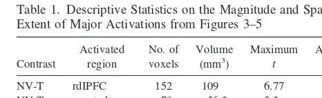
As mentioned in Methods and Materials, the statistical maps presented in Figures 3– 8 are uncorrected for multi-ple comparisons. In view of the large number of statistical tests performed for these contrasts, the results are vulner-able to type I error and falsely reporting an area as activated; however, we believe it is unlikely that the areas emphasized (rdlPFC, right ventral temporal/occipital lobe) represent false positives for the following three reasons:1) separate analyses were conducted for the visual and nonvisual separation conditions, each contrasted to the together condition, and these areas were activated in both contrasts; 2) neural activity in the same areas were significantly positively correlated with the stress hormone cortisol; and 3) the magnitude and spatial extent of the activations suggest that they are unlikely to be due to chance. In support of this third point, we present Table 1, which provides descriptive statistics on the activations observed in the nonvisual and visual separation conditions. The sites of relative activation are large in terms of both spatial extent and magnitude. Our maximumt-values fall well beyond the critical values of at distribution with 4 degrees of freedom (3.75 forp5.01; 4.60 forp5.005). Thus, we consider type I error to be an unlikely explana-tion for these activaexplana-tions.
Our study design involved two different separation conditions in an attempt to vary the magnitude of juvenile distress. Based on earlier studies (Levine et al 1985), we
expected that the nonvisual separation condition would be the most stressful; however, both physiologic (i.e., cortisol concentrations) and behavioral (“coo” calls) data suggest that the two separation conditions were about equally stressful. Therefore, difference images between the two separation conditions are uninformative with respect to the neural correlates of distress/anxiety. The difference be-tween the current and previous studies may depend on our use of juvenile compared to infant monkeys. The offspring used in this study were postweaning age and consequently likely to have very different relationships and dependen-cies on their mothers than preweaning-aged offspring. Indeed, the response of a postweaning-age animal to separation or presence of its mother may be most compa-rable to separation and presence of a peer (Gust et al 1993, 1994, 1996). Future studies will examine potential age-related differences in the response to maternal compared to peer separations in an effort to define neural activation patterns specific to maternal separation and comfort. It is possible that the neural activations reported here relate to stress in a more general sense, rather than to separation stress, specifically. The pattern of neural activity observed in this study will need to be compared with that in response to other stressors to determine its specificity.
Our findings can also be compared with the known functional neuroanatomy of stress and anxiety in rodents. Amygdaloid and hypothalamic corticotropin-releasing factor (CRF) projections to brainstem nuclei appear to be involved in rodent stress and anxiety (Davis 1997; Ladd et al 2000; Steckler and Holsboer 1999). In our study, difference images did not reveal stress-related activation in any of these areas; however, voxel-by-voxel correla-tional analyses with cortisol revealed widespread brain-stem activation that included the midbrain central gray matter, and areas containing the locus coeruleus and raphe nuclei (Figure 6), regions known to receive dense CRF projections in rodents (Price et al 1998; Swanson et al 1983). Because of the limited spatial resolution of our PET images (about 11 mm FWHM), the brainstem activation in Figure 6 could either represent a single activation with a large spatial extent or several indistinguishable smaller activations. Of greater relevance than the distribution of
Table 1. Descriptive Statistics on the Magnitude and Spatial Extent of Major Activations from Figures 3–5
Contrast
NV-T rdIPFC 152 109 6.77 4.47 NV-T rventraltemp 78 56.2 5.2 4.06
V-T rdIPFC 18 13 4.96 4.2
V-T rventraltemp 55 39.6 4.78 3.8
rodent CRF projections is recent evidence that primate brains have moderate levels of CRF1and CRF2receptors in the locus coeruleus and the raphe nuclei, respectively (Sanchez et al 1999).
The lack of amygdala and hypothalamic activation is not surprising, given that rodent 2-deoxyglucose (2-DG) studies have not consistently found increased local cere-bral glucose utilization (LCGU) in these areas with stress (e.g., swim stress, white noise, foot shock; Ableitner and Herz 1987; Caldecott-Hazard et al 1988; Duncan et al 1993, 1996; Justice et al 1989). Given functional MRI evidence that fear-related amygdala activity rapidly atten-uates with time (Breiter et al 1996; LaBar et al 1998; Phelps et al 2000), the absence of amygdala and hypotha-lamic activation may be a consequence of the extended time interval (30 – 45 min) over which neural activity was integrated with both the18F-FDG and 2-DG techniques.
Correlation analyses also revealed prominent cortisol-related activations in the anterior cerebellar vermis, as well as the lateral cerebellum. The primate cerebellar cortex has a very high density of CRF1 receptors (Sanchez et al 1999). Thus, it is possible that CRF is involved in some separation-induced neural activations, though it should be noted that some regions of the primate brain that are rich in CRF receptors (e.g., amygdala and hippocampal forma-tion) were not related to cortisol (Figure 6).
A limitation of this study is that the average plasma cortisol concentration in the together condition was higher than that reported in other studies that examined baseline nonstressed cortisol levels in rhesus monkeys (Kalin et al 1983, 1998; Sanchez et al 1997). The latter are typically between 10 and 30 mg/dL. In our study, group-housed subjects were used to ensure normal social behavior. Because of the risks involved with reintroducing animals to their social groups after a prolonged absence, mother– juvenile pairs could only be removed from their groups for 3 weeks. We suspect that the elevated baseline cortisol concentrations reflect a lack of complete habituation to the experimental procedures used, and so this study may involve comparison of high and moderate stress conditions rather than high and low stress conditions.
If subjects were already moderately stressed in the together condition (as suggested by the cortisol data), then it is possible that areas involved with stress (perhaps the amygdala and hypothalamus) were activated in both the together and the separation conditions but that this activa-tion was subtracted in the difference images. Inspecactiva-tion of raw [18F]-FDG images revealed low levels of activity in the amygdala compared with other brain areas. Normal-ized amygdala activity was quantified and found to be well below the global image mean in both the separation and the together conditions. Despite this evidence, it may be premature to conclude that the amygdala was not activated
in both conditions because some experimental interven-tions that activate the amygdala yield amygdala to whole brain activity ratios of less than 1.
The neural response of juvenile monkeys to maternal separation might be expected to vary as a function of the quality of the mother–juvenile bond. Maternal–juvenile affil-iation was used to predict neural activity in both the together and the separation conditions. Regions showing positive correlations in both the separation and together conditions are not related to recent maternal–juvenile interactions (because there were none in the nonvisual condition). That is, juveniles from highly affiliative pairs consistently show more activity in these areas than do juveniles from less affiliative pairs, regardless of which condition they are in. On the other hand, regions that show positive correlations uniquely in the to-gether condition (e.g., ldlPFC) are interpreted here as regions that are activated by recent maternal–juvenile affiliation. Regions where activity was positively correlated with affili-ation only in the separaffili-ation conditions, and not in the together condition (e.g., bilateral temporal poles) are interpreted here as regions that were activated by the loss of maternal affiliation.
Brain regions that were deactivated in the separation conditions (blue regions in Figures 3 and 5) represent regions that were more active in the together condition. The analysis depicted in Figure 7 in which affiliation was correlated with brain activity identifies brain regions that were activated by affiliative behavior with the mother, not just by her presence. The results indicate that affiliation with the mother activates the ldlPFC. This finding supports the hypothesis that left frontal lobe activity is associated with approach-related mo-tivation (Davidson 1992). A similar region of the left frontal lobe was negatively correlated with cortisol and showed reduced activity in the visual separation condition, suggesting the possibility that ldlPFC activation may mediate the reduc-tion in cortisol provoked by maternal–juvenile affiliareduc-tion in the together condition.
Another marker of the mother–juvenile bond is the amount of aggression the juvenile received from his mother in the together condition. Activity in the medial cerebellum (vermis) was correlated with both cortisol and received aggression (Figures 6 and 8). This finding is consistent with a growing body of evidence that implicates the medial cerebellum in emotion regulation and the lateral cerebellum in cognition (Schmahmann 1996).
correla-tional analyses that showed activity in the activated regions (rdlPFC and cuneus, lingual, posterior fusiform gyrus) to be positively correlated with cortisol and that in the deactivated region (ldlPFC) to be negatively corlated. Correlational, but not contrast, analyses also re-vealed activation in brainstem regions known to be in-volved in rodent stress and anxiety, however neither the amygdala nor the hypothalamus was activated in any of the analyses. Finally, there is a suggestion that maternal affiliation activates the ldlPFC. Future studies will involve separation at an earlier age when the mother-infant bond is stronger, pharmacological investigations of the role of CRF or other neurotransmitter systems in the observed patterns of activation, and the use of higher resolution PET cameras (e.g., MicroPET; Chatziioannou et al 1999) to more specifically localize stress-related activations.
This study was supported by National Institutes of Health (Grant No. P51-RR0165-36), the Markey Center for Neurological Sciences, and the Emory Center for PET.
The authors thank the following people for assistance with various aspects of the manuscript: Greg Berns, Tim Ely, Jennifer Fergusson, Kelly Hartline, Jim Herndon, Leonard Howell, Tom Insel, Mar Sanchez, and John Votaw. They also thank Mark Wilson and the Yerkes Assay Lab for performing the endocrine assays and for guidance in interpreting these hormonal data.
References
Ableitner A, Herz A (1987): Changes in local cerebral glucose utilization induced by the beta-carbolines FG 7142 and DMCM reveal brain structures involved in the control of anxiety and seizure activity.J Neurosci7:1047–1055. Bowlby J (1982):Attachment and Loss,2nd ed. New York: Basic
Books.
Breiter HC, Etcoff NL, Whalen PJ, Kennedy WA, Rauch SL, Buckner RL, et al (1996): Response and habituation of the human amygdala during visual processing of facial expres-sion.Neuron17:875– 887.
Brodmann K (1909): Vergleichende Lokalisationslehre der Grosshirnrhine.Leipzig, Germany: Barth.
Caldecott-Hazard S, Mazziotta J, Phelps M (1988): Cerebral correlates of depressed behavior in rats, visualized using 14C-2-deoxyglucose autoradiography. J Neurosci 8:1951– 1961.
Chatziioannou AF, Cherry SR, Shao Y, Silverman RW, Meadors K, Farquhar TH, et al (1999): Performance evaluation of microPET: A high-resolution lutetium oxyorthosilicate PET scanner for animal imaging.J Nucl Med40:1164 –1175. Davidson R, Fox N (1989): Frontal brain asymmetry predicts
infants’ response to maternal separation.J Abnorm Psychol
98:127–131.
Davidson RJ (1992): Anterior cerebral asymmetry and the nature of emotion.Brain Cogn20:125–151.
Davis M (1997): Neurobiology of fear responses: The role of the amygdala.J Neuropsychiatr Clin Neurosci9:382– 402.
Dawson G, Panagiotides H, Klinger LG, Hill D (1992): The role of frontal lobe functioning in the development of infant self-regulatory behavior.Brain Cogn20:152–175.
De Cristofaro MTR, Sessarego A, Pupi A, Biondi F, Faravelli C (1993): Brain perfusion abnormalities in drug-naive, lactate-sensitive panic patients: A SPECT study. Biol Psychiatry
33:505–512.
Duncan GE, Johnson KB, Breese GR (1993): Topographic patterns of brain activity in response to swim stress: Assess-ment by 2-deoxyglucose uptake and expression of Fos-like immunoreactivity.J Neurosci13:3932–3943.
Duncan GE, Knapp DJ, Johnson KB, Breese GR (1996): Func-tional classification of antidepressants based on antagonism of swim stress-induced fos-like immunoreactivity.J Pharma-col Exp Ther277:1076 –1089.
Gottschalk LA, Buchsbaum MS, Gillin JC, Wu J, Reynolds CA, Herrera DB (1991): Positron-emission tomographic studies of the relationship of cerebral glucose metabolism and the magnitude of anxiety and hostility experienced during dream-ing and wakdream-ing.J Neuropsychiatr Clin Neurosci3:131–142. Gottschalk LA, Buchsbaum MS, Gillin JC, Wu J, Reynolds CA, Herrera DB (1992): The effect of anxiety and hostility in silent mentation on localized cerebral glucose metabolism.
Compr Psychiatry33:52–59.
Gust DA, Gordon TP, Brodie AR, McClure HM (1994): Effect of a preferred companion in modulating stress in adult female rhesus monkeys.Physiol Behav55:681– 684.
Gust DA, Gordon TP, Brodie AR, McClure HM (1996): Effect of companions in modulating stress associated with new group formation in juvenile rhesus macaques. Physiol Behav 59: 941–945.
Gust DA, Gordon TP, Hambright MK (1993): Response to removal from and return to a social group in adult male rhesus monkeys.Physiol Behav53:599 – 602.
Justice A, Feldman SM, Brown LL (1989): The nucleus locus ceruleus modulates local cerebral glucose utilization during noise stress in rats.Brain Res490:73– 84.
Kalin NH, Larson C, Shelton SE, Davidson RJ (1998): Asym-metric frontal brain activity, cortisol, and behavior associated with fearful temperament in rhesus monkeys.Behav Neurosci
112:286 –292.
Kalin NH, Shelton SE, Kraemer GW, McKinney WT (1983): Associated endocrine, physiological and behavioral changes in rhesus monkeys after intravenous corticotropin-releasing factor administration.Peptides4:211–215.
Kimbrell TA, George MS, Parekh PI, Ketter TA, Podell DM, Danielson AL, et al (1999): Regional brain activity during transient self-induced anxiety and anger in healthy adults.
Biol Psychiatry46:454 – 465.
LaBar KS, Gatenby JC, Gore JC, LeDoux JE, Phelps EA (1998): Human amygdala activation during conditioned fear acquisi-tion and extincacquisi-tion: A mixed-trial fMRI study. Neuron
20:937–945.
Ladd CO, Huot RL, Thrivikraman KV, Nemeroff CB, Meaney MJ, Plotsky PM (2000): Long-term behavioral and neuroen-docrine adaptations to adverse early experience.Prog Brain Res122:79 –101.
Lovejoy J, Wallen K (1990): Adrenal suppression and sexual initiation in group-living female rhesus monkeys. Horm Behav24:256 –269.
Mountz JM, Modell JG, Wilson MW, Curtis GC, Lee MA, Schmaltz S, Kuhl DE (1989): Positron emission tomographic evaluation of cerebral blood flow during state anxiety in simple phobia.Arch Gen Psychiatry46:501–504.
Nordahl TE, Semple WE, Gross M, Mellman TA, Stein MB, Goyer P, et al (1990): Cerebral glucose metabolic differences in patients with panic disorder.Neuropsychopharmacology
3:261–272.
Phelps EA, O’Connor KJ, Gatenby JC, Gore JC, Grillon C, Davis M (1998): Activation of the human amygdala by a cognitive representation of fear.NeuroimageS8:9.
Preuss TM (1993): The role of the neurosciences in primate evolutionary biology. In: MacPhee RDE, editor. Primates and Their Relatives in Phylogenetic Perspective.New York: Plenum Press, 333–362.
Price ML, Curtis AL, Kirby LG, Valentino RJ, Lucki I (1998): Effects of corticotropin-releasing factor on brain serotonergic activity.Neuropsychopharmacology18:492–502.
Rauch SL, Savage CR, Alpert NM, Fischman AJ, Jenike MA (1997): The functional neuroanatomy of anxiety: A study of three disorders using positron emission tomography and symptom provocation.Biol Psychiatry42:446 – 452. Rauch SL, van der Kolk BA, Fisler RE, Alpert NM, Orr SP,
Savage CR, et al (1996): A symptom provocation study of posttraumatic stress disorder using positron emission tomog-raphy and script driven imagery.Arch Gen Psychiatry 53: 380 –387.
Reiman EM, Raichle ME, Robins E, Mintun MA, Fusselman MJ, Fox PT, et al (1989): Neuroanatomical correlates of a lactate-induced anxiety attack.Arch Gen Psychiatry46:493– 500.
Sanchez MM, Hearn EF, Rilling JK, Insel TR, Plotsky PM (1997): The hypothalamo-pituitary-adrenal function and be-haviour in infant rhesus monkeys. Psychoneuroendocrinol-ogy22(suppl 2):S198.
Sanchez MM, Young LJ, Plotsky PM, Insel TR (1999): Autora-diographic and in situ hybridization localization of cortico-tropin-releasing factor 1 and 2 receptors in nonhuman primate brain.J Comp Neurol408:365–377.
Schmahmann JD (1996): From movement to thought: Anatomic substrates of the cerebellar contribution to cognitive process-ing.Hum Brain Mapp4:174 –198.
Sokoloff L, Reivich M, Kennedy C, Des Rosiers MH, Patlak CS, Pettigrew KD, et al (1977): The [14C]deoxyglucose method
for the measurement of local cerebral glucose utilization: Theory, procedure and normal values in the conscious and anesthetized albino rat.J Neurochem28:897–916.
Stapleton JM, Morgan MJ, Liu X, Yung BC, Phillips RL, Wong DF, et al (1997): Cerebral glucose utilization is reduced in second test session.J Cereb Blood Flow Metab17:704 –712. Steckler T, Holsboer F (1999): Corticotropin-releasing hormone receptor subtypes and emotion. Biol Psychiatry 46:1480 – 1508.
Swanson LW, Sawchenko PE, Rivier J, Vale WW (1983): Organization of ovine corticotropin-releasing factor immuno-reactive cells and fibers in the rat brain: An immunohisto-chemical study.Neuroendocrinology36:165–186.
Walker AE (1940): A cytoarchitectural study of the prefrontal area of the macaque monkey.J Comp Neurol73:59 – 86. Woods RP, Grafton ST, Watson JD, Sicotte NL, Mazziotta JC
(1998): Automated image registration: II. Intersubject valida-tion of linear and nonlinear models.J Comput Assist Tomogr
22:153–165.
Woods RP, Mazziotta JC, Cherry SR (1993): MRI-PET regis-tration with automated algorithm.J Comput Assist Tomogr
17:536 –546.
Wu JC, Buchsbaum MS, Hershey TG, Hazlett E, Sicotte N, Johnson JC (1991): PET in generalized anxiety disorder.Biol Psychiatry29:1181–1199.
![Figure 1. Blood and brain time activity curves for three adult rhesus monkeys following injection of [18F]-fluorodeoxyglucose.](https://thumb-ap.123doks.com/thumbv2/123dok/3142703.1383370/2.612.61.541.86.274/figure-blood-activity-curves-monkeys-following-injection-fluorodeoxyglucose.webp)