10
Antihyperpigmentation Effect of The Combination of Turmeric (Curcuma
domestica Val.) and Bitter Melon Leaves (Momordica Charantia L.)
Ethanol Extracts on Guinea Pig Skin
Efek Antihiperpigmentasi Kombinasi Ekstrak Etanol Rimpang Kunyit (Curcuma Domestica Val.) dan Daun Pare (Momordica Charantia L.) pada Kulit Marmut
Risha Fillah Fithria*, Yance Anas, and Erika Indah Safitri
Department of Pharmacology and Clinical Pharmacy, Faculty of Pharmacy, Wahid Hasyim University, Semarang, Indonesia
*E-mail: [email protected]
Diterima: 13 Oktober 2017 Direvisi: 13 November 2017 Disetujui: 21 November 2017
Abstract
Turmeric (T) and Bitter Melon leaves (BM) extract has been proven in decreasing melanin contents in in vitro study, but their single extracts effects were lower than the positive control. A study confirmed the combination of plants extracts had melanogenic effect better than the positive control and their single extracts. This study aimed to investigate the anti-hyperpigmentation effect of the combination of T and BM extract on guinea pig skin and compared with the positive control group. This study used a post-test control design. Twenty-five guinea pigs were divided into 5 groups. The negative control group was given by dimethyl sulfoxide; the positive control group was given by a pharma cream that consists of hydroquinone, tretinoin, and fluocinolone acetonide. The combination of extracts was given to experimental groups with doses 500 µg/mL of T and 200 µg/mL of BM; 750 µg/mL of T and 400 µg/mL of BM; 1.000 µg/mL of T and 600 µg/mL of BM, respectively. All groups exposed to UV-B light in 2 minutes/day for 2 weeks. Each experimental group was given 1 ml combination extract once a day for 2 weeks and in the last step, skin biopsies were done. The histopathological examination was conducted by staining with Fontana-Masson and Nuclear Fast Red. The average percentage of melanin area were compared in all group and analyzed with the Kruskal Wallis test followed by Mann-Whitney test with 95% of confidence level. The result showed group-2 and 3 had the better effect than pharma cream. Keywords: Antihyperpigmentation; Turmeric; Bitter melon; Guinea pig; Melanin
Abstrak
Ekstrak rimpang kunyit dan ekstrak daun pare terbukti mampu menurunkan kandungan melanin secara in vitro namun efek keduanya lebih rendah dibandingkan kelompok kontrol positif. Penelitian sebelumnya membuktikan kombinasi ekstrak tanaman mempunyai efek antimelanogenik yang lebih besar dibandingkan kelompok kontrol positif dan masing-masing ekstrak tunggal. Penelitian ini bertujuan membuktikan adanya efek antihiperpigmentasi kombinasi ekstrak etanol rimpang kunyit dan daun pare pada kulit marmut jantan serta membandingkan dengan krim farma. Desain penelitian ini menggunakan post-test only control group design. Ekstraksi menggunakan metode maserasi dengan etanol 70%. Dua puluh lima ekor marmut dipaparkan sinar UVB 2 menit/hari selama 2 minggu kemudian dibagi menjadi 5 kelompok. Kelompok kontrol negatif diberi dimetil sulfoksida sedangkan kontrol positif diberi krim farma yang mengandung hidrokuinon 4%; tretinoin 0,05%; dan fluocinolone acetonide 0,01%. Kelompok perlakuan diberi 1 mL ekstrak per hari selama 2 minggu dengan kombinasi ekstrak pada kelompok 1 sebesar 500 µg/mL kunyit dan 200 µg/mL daun pare, kelompok 2 sebesar 750 µg/mL kunyit dan 400 µg/mL daun pare, dan kelompok 3 sebesar 1000 µg/mL kunyit dan 600 µg/mL daun pare. Setelah perlakuan dilakukan biopsi jaringan. Pemeriksaan histopatologi dilakukan dengan pewarnaan Fontana-Masson dan Nuclear Fast Red. Perbedaan rata-rata persentase luas melanin pada seluruh kelompok penelitian diuji menggunakan Kruskal-Wallis test yang dilanjutkan Mann-Whitney test dengan taraf kepercayaan 95%. Data menunjukkan bahwa kelompok perlakuan kombinasi 2 dan 3 mempunyai efek antihiperpigmentasi yang lebih baik dibandingkan krim farma.
Kata kunci: Antihiperpigmentasi; Kunyit; Pare; Marmut; Melanin
11
INTRODUCTION
Indonesia is a tropical country that many obtain exposure to ultraviolet light. This exposure will trigger the skin to produce Reactive Oxygen Species (ROS). If ROS continue to be produced, it will activate DNA transcription nuclear factor which triggering tyrosinase enzyme to convert tyrosine in several steps into melanin. The excessive melanin production can lead into hyper-pigmentation in the epidermal layer skin and cause the skin become darker.1
The melanin formation can be prevented with synthetic substances that are usually contained in cosmetics, for the example are kojic acid and hydroquinone. hydroquinone can cause some side effects, such as contact dermatitis, irritation, hyperpigmentation post-inflammation, and okronosis.3,4
These side effects are the reason for finding inhibitors of melanin production that is safe for skin, for example from natural compounds which have lower side effects.5 The natural antioxidant com-pounds have been shown could inhibit melanogenesis or melanin formation, for examples are curcumin and flavonoids. One of the plants containing curcumin is Turmeric (T) and containing flavonoids is Bitter Melon leaves (BM).5,6
The anti-hyperpigmentation research with curcumin and bitter melon leaves showed anti-hyperpigmentation effect in in vitro study. Last research by Sugiharto, et al6 showed that turmeric extract at a concentration of 25 mg/mL was able to degrade melanin by 45.67-53.87% in B16-F1, which is lower than kojic acid’s effect (59.03-66.76%). Tsai, et al showed the bitter melon leaves extracts at a concentration of 200 ug/mL was able to reduce melanin by 11.29% in the B16-F10
cell culture, which is lower than kojic acid’s effect (41.20%).7
The other research by Rizza et al showed that several Mediterranean plant extracts possessed an inhibitory effect on tyrosinase enzyme. Each extract showed a similar inhibiting activity but less intensive than kojic acid and hydroquinone. Otherwise, when the extracts were combined, it could produce a significant higher activity than kojic acid and hydroquinone.8
The in vitro studies based on the provision of treatment outside of a living organism without describing the effects on living organisms. So, in this study we conducted research on guinea pig skin with expectations to describe the activity of the substances in humans by the biological similarity. This study aimed to investigate antihyperpigmentation combi-nation of T and BM extract on guinea pig skin and compared to pharma cream as the positive control group. The anti-hyperpigmentation effect is based on the observation of the amount of melanin in the epidermis layer.
METHOD
12 calculating the amounts of melanin.
Materials and Tools
Bitter melon leaves and turmeric were purchased from the bitter melon and turmeric plant cultivation at Boja, Central Java. Their fresh and dry parts used as the requirement for this study.
This research used ethanol (Brataco, 70%), dimethyl sulfoxide (Sigma, p.a), Formalin (Hepilab), formalin buffer (Hepilab), xylene (Merck, 99%), sodium thiosulfate solution (Merck, 0,1 mol/L), nuclear fast red (Sigma, nuclear fast red in 5% of aluminum sulfate).
The amount of melanin’s analysis used Opti-lab Pro camera and Olympus CX41 microscope with 400x magnification. The image results were edited using Adobe Photoshop CS3 software version 10:01.
The extraction process extraction process was done by maceration method with ethanol 70%. The simplicia powder was weighed approximately 500 grams put in a jar and added 3,75 L of 70% ethanol, closed and left for 5 days, protected from light while stirring
occasionally at least three times a day for 5 days. The filtrate was mixed and concentrated by using a rotary evaporator at temperatures <50ºC to obtain a thick extract.
Histological Preparation
The guinea pig skin tissues inserted into 10% formalin phosphate buffer solution for 1 day. Then, the tissue sections were trimmed and soaked consecutively in alcohol concentration of 30%, 40%, 50%, 70%, 80%, 90%, 96%, respectively 3 times for 25 minutes. The third step is clearing phase, the skin tissue was soaked in clearing agent (alcohol: xylene 1:1) for 30 minutes and dipped in pure xylene until transparent. After infiltrating 4 times with pure paraffin, the skin tissue was embedded in liquid paraffin until it shaped into a block (± 1 day) so could be easily slice with a microtome. Tissue cutting process was done using a Leica microtome 820 with 5 μ thickness, taken sliced serially into 5 μ, 10 μ, 15 μ, and then the tissue slice was stucked on sticker-smeared object glass.9
Staining with Fontana-masion and nuclear fast red
The slides were soaked twice in xylene, each for 5 minutes, then soaked in 100%, 95%, and 70% ethanol, and in dH2O
respectively for 2 minutes. After that, the slides were soaked again in Silver Nitrate Fontana solution for 2 hours and incubated at 56ºC in an oven. The slides were washed three times with dH2O, dropped by 1%
Gold Chloride solution and left in for 5 minutes. The slides were rewashed three times with dH2O and dropped 5% Sodium
thiosulfate solution, left in for a minute. Then slides were washed with dH2O and
stained with Nuclear Fast Red for 5 minutes. After that, the slides were washed twice with dH2O and dehydrated with
13 preparation was photographed three times using an Optilab Pro camera and Olympus CX41 microscope with 400x magnification and saved in JPEG format. The image was edited using Adobe Photoshop CS3 software version 10:01, to pick epidermal tissue at the left, middle and right side of the preparation with the Polygonal Lasso tool. The field of view was taken, an area which indicated most melanin was marked with a black area.9
Calculation procedure of the amount of melanin
The calculation of the amount of melanin in pixel units was done with the Image version 1,47t software using the red channel with threshold adjustment. The amount of normal melanin was calculated per field of view as follows:9
Data analysis
The number of melanin differences in the entire test group was analyzed with the Kruskal Wallis test followed by Mann-Whitney test with 95% of confidence level.
RESULTS AND DISCUSSION
Guinea pigs were used in this study because they were easy to obtain, inexpensive, easy to handle, had many biological similarities with humans, and had some kinds of melanin, i.e. eumelanin, pheomelanin, and albino. Male Guinea pigs were used to avoid the influence of Melanocyte Stimulating Hormone (MSH), estrogen, and progesterone that are widely available in females than males. Therefore, production of melanin is not much
influenced by the internal triggers include hormonal stimulation but due to UVB rays.6
The guinea pigs were exposed to UVB rays during the day for conditioning such as exposure to sunlight. UVB radiation is intended to stimulate the production of melanin by melanocytes in the basal stratum (germ). The treatment conducted for 2 weeks at night to avoid the sunlight because of hyperpigmentation treatment should avoid sunlight in order to achieve maximum results. Anti-hyperpigmentation effects in this study reflected by the average percentage of melanin area on each group at the end of the treatment. The histopathological results of the combina-tion of turmeric and bitter melon extract can be seen in figure 1.
This study reinforces the previous study conducted by Sugiharto et al who
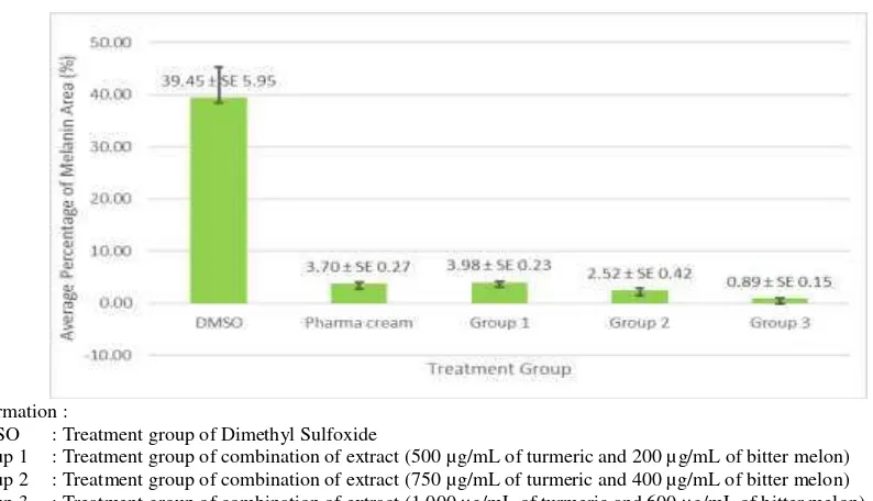
The average percentage of melanin area (figure 2) of combination treatment group-1 was 3,98%, combination group 2 was 2,52%, and combination group 3 was 0,89%. The higher concentration of the extract combination has better ability of melanin formation inhibitor which characterized by smaller percentage of melanin area.
14
Information :
a. A cross section of the epidermis layer after the administration of DMSO b. A cross section of the epidermis layer after the administration of pharma cream c. A cross section of the epidermis layer after the administration of combination group 1 d. A cross section of the epidermis layer after the administration of combination group 2 e. A cross section of the epidermis layer after the administration of combination group 3 The yellow arrow indicates the location of melanin marked with the black area
Figure 1. Histopathological results
Information :
DMSO : Treatment group of Dimethyl Sulfoxide
Group 1 : Treatment group of combination of extract (500 µg/mL of turmeric and 200 µg/mL of bitter melon) Group 2 : Treatment group of combination of extract (750 µg/mL of turmeric and 400 µg/mL of bitter melon) Group 3 : Treatment group of combination of extract (1,000 µg/mL of turmeric and 600 µg/mL of bitter melon).
Figure 2. Average percentage of melanin area of turmeric and bitter melon leaves extract combination compared with the control group
control that statistically significant, which meant that both combination groups revealed a better effect than the pharma cream (p <0,05).
The anti hyperpigmentation effect result of ethanol extract of turmeric on guinea pig skin contained in the research
report.12 The combination group 1 showed anti hyperpigmentation effect higher than 1000 μg/ml of turmeric extract (2,01%) (p <0,05). Meanwhile, the combination group-3 had the average percentage of melanin area which was significantly smaller than 500 μg/ml of turmeric extract
e
c d
15
(p <0,05), which meant that the combination group 3 has a better anti hyperpigmentation effect than 500 μg/ml of turmeric extract.
The anti-hyperpigmentation effect of ethanol extract of bitter melon leaves on guinea pig skin has been published in Cendekia Eksata Journal.13 The combination group 1 showed higher anti hyperpigmentation effect than 200, 400, and 600 μg/ml of bitter melon leaves extract group which reduced melanin by 2,01; 1,06; and 0,62 % respectively (p <0,05). The combination group 2 showed higher anti hyperpigmentation effect than 400 μg/ml of bitter melon leaves extract (0,62%) (p <0,05).
These findings were consistent with the results of Rizza et al (2012) that evaluated skin whitening effects of Mediterranean herbal extracts by in vitro and in vivo models.8 The single extract of caper buds, blood orange, rice grains, and olive leaf showed less intensive inhibiting activity than kojic acid and hydroquinone. Otherwise, when all the extracts were combined, it has the higher activity significantly than kojic acid and hydroquinone.
The possibility of antihyper-pigmentation effect of turmeric and bitter melon leaves extract combination is due to compounds contained in the extract combination. The compounds are flavonoids, saponins, alkaloids, terpenoids, phenols, and tannins.14 The ethanol extract of bitter melon leaves contains phenolic compounds, polyphenols, tannins, saponins, alkaloids, vitamin C, gallic acid and catechin.15 The active compound suspected to play a role in decreasing the amount of melanin is curcumin through the antioxidant mechanism.6 Whereas the active compound suspected to play a role in decreasing the amount of melanin is gallic acid, salicylic acid, cinnamic acid, myricetin, quercetin, and lutein through the antioxidant activity, cell protection, and anti melanogenic activities.7
CONCLUSIONS
The combination of turmeric and bitter melon leaves extract (group-2 and 3) showed better effect than the pharma cream.
RECOMMENDATIONS
This study proved that the combination of natural ingredients had a better effect than the pharmaceutical cream, in the hope of having fewer side effects, so it needs to be tested further on the toxicity test on Director General of Development and Research Enhancement, The Ministry of Research, Technology and Higher Education.
REFERENCES
1. Fitrie, AA. Histologi dari melanosit, Sumatera Utara: e-USU Repository Universitas; 2004.
2. Miyazawa M. Inhibitory compound of tyrosinase activity from the sprout of
Polygonium hydropiper L. (Benitade),
Biology Pharmaceutical Bulletin. 2007; 30:595-97.
3. Baumann L, Saghari S, Weisberg E, editors. Cosmetic dermatology, 2nd edition. New York: McGraw Hill; 2009.
4. Momtaz S, Mapunya BM, Houghton PJ, Edgerly C, Hussein A, Naidoo S, Lall N. Tyrosinase inhibition by extracts and constituents of Sideroxylon inerme L. stem bark, used in South Africa for skin
Total [Skripsi]. Makassar: Universitas Hassanudin; 2015.
B16-16 F1. Jurnal Berkala Penelitian Hayati.
2012;17:173–76.
7. Tsai TH, Huang CJ, Wu WH, Huang WC, Chyuan JH, Tsai PJ. Antioxidant, cell-protective, and anti-melanogenic activities of leaf extracts from wild bitter melon
(Momordica charantia Linn. var.
abbreviata Ser.) cultivars. Botanical Studies. 2014 Dec;55(1):1-7.
8. Rizza L, Bonina C, Frasca G, Puglia C. Skin-whitening effects of Mediterranean herbal extracts by in vitro and in vivo models. Journal of cosmetic science. 2012;63(5):311-20.
9. Hastiningsih I. Krim ekstrak etanol kulit batang pohon nangka (Arthocarpus
heterophilus) sama efektifnya dengan krim
hidrokuinon dalam mencegah peningkatan jumlah melanin pada kulit marmut (Cavia
porcellus) yang Dipapar Sinar UVB
[Tesis]. Denpasar: Universitas Udayana; 2015.
10. Shankar K, Godse K, Aurangabadkar S, Lahiri K, Mysore V, Ganjoo A, Vedamurty M, Kohli M, Sharad J, Kadhe G, Ahirrao P. Evidence-based treatment for melasma: expert opinion and a review. Dermatology and therapy. 2014 Dec; 4(2): 165-86.
11. Junqueira LC, Mescher AL. Basic histology: Text and Atlas, 13th edition. New York; Mc-Graw Hill Education; 2013.
12. Fithria RF, Anas Y, Utami HM. Aktivitas Antihiperpigmentasi ekstrak etanol rimpang kunyit (Curcuma domestica L.) pada kulit marmut Belanda (Cavia
porcellus) jantan [Research Report].
Semarang: Universitas Wahid Hasyim; 2016.
13. Fithria RF, Anas Y, Putri FAW. The antihyperpigmentation effect pare leaves
(Momordica charantia L.) ethanol extract
on guinea pig (Cavia porcellus) Skin. Jurnal Cendekia Eksata. 2017:2(1);47-53. 14. Dutta B. Study of secondary metabolite
constituents and curcumin contents of six different species of genus Curcuma.
Journal of Medicinal Plants. 2015;3(5):116-9.