Summary We compared root system morphogenesis of micropropagated transplants of Prunus cerasifera L. inocu-lated with either of the arbuscular mycorrhizal (AM) fungi Glomus mosseae or Glomus intraradices or with the ericoid mycorrhizal species Hymenoscyphus ericae. All plants were grown in sand culture, irrigated with a nutrient solution that included a soluble source of phosphorus, for 75 days after transplanting. Arbuscular mycorrhizal colonization increased both the survival and growth (by over 100%) of transplants compared with either uninoculated controls or transplants in-oculated with H. ericae. Arbuscular mycorrhizal colonization increased root, stem and leaf weights, leaf area, root length and specific leaf area, and it decreased root length/leaf area ratio, root/shoot weight ratio and specific root length.
Both uptake of phosphorus and its concentration in leaves were increased by AM infection, although the time course of the relationships between intensity of AM infection and P nutrition were complex and suggested a role for factors other than nutrition. The time course for the development of infec-tion varied. It was most rapid with G. mosseae, but it was ultimately higher with G. intraradices.
None of the treatments significantly affected the lengths of adventitious roots or the first-, second- or third-order laterals that developed from them. Arbuscular mycorrhizal coloniza-tion increased the intensity of branching in all root orders with the effect being most obvious on first-order lateral roots where the number of branches increased from under 100 to over 300 branches m−1. As a result, although first-order laterals made up 55% of the root systems of control plants, the comparable value was 36% in AM-infected plants. In contrast, second-or-der laterals represented 25% of control root systems, but 50% of AM-colonized root systems. Glomus intraradices but not G. mosseae increased root diameter. Anatomical studies re-vealed no changes in the overall form of the root tip, although there were changes in the diameter of the root cap, cell num-bers and cell size. Hymenoscyphus ericae increased the
dura-tion of the metaphase index. Both AM fungal treatments in-creased the concentrations of soluble proteins in root extracts and modified the protein profiles by the elimination and addi-tion of protein bands detected by PAGE analysis. We conclude that AM fungal inoculation influenced processes in the root system at different levels, but not all effects were due to improved P nutrition or increased physiological age.
Keywords: adventitious roots, colonization, fungi, Glomus in-traradices, Glomus mosseae, Hymenoscyphus ericae, infec-tion, lateral roots, phosphorus.
Introduction
Root system morphology is genetically determined and varies among species and individuals (Harper et al. 1991). It can be modified by many environmental factors, including nutrient and water availability and temperature (Drew and Saker 1978, Scott Russell 1982). Root plasticity is also influenced by arbuscular mycorrhizal (AM) fungi; although variable effects have been reported (see review by Berta et al. 1993), the most common effect is increased root branching.
The rapid development of a functioning root system is criti-cal to the successful establishment of many horticultural and other woody species. In addition, because root system mor-phology influences root function (Harper et al. 1991), it is of interest to understand the effects of mycorrhizal fungi on the first phases of root development, particularly in rootstocks that are frequently micropropagated to obtain homogeneous and disease-free clones. It has been found that early AM infection is beneficial to the performance of micropropagated plants (Ravolanirina et al. 1989a,1989b, Branzanti et al. 1992, Vest-berg 1992), but the mechanism of this effect is unclear.
We investigated the effects of mycorrhizal colonization dur-ing the first phases of root development after outplantdur-ing of micropropagated Prunus cerasifera L. clone MrS2/5, an
im-Arbuscular mycorrhizal induced changes to plant growth and root
system morphology in
Prunus cerasifera
G. BERTA,
1A. TROTTA,
1A. FUSCONI,
1J. E. HOOKER,
2M. MUNRO,
2D. ATKINSON,
2M. GIOVANNETTI,
3S. MORINI,
4P. FORTUNA,
4B. TISSERANT,
5V. GIANINAZZI-PEARSON
5and S. GIANINAZZI
51 Università di Torino, Dipartimento di Biologia Vegetale, viale Mattioli 25, 10125 Torino, Italy
2 Soil Biology Unit, Scottish Agricultural College, Mill of Craibstone, Aberdeen AB2 9TS, U.K.
3 Università di Pisa, Istituto di Microbiologia Agraria, Centro di Studio per la Microbiologia del Suolo, via del Borghetto 80, Pisa, Italy
4 Università di Pisa, Dipartimento di Coltivazione e Difesa delle Specie Legnose, Sezione di Coltivazioni Arboree, Pisa, Italy
5 Station de Génétique et Amélioration des Plantes, Laboratoire de Phytoparasitologie, INRA/CNRS, BV 1540-21034 Dijon Cedex, France
Received March 17, 1994
portant rootstock for peach, infected with either of the AM fungi, Glomus mosseae or G. intraradices. Because AM-in-duced enhancement of phosphate assimilation in mycorrhizal plants influences plant development, we also determined the extent to which morphological and anatomical changes in roots were due to changes in P content. However, hormonal mechanisms may also underlie variations in root system mor-phology in endomycorrhizal systems (Berta et al. 1993), and Hooker et al. (1992) have shown that major morphological changes occur in the root system under conditions where P nutrition is unlikely to be a major factor. To assess alternative mechanisms underlying root system morphology, therefore, Prunus plants, well supplied with soluble phosphate, were grown in the presence of the ericoid mycorrhizal fungus Hymenoscyphus ericae, which is not able to infect Prunus plants symbiotically but synthesizes IAA in pure culture (Berta et al. 1988).
Because root system structure depends on meristematic ac-tivity of root apices, all factors that affect root development must directly or indirectly affect root apical meristem activity and structure. Therefore, we have related morphology and development of the root system to the structure of the apex of lateral roots. Furthermore, because modifications in rooting patterns and physiology imply changes in gene expression, and the presence of AM fungi within roots is known to modify protein metabolism (Dumas et al. 1989, Wyss et al. 1990, Schellenbaum et al. 1992), we also studied changes in the amounts and types of such gene products in mycorrhizal roots of Prunus.
Materials and methods
Micropropagation
Shoot tips (0.5--0.7 cm in length) of the micropropagated Prunus cerasifera L. clone ‘‘MrS2/5’’weremultiplied accord-ing to Morini et al. (1990). For shoot proliferation, MS growth medium was supplemented with 0.4 mg l−1 thiamine, 100 mg l−1 myoinositol, 60 mg l−1 NaFeEDTA, 30 g l−1 sucrose and 0.6 mg l−1 BA. Indolebutyric acid (IBA) was used at 0.4 mg l−1 for rooting. The pH of the medium was adjusted to 5.2, 6 g l−1 of Difco Bacto Agar was then added and the medium autoclaved. Both shoot proliferation and rooting were carried out in 500-ml glass vessels that were wrapped in polyethylene film and contained 120 ml of growth medium. The microplants were cultured in a growth cabinet with a 16-h photoperiod at 40 µmol m−2 s−1 photon irradiance at 21 °C. Shoot cultures were transferred to rooting medium after 2 weeks.
Plant growth and infection
Three-week-old rooted microplants were transplanted to indi-vidual 1.5-l polyethylene pots filled with quartz sand and divided into four groups. One group of pots was not inoculated and served as the nonmycorrhizal (NM) controls. The remain-ing three groups were inoculated with either Glomus in-traradices Schenck & Smith (isolate LPA8), Glomus mosseae (Nicol. & Gerd.) Gerdemann & Trappe (isolate BEG/2), or Hymenoscyphus ericae (Read) Korf & Kernan (= Pezizella
ericae Read) (isolate Lpae25). The crude AM inoculum con-sisted of external mycelium and infected root fragments ob-tained from Allium porrum L. pot cultures inoculated with G. mosseae, and from Trifolium pratense L. pot cultures inocu-lated with G. intraradices (3 g and 0.8 g of root fragments per pot were used, respectively). The inoculum of H. ericae con-sisted of mycelium maintained on malt agar and each pot received three 8-mm agar disks of mycelium taken from the edge of the colony. Plants were maintained in a growth cham-ber at a day/night temperature of 22/20 °C, 75% RH and a 16-h photoperiod with 150 µmol m−2 s−1 photon irradianceat pot height (fluorescent lamps, Osram L 40W/20R cool white and L 40W/10 day light). During the first week, pots were covered with plastic film, which afterward was pierced to permit plant acclimatization. On alternate days, the pots were watered to saturation with Long Ashton nutrient formula ‘‘nitrate type’’ (Hewitt 1966) containing 2 mM KN03, 2 mM Ca(N03)2, 0.75 mM MgSO4, 32 µM Na2HPO4 plus recommended micronutri-ents.
Harvests
Six plants from each of the four treatments were harvested at 18, 32, 47 and 75 days after transplanting and assessed for changes in biomass, root morphological development and leaf P content. At each harvest, the degree of root colonization was evaluated microscopically after staining with trypan blue in lactophenol (Phillips and Hayman 1970), and percentage colo-nization of the root system was quantified by the grid intersect method (Giovannetti and Mosse 1980).
Seventy-five days after transplanting, root tip morphology, and mitotic and metaphase indices were evaluated on first-or-der laterals because mycorrhizal infection in woody plants affects mainly lateral roots (Hooker et al. 1992). Eighteen, 32 and 47 days after outplanting, root proteins were determined by SDS-gel electrophoresis.
The data on mycorrhizal colonization, fresh and dry weights, leaf area, P content, root tip morphology and mer-istem activity were compared by ANOVA, and P ≤ 0.05 was adopted as the level of significance. The least significant dif-ferences between values of the morphometric parameters were calculated (P < 0.05). Standard errors were calculated for all data.
A regression analysis of plant growth was carried out with the data on degree of colonization, total root length, mean length of adventitious roots, intensity of branching of first-or-der lateral roots and relative leaf area (RLA). The following linear and nonlinear regressions were used (Causton and Venis 1981, Berta et al. 1990):
y = mt + c (linear function)
y = a
1+ be−kt (logistic function)
Biomass assessments
Shoot and root fresh weights were determined by weighing, after washing and blotting the samples with filter paper. Leaves were photocopied, to allow leaf area measurements, before drying for 24--48 h at 80 °C.
Morphometric analyses
Measurements of root morphology were carried out on the six replicate plants at each harvest. Roots were preserved in 50% ethanol. Roots were separated into branching orders and meas-ured from video images (Image Analysis System, Magiscan, Joyce-Loebl Ltd.) (Hooker and Atkinson 1992). At each har-vest, the following morphometric parameters were deter-mined: number of roots of each order, number of axes, mean root length, total root length, the degree of branching of the root system (evaluated as total root number divided by total root length), lateral root frequency (represented by the number of roots of order n, divided by the length of roots of order n − 1), and the percentage of roots present in each order. At Day 75, the mean diameter of roots in each order was also determined.
At each harvest, leaf area was determined by digitized image analysis using Image Grabber 2.3 Software ( Macintosh IIx) and a CCD KP-C503 Hitachi camera. The digitized images were analyzed with OptiLabTM/x 1.4 software.
Phosphorus content
Dry material was wet-digested (Kuttner and Lichtenstein 1932) and then analyzed for total phosphorus content (Fogg and Wilkinson 1958). Samples (30--40 mg) of dry material were weighed into 100-ml Kjeldahl flasks, and 1 ml of 10 M H2SO4 was added. The samples were then digested on a Kjeldahl microburner by adding 10--20 drops of 30% H2O2. After neutralization and addition of ammonium molybdate--sulfuric acid solution, samples were boiled for 1 min with 100 mg of ascorbic acid. After color development, the samples were diluted to 50 ml, and the optical density at 660 nm determined with a Varian DMS 100 spectrophotometer. Values of P content were obtained from a 10--350 µg P calibration curve.
Root tip morphology of secondary roots
Root tips of secondary roots of uninoculated P. cerasifera plants and plants inoculated with G. mosseae, G. intraradices or H. ericae were fixed in 4% formaldehyde in 0.1 M phos-phate buffer, pH 7, for 2 h, postfixed in 1% OsO4, dehydrated and embedded in Durcupan ACM (Fluka). Median longitudi-nal sections were cut on a Reichert ultramicrotome, and root tip morphology was examined after staining with 1% toluidine blue in 1% sodium tetraborate. The diameters of 10 root apices per sample were measured at the base of the meristem on median longitudinal sections with the aid of an ocular mi-crometer.
Determination of meristematic activity
Randomly chosen parts of root systems were fixed as de-scribed by Greilhuber (1988) for tannin-rich plant material.
Fixed root tips of first-order laterals were cut and treated with a pectinase-cellulase mixture for 3 h at room temperature, followed by 24 vol of H2O2 for 10 min, to reduce the quantity of tannins. The samples were then stained with Feulgen, smeared and mounted on glass slides.
The mitotic index (MI) and percentage of mitotic cells in metaphase (MeI) for roots of equal length (1.5 cm) were determined. Each value for MI was based on a total of 3000 cells, 1000 being scored from each of three slides.
Quantitative protein analysis
Root pieces were placed in liquid nitrogen and soluble root proteins were extracted with 0.1 M Tris-HCl buffer, pH 7.6, containing 1% Na-thioglycolate, 0.4% NaCl and 10 mM mer-captoethanol (1/1, v/v). The macerate was centrifuged at 48,000 g for 20 min, and protein content of the supernatant was determined by the Bradford (1976) method.
Qualitative protein analysis
Twenty-five µl of soluble root protein extract was analyzed by electrophoresis in 15% SDS polyacrylamide gel by the method described by Davis (1964) and proteins were detected by silver nitrate staining (Blum et al. 1987). Qualitative analysis of alkaline phosphatase isozymes in root tissues was realized after protein extraction in 0.1 M borate buffer, pH 8.8, (2/1, w/v) by native PAGE electrophoresis as described by Giani-nazzi-Pearson and Gianinazzi (1978).
Results
Plant biomass and development
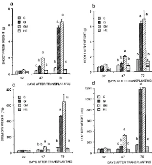
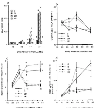
All of the micropropagated plants of P. cerasifera were about the same size when transplanted (Figure 1). Eighteen days after transplanting, the arbuscular mycorrhizal (AM) plants were uniformly healthy, whereas only 70% of both the nonmy-corrhizal (NM) controls and plants inoculated with H. ericae were healthy. At this time, no differences were observed in shoot and root fresh weights, or in stem and leaf dry weights of the healthy plants (Figure 2). By Day 75, plant weights had increased only slightly in the NM controls and plants inocu-lated with H. ericae, e.g., root weight in the NM controls was only 5.5 times that at Day 18. In contrast, shoot and root fresh weights and leaf area of AM plants (Figure 3a) were all significantly higher than those of NM controls by Day 32 (Figures 2a and 2b), and the differences increased with time (cf. Days 47 and 75). Stem and leaf dry weights (Figures 2c and 2d) were significantly higher in the AM plants than in the NM controls by Day 47. Growth of plants infected with G. in-traradices was delayed compared with growth of plants in-fected with G. mosseae. Differences between the two AM treatments were generally significant at Day 47, but not at Day 75.
those of NM controls (Figure 3b). At Day 18, the fresh weight/dry weight ratios of shoots and leaves were similar for all treatments (3.8 for shoots and 3.3 for leaves). The ratios decreased in all treatments by Day 32 and then began to increase again around Day 47. At the final harvest, no treat-ment differences were evident (values of 3.0 for shoots and 3.0 for leaves).
The root/shoot fresh weight ratio (R/S) was similar in all treatments until Day 47 when it was reduced in AM plants (Figure 3c). Effects of the two AM species on R/S were not significantly different. Initially, the root length/leaf area ratio (RLA) increased linearly with time in all treatments, although there was considerable variability in plants inoculated with H. ericae and the increase occurred at a slow rate in AM plants (data not shown).
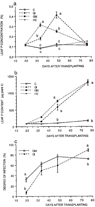
Leaf phosphorus concentrations (Figure 4a) were similar in all treatments on Day 18, and they remained constant in NM controls and plants inoculated with H. ericae throughout the experiment. Leaf phosphorus concentrations were signifi-cantly higher in the AM plants on Days 32 and 47 than on Day 18, but by Day 75, foliar P concentrations were similar in plants in all treatments. The increase in foliar P concentration in AM plants, which paralleled the increase in growth,
oc-curred earlier in plants infected with G. mosseae than in plants infected with G. intraradices.
Total leaf phosphorus content was also similar among the treatments at Day 18. It increased in all treatments with time, but it increased more quickly in the AM plants than in NM controls and plants inoculated with H. ericae (Figure 4b).
Development of colonization
Staining of root samples indicated that AM infection was absent from NM controls and plants inoculated with H. ericae (Figures 5a and 5b), but well established in plants in the AM treatments (Figures 5c--e). The pattern of colonization devel-opment varied between the two AM treatments. During the initial two harvests, the degree of colonization was higher in plants infected with G. mosseae than in plants infected with G. intraradices (Figure 4c), but by Day 75, it was higher in plants infected with G. intraradices (Figure 4c).
Root system morphology
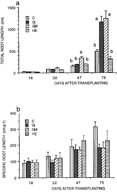
Total root length Total root length increased with time in all treatments, but the increase was greatest in AM plants (Fig-ure 6a). Application of a logistic model of root extension growth gave higher asymptotic values in AM plants than in NM controls and plants infected with H. ericae; the asymptotic value was particularly high in plants colonized with G. in-traradices (Table 1).
At Day 18, the total root length/root mass ratio (specific root length, SRL) was similar in all treatments; thereafter, it in-creased in all treatments, but reached higher values in NM controls and plants inoculated with H. ericae than in AMplants (Figure 6b).
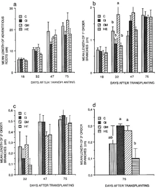
The growth of root axes The root system of the micropropa-gated plants consisted of a series of adventitious roots, which developed from the base of the shoot, together with branched roots that developed from the adventitious roots. Mean root length and the average length of an individual root axis in-creased with time, and there were no differences among treat-ments (Figure 7a). Application of a logistic model of root extension growth confirmed that there were no effects of the fungi on growth of adventitious roots.
Throughout the study, mean length of first-order laterals increased in all treatments, except in plants infected with G. mosseae, where mean length of first-order lateral roots only increased until Day 32 (Figure 7b). In all treatments, second-order lateral roots, which were present only after the first harvest, increased in length after Day 32 (Figure 7c). Third-or-der lateral roots were present only at the final harvest. There were no significant differences in third-order lateral root length among treatments, except in plants inoculated with H. ericae, which had significantly shorter third-order lateral roots than plants in either of the AM treatments (Figure 7d).
Effects on root branching At Days 18 and 47, there were no significant differences among treatments in degree of branch-ing of adventitious roots, but at Days 32 and 75, branchbranch-ing frequency was significantly higher in AM plants than in NM controls and plants inoculated with H. ericae (Figure 8a). Figure 1. The effects of various mycorrhizal treatments on the growth
Frequency of branching was highest in AM plants at Day 75. The intensity of branching of first-order lateral roots in-creased with time for all treatments, and there were significant differences between AM plants and NM controls and plants inoculated with H. ericae (Figure 8b, Tables 1 and 2). The rate of increase was approximately linear; however, the slope var-ied among treatments decreasing in the order of G. mosseae, G. intraradices, NM controls and H. ericae (6.79, 5.47, 1.93 and 1.29, respectively). Of the parameters tested, the greatest morphological effect of AM infection was on intensity of branching of first-order lateral roots. The intensity of branch-ing of second-order lateral roots was significantly higher in plants infected with G. intraradices than in plants in the other treatments (Figure 8c).
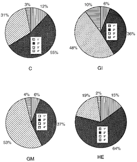
Percentage contribution of the different root orders In all treatments, the percentage of the root system present as adven-titious roots decreased with time, whereas the proportion of the root system present as second-order lateral roots increased. First-order lateral roots made up a relatively constant propor-tion of the root system in NM controls and plants inoculated with H. ericae, but the proportion decreased markedly in AM plants with time. After Day 32, plants colonized with G. mosseae had a significantly smaller proportion of the root system as adventitious roots and a higher proportion as first-order lateral roots. In plants inoculated with H. ericae, the greatest proportion of the root system was adventitious roots
and the smallest proportion was second-order lateral roots. At Day 47, plants in both AM treatments had a lower proportion of adventitious roots and a higher proportion of second-order lateral roots than NM controls. At Day 75, (Figure 9 ), the root system in NM controls and plants inoculated with H. ericae were dominated by first-order lateral roots (55 and 64%, re-spectively), whereas in plants inoculated with G. mosseae and G. intraradices,the roots systems were dominated by second-order lateral roots (53 and 48%, respectively) (Figure 9).
Root diameter
Diameters of primary, secondary and tertiary roots were sig-nificantly higher in plants colonized by G. intraradices than in plants in the other treatments, whereas diameters of quaternary roots were higher in plants colonized by G. mosseae. The diameters of some root orders were smaller in plants colonized by H. ericae than in NM controls and AM plants.
Root tip morphology of second-order roots and meristematic activity
Median longitudinal sections of root apices of second-order lateral roots showed a closed organization of the meristems. They possessed discrete caps, i.e., there was a clear boundary between the initials of the cortex and the initials of the cap. The epidermis differentiated as the inner cell layer of the lateral cap (Figure 10).
Figure 2. The effects of various mycorrhi-zal treatments on (a) shoot fresh weight, (b) root fresh weight, (c) stem dry weight, and (d) leaf dry weight of
There was no variation in root tip morphology among treat-ments, but root apex diameter, measured at its base, was significantly larger in plants infected with G. intraradices (355.7 ± 16.7 µm, P < 0.01) and smaller in plants infected with H. ericae (190.8 ± 16.4 µm, P < 0.01) than in NM controls and plants infected with G. mosseae (246.0 ± 14.8 µm and 267.6 ± 13.9 µm, respectively, P > 0.25). Variations in size depended on both the number and size of the constituent cells. Root apices of plants inoculated with H. ericae were more inten-sively stained than root apices of plants in the other treatments (Figure 10e).
The mitotic index (MI) of second-order roots was similar in all treatments (Table 3). In contrast, there were significant differences between metaphase indices (MeI) of NM controls and plants inoculated with H. ericae (P < 0.05), and between plants inoculated with H. ericae and plants inoculated with G. mosseae (P < 0.01) (Table 3).
Qualitative and quantitative analyses of proteins
Root extracts of AM plants contained more soluble proteins than NM controls or plants inoculated with H. ericae. Protein content was higher in plants colonized by G. intraradices than in plants in the other treatments, and the differences increased with time (Table 4). One protein band, which was present in
root extracts of plants in all treatments on Day 32, was only present in NM controls and plants inoculated with H. ericae on Day 47. Some protein bands were enhanced in root extracts of AM plants, and three bands were detected only in AM plants (Figure 11). At Day 47, an alkaline phosphatase isozyme was present in root extracts of plants colonized with G. mosseae (Figure 12).
Discussion
The survival of micropropagated plants during their transition from in vitro culture to growth in soil is a significant horticul-tural problem (Fortuna et al. 1992). We found that colonization of plants with AM fungi immediately on transplanting elimi-nated losses, regardless of the state of root development at transplanting (Figure 1). At the time of transplanting, the plants only had primary roots, which are known to be less susceptible to colonization with AM fungi than the higher-or-der laterals which are produced during ex vitro development. Thus the early ex vitro stage is probably the most appropriate time to apply AM fungi. The mechanisms of the effects of AM fungi on early growth and survival of microplants are unclear. At Day 18, shoot weight, root weight and length (Figure 2), leaf area (Figure 3) and the concentration of P in leaves Figure 3. The effects of various mycorrhi-zal treatments on (a) leaf area, (b) spe-cific leaf area, (c) root/shoot fresh weight ratio, and (d) root length/leaf area ratio of
P. cerasifera. Treatments were: C = non-mycorrhizal control, HE = inoculated with H. ericae, GI = inoculated with
(Figure 4), usually the first tissue to indicate impeded nutrient supply, were similar in all treatments. At this time, however, the extent of infection by G. mosseae was substantial (35%) and comparable to that obtained with similar sized plants by Fortuna et al. (1992), whereas colonization by G. intraradices was low (5%), although sufficient to improve survival from 70% (for NM controls) to 100%.
Modifications in the growth pattern of AM plants may be the result of the plant being colonized by the fungus or they may be due solely to the presence of the fungus (Berta et al. 1993). To resolve this question, we included H. ericae, an ericoid mycorrhizal fungus, because it is known to produce auxins when grown in pure culture (Berta et al. 1988), but it is not
capable of colonizing and developing arbuscles (Figure 5b). We found plants inoculated with H. ericae behaved similarly to the NM controls indicating that colonization by an AM fungus is needed for morphogenetic and other effects to be initiated, and that the mechanisms underlying the changes were probably not mediated by auxins (Berta et al. 1988).
In most studies, the growth of AM and nonmycorrhizal plants differ, and so the effects of the AM fungi are confounded with effects due to plant size or physiological age. It is also important to separate direct effects that are due to improved AM-fungi-mediated nutrient access and supply, resulting from the development of extra matrical hyphae, from indirect effects of improved growth resulting in increased nutrient uptake. Although many studies of AM fungi have shown direct nutri-ent-mediated effects, Hooker et al. (1992) were the first to demonstrate that both direct nutrient and indirect growth ef-fects occur in Populus, i.e., they observed morphogenetic ef-Figure 4. The effects of various mycorrhizal treatments on (a) foliar
phosphorus concentration, (b) foliar phosphorus content, and (c) de-gree of AM fungal infection in plants inoculated with G. intraradices
(GI) and G. mosseae (GM). Means followed by the same letter are not significantly different (P≤ 0.05).
Figure 5. Root squashes of P. cerasifera. Squashes were stained with trypan blue in lactophenol to indicate fungal mycelium. (a) Nonmy-corrhizal control showing no hyphae present; (b) roots inoculated with
fects even though they had manipulated nutrient supply to ensure that the growth and nutrition of nonmycorrhizal control and AM plants were identical.
We used a similar experimental design to show that AM-in-duced mycorrhizal growth effects occurred in P. cerasifera even though the plants were grown in the presence of excess P. In our study, plants were grown in quartz sand, which has a low P binding capacity, and were supplied with P as the soluble salt Na2HPO4 at a concentration of 32 µmol. By the end of the experiment, 2600 mg of phosphorus had been added to each pot. Most of this phosphorus would have been directly avail-able in the sand solution. The total phosphorus removed was estimated to be 300 mg for NM controls and 2250 and 2560 mg for plants inoculated with G. intraradices and G. mosseae, respectively. Thus, the NM controls were not P deficient.
Internal P content influences root geometry (Amijee et al. 1989) and assimilate partitioning to lateral roots, and an ele-vated P content enhances lateral root formation (Adalsteinsson and Jensen 1989). In a study with Andropogon gerardii Vitm., both AM fungi and P enhanced growth and increased the number and diameter of primary, secondary and tertiary roots, but decreased SRL (Hetrick et al. 1988). In Gossypium hirsu-tum L., AM fungi and P increased plant weight, but only AM colonization decreased SRL (Price et al. 1989). In A. porrum (Trotta et al. 1991a), AM effects on growth, individual root lengths, root numbers and branching occurred at low but not at high external P concentration suggesting that mycorrhizal ef-fects resulted from enhanced P nutrition. However, in a further study of A. porrum, with a wider range of P concentrations and more detailed measurements of root development, Trotta et al. (1991b) found that first- and second-order lateral roots were shorter in mycorrhizal plants than in control plants at every P concentration tested, indicating an effect independent of P nutrition. The authors concluded that some fungal effects on morphogenesis might be modified by metabolites or hor-mones. This concurs with the findings of Hooker et al. (1992), who also found effects of AM infection that were independent of external nutrient supply.
We found that, although the two AM treatments increased plant P concentrations between Days 32 and 47, changes in the extent of AM infection were poorly correlated with changes in plant P concentration and growth. An increase in colonization by G. mosseae from 30 to 60% between Days 18 and 32 was associated with an increase in plant P concentration from 0.1 to 0.3% but a relatively small increase in total P uptake (Fig-ure 4), whereas an increase in colonization from 60 to 70% between Days 32 and 47 was associated with a decrease in plant P concentration but a large increase in total P uptake. An increase in colonization by G. intraradices from 5 to 45% between Days 18 and 32 was associated with a only small increase in plant P concentration from 0.11 to 0.17% and with little change in total P uptake. Increases in colonization from 45 to 50% and 50 to 80% were associated with changes in plant P concentration from 0.17 to 0.40% and from 0.40 to 0.11%, respectively, and with nonlinear increases in the rate of total P uptake. Even allowing for root extension growth during this period and for effects associated with the length of infected Figure 6. The effects of various mycorrhizal treatments on (a) total
root length per plant and (b) specific root length of P. cerasifera. Treatments were: C = nonmycorrhizal control, HE = inoculated with
H. ericae, GI = inoculated with G. intraradices, and GM = inoculated with G. mosseae. Means followed by the same letter are not signifi-cantly different (P≤ 0.05).
Table 1. Values of the parameters used in the best fits of total root length of nonmycorrhizal (NM) controls and plants inoculated with
H. ericae (HE), G. intraradices (GI) or G. mosseae (GM) with the values of F and R2. The data were fitted by logistic functions.
Parameters F R2
NM Controls a = 70.00 55 0.962
b = 9684.30
k = −2.41
H. ericae a = 55.00 31 0.939
inoculated plants b = 6668.24
k = −2.46
G. intraradices a = 500.00 13 0.931
infected plants b = 3569.70
k = −1.42
G. mosseae a = 110.00 92 0.975
infected plants b = 3473.90
roots, which increased most between Days 47 and 75, there was no simple relationship between AM colonization and P nutrition. In both AM treatments, increased P uptake, linked to increased growth, occurred after Day 32 and was associated with significant (40%) colonization of the root system. The form of P supply and the inert growing medium used suggest that limited uptake as a result of poor root development in NM plants was the probable cause of these effects.
The patterns of colonization by the two AM fungi differed with time and with P concentration, and the two fungi had different effects on shoot growth. At Day 75, leaf weights of plants in the two treatments were similar, but stem weight was higher in plants inoculated with G. mosseae than in plants inoculated with G. intraradices. In NM controls, the intensity of root branching decreased from the adventitious roots to the secondary lateral roots (cf. Pagès et al. 1992), whereas AM fungi increased the degree of branching of adventitious and primary lateral roots, but only G. intraradices increased branching frequency of secondary lateral roots. The noncolo-nizing H. ericae fungus decreased root branching.
Both AM fungi induced significant decreases in specific root length (SRL). Reduced SRL can result from: (1) an increase in
root diameter leading to more material per unit length of root axis, (2) a decrease in cell size leading to increased tissue density and thus weight per unit length, and (3) an increase in cellular or intracellular constituents leading to increased weight per unit length. Data on root diameter (Table 3) indicate that G. intraradices increased the root diameter of all orders of roots, whereas G. mosseae had no effect on diameter. Because cell size in AM roots was increased compared to that in NM control tissue, we conclude that the effects were not caused by carbohydrate starvation. The decrease in SRL in G. mosseae -colonized roots was probably caused by an increase in tissue contents because tissue sections indicated an increased amount of cellular inclusions in AM-infected tissue (Figure 5). Roots colonized by G. mosseae contained a high amount of fungal structures (Figure 5). Thus the decreases in SRL caused by the two AM fungi appear to be caused by different mechanisms.
was higher in the AM plants; however the difference was less than that based on a comparison of NM controls and AM plants of similar age. Therefore, we conclude that about half of the modification in branching was related to plant size and the rest to a specific influence of the AM fungus. Re-assessment of the data of Schellenbaum et al. (1991) gave a similar result; how-ever, this was not the case when the data for Platanus acerifo-lia (Ait.) Willd. (Tisserant et al. 1992) were re-evaluated.
Contrary to findings for endomycorrhizal A. porrum plants (Berta et al. 1990), meristematic activity was not influenced by AM fungi. Metaphase indices were higher, however, in plants inoculated with H. ericae than in NM controls or AM plants, indicating a tendency for this fungus to inhibit mitotic activity. The AM fungi had no effect on the structure of the root apex of first-order lateral roots; however, the endomycorrhizal root apices were larger, which agrees with the findings of Fusconi et al. (1994) for A. porrum plants. The large reduction in size Figure 8. The effects of mycorrhizal treatments on number of branch
roots emerging from (a) adventitious roots, (b) first-order branches, and (c) second-order branches. Treatments were: C = nonmycorrhizal control, HE = inoculated with H. ericae, GI = inoculated with G. in-traradices, and GM = inoculated with G. mosseae. Means followed by the same letter are not significantly different (P≤ 0.05).
Table 2. Effects of inoculation with H. ericae (HE), G. intraradices (GI) or G. mosseae (GM) on the mean diameter (mm) of roots of P. cerasifera
plants at Day 75. Values in columns followed by the same letters are not significantly diferent from each other (P = 0.05).
Inoculum First-order roots Second-order roots Third-order roots Fourth-order roots
Control 1.37 ± 0.13 a 0.51 ± 0.04 a 0.31 ± 0.04 a 0.21 ± 0.01 a HE 1.05 ± 0.08 b 0.47 ± 0.03 a 0.27 ± 0.01 a 0.24 ± 0.01 b GI 2.11 ± 0.14 c 0.66 ± 0.05 b 0.43 ± 0.06 b 0.22 ± 0.01 ab GM 1.47 ± 0.15 a 0.46 ± 0.03 a 0.30 ± 0.03 a 0.28 ± 0.01 c
Figure 9. The effects of mycorrhizal treatments on proportions of root system present as adventitious roots (1°), first-order laterals (2°), second-order laterals (3°), and third-order laterals (4°) at Day 75. Treatments were: C = nonmycorrhizal control, HE = inoculated with
of the root apices of plants inoculated with H. ericae suggests a parasitic interaction between the fungus and P. cerasifera.
Both qualitative and quantitative differences in protein com-position were found between the NM controls and AM plants, confirming that AM colonization results in the synthesis of Figure 10. Median longitudinal sections of root apices of second-order
laterals. (a) NM Control apex showing a closed organization with a row of cells (initials of the outer cortex layer) sandwiched between the initials of the cap (CI) and a group of stele (S) and cortical cells (C). Vacuoles containing tannins are evident in the outer cap, epidermis and endodermis (arrows); magnification 150×. Initial zone (b)--(e): (b) NM control, (c) inoculated with G. mosseae, (d) inoculated with G. in-traradices, and (e) inoculated with H. ericae. Note the treatments resulted in variations in cell number and size but not structure; magni-fication 30×.
Figure 11. (a) Soluble protein patterns on 15% SDS-PAGE of 4- and 6-week-old roots of P. cerasifera uninfected (C), or inoculated with
G. intraradices (GI), G. mosseae (GM) or H. ericae (HE). (b) Soluble protein patterns on 10% SDS-PAGE of 6-week-old roots of P. cerasif-era uninfected (C), or inoculated with G. intraradices (GI),
G. mosseae (GM) or H. ericae (HE). One band (Rf = 0.78) was only present in root extracts of NM controls and plants infected with
H. ericae, whereas three additional bands (Rf values = 0.011, 0.60 and 0.85) were present in root extracts of AM plants.
Table 3. Effects of inoculation with H. ericae (HE), G. intraradices
(GI) or G. mosseae (GM) on mitotic index (MI) and metaphase index (MeI) of second-order laterals of P. cerasifera plants at Day 75. Values in columns followed by the same letters are not significantly different from each other (P = 0.05).
MI MeI
Control 3.1 ± 0.4 a 17.1 ± 1.8 a HE 3.2 ± 0.2 a 24.8 ± 1.5 abc GI 3.5 ± 0.3 a 20.7 ± 1.4 ac GM 3.9 ± 0.3 a 19.2 ± 1.6 b
Table 4. The effect of inoculation with H. ericae (HE), G. intraradices
(GI) or G. mosseae (GM), as compared to controls (C), on the protein content (ng) of root extracts of P. cerasifera.
Inoculum 18 Days 32 Days 47 Days
C 80 145 220
HE 82 135 163
GI 80 160 250
GM 82 286 583
Figure 12 Alkaline phosphatase activity present in 6-week-old root extracts of NM controls and plants inoculated with G. intraradices
specific new soluble proteins and enhances the production of some proteins present in noncolonized roots (cf. Gianinazzi-Pearson and Gianinazzi 1978, Dumas et al. 1989, Pacovsky 1989, Abdel-Fattah 1990, Wyss et al. 1990, Schellenbaum et al. 1992).
We conclude that infection of roots by AM fungi modifies plant morphogenesis at different levels ranging from whole plants to gene products, and that improved mineral nutrition does not account entirely for the improved growth or for the major morphogenetic changes observed.
Acknowledgments
We thank COST Action 810 for providing funds to assist in the coordination of the experiment. The cost of the experimental work was funded by several national agencies. We are particularly grateful to the Italian Murst, the Scottish Office Agriculture and Fisheries Depart-ment (SOAFD) and the French INRA.
References
Abdel-Fattah, G. 1990. Some ecological and physiological studies on vesicular arbuscular (VA) mycorrhizal fungi. Ph.D. Thesis. Man-soura University, Egypt, 202 p.
Adalsteinsson, S. and P. Jensen. 1989. Modifications of root geometry in winter wheat by phosphorus deprivation. J. Plant Physiol. 135:513--517.
Amijee, F., P.B. Tinker and D.P. Stribley. 1989. Effects of phosphorus on the morphology of VA mycorrhizal root system of leek (Allium porrum). Plant Soil 119:334--336.
Berta, G., V. Gianinazzi-Pearson, G. Gay and G. Torri. 1988. Morpho-genetic effects of endomycorrhiza formation on root system of
Calluna vulgaris (L.) Hull. Symbiosis 5:33--34.
Berta, G., A. Fusconi, A. Trotta and S. Scannerini. 1990. Morphoge-netic modifications induced by the mycorrhizal fungus Glomus
strain E3 in the root system of Allium porrum L. New Phytol. 114:207--215.
Berta, G., A. Fusconi and T. Trotta. 1993. VA Mycorrhizal infection and the morphology and function of root systems. Environ. Exp. Bot. 33:159--173.
Blum, H., H. Beier and H.J. Gross. 1987. Improved silver staining of plant proteins, RNA and DNA in polyacrylamide gels. Electropho-resis 8:93--99.
Bradford, M.M. 1976. A rapid and sensitive method for the quantifi-cation of microgram quantities of protein utilizing the principle of protein--dye binding. Anal. Biochem. 72:248--254.
Branzanti, B., V. Gianinazzi-Pearson and S. Gianinazzi. 1992. Influ-ence of phosphate fertilization on the growth and nutrient status of micropropagated apple infected with endomycorrhizal fungi during the weaning stage. Agronomie 12:841--846.
Causton, D.R. and J.C. Venis. 1981. The biometry of plant growth.
Arnold, London, pp 307.
Davis, B.J. 1964. Disc electrophoresis. II. Method and application to human serum proteins. Ann. N.Y. Acad. Sci. 121:404--427. Drew, M.C. and L.R. Saker. 1978. Nutrient supply and the growth of
the seminal root system in barley. III. Compensatory increases in growth of lateral root system, and in rates of phosphate uptake, in response to a localized supply of phosphate. J. Exp. Bot. 29:435--451.
Dumas, E., V. Gianinazzi-Pearson and S. Gianinazzi. 1989. Production of new soluble proteins during VA endomycorrhiza formation. Agric. Ecosyst. Environ. 29:111--114.
Fogg, D.N. and N.T. Wilkinson. 1958. The colorimetric determination of phosphorus. Analyst 83:406--414.
Fortuna, P., S. Citernesi, S. Morini, M. Giovannetti and F. Loreti. 1992. Infectivity and effectiveness of different species of arbuscular my-corrhizal fungi in micropropagated plants of Mr S 2/5 plum root-stock. Agronomie 12:825--830.
Fusconi, A., G. Berta, A.M. Tagliasacchi, S. Scannerini, A. Trotta, E. Gnavi and S. De Padova. 1994. Root apical meristems of arbuscular mycorrhizae of Allium porrum L. Environ. Exp. Bot. 34:181--193. Gianinazzi-Pearson, V. and S. Gianinazzi. 1978. Enzymatic studies on
the metabolism of vesicular arbuscular mycorrhiza. II. Soluble alkaline phosphatase specific to mycorrhizal infection in onion roots. Physiol. Plant Pathol. 12:45--53.
Giovannetti, M. and B. Mosse. 1980. An evaluation of techniques for measuring vesicular-arbuscular mycorrhizal infection in roots. New Phytol. 84:489--500.
Greilhuber, J. 1988. ‘‘Self-tanning’’----a new important source of stoichiometric error in cytophotometric determination of nuclear DNA content in plants. Plant Syst. Evol. 158:87--96.
Harper, J.L., M. Jones and N.R. Hamilton. 1991. The evolution of roots and the problems of analyzing their behavior. In Plant Root Growth. An Ecological Perspective. Ed. D. Atkinson. Blackwell Scientific Publications, Oxford, pp 3--24.
Hetrick, D.B.A., J.F. Leslie, G. Thompson Wilson and D. Gerschefske Kitt. 1988. Physical and topological assessment of effects of a vesicular-arbuscular mycorrhizal fungus on root architecture of big blue stem. New Phytol. 110:85--96.
Hewitt, E.J. 1966.Sand and water culture methods used in the study of plant nutrition. Tech. Comm., CAB, London, 347 p.
Hooker, J.E. and D. Atkinson. 1992. Application of computer-aided image analysis to studies of arbuscular endomycorrhizal fungi ef-fects on plant root system morphology and dynamics. Agronomie 12:821--824.
Hooker, J.E., M. Munro and D. Atkinson. 1992. Vesicular-arbuscular mycorrhizal fungi induced alteration in poplar root system mor-phology. Plant Soil 145:207--214.
Kuttner, T. and L. Lichtenstein. 1932. Micro colorimetric studies. III. Estimation of organically bound phosphorus. A system of analysis of phosphorus compounds in blood. J. Biol. Chem. 95:661--670. Morini, S., P. Fortuna, R. Sciutti and R. Muleo. 1990. Effect of
different light-dark cycles on growth of fruit tree shoots cultured in vitro. Adv. Hortic. Sci. 4:163--166.
Pagès, L., J. Chadoeuf and J. Kervella. 1992. Modélisation sto-chastique de la croissance et du developpement du système raci-naire de jeunes pechers. I. Estimation et validation du modèle. Agronomie 12:447--458.
Packovsky, R.S. 1989. Carbohydrate, protein and amino acid status of
Glycine--Glomus--Bradyrhizobium symbioses. Physiol. Plant. 75:346--354.
Philipps, J.M. and D.S. Hayman. 1970. Improved procedures for clearing roots and staining parasitic and vesicular-arbuscular my-corrhizal fungi for rapid assessment of infection. Trans. Br. Mycol. Soc. 55:158--161.
Price, N.S., R.W. Roncadori and R.S. Hussey. 1989. Cotton root growth as influenced by phosphorus nutrition and vesicular-arbus-cular mycorrhizas. New Phytol. 111:61--66.
Ravolanirina, F., L. Blal, S. Gianinazzi and V. Gianinazzi-Pearson. 1989a. Production of endomycorrhizal explants of micropropa-gated grapevine rootstocks. Agric. Ecosyst. Environ. 29:323--327. Ravolanirina, F., L. Blal, S. Gianinazzi and V. Gianinazzi-Pearson.
Schellenbaum, L., G. Berta, F. Ravolanirina, B. Tisserant, S. Giani-nazzi and A.H. Fitter. 1991. Influence of endomycorrhizal infection on root morphology in a micropropagated woody plant species (Vitis vinifera L.). Ann. Bot.68:135--141.
Schellenbaum, L., S. Gianinazzi and V. Gianinazzi-Pearson. 1992. Comparison of acid soluble protein synthesis in roots of endomy-corrhizal wild type Pisum sativum and corresponding isogenic mutants. J. Plant Physiol. 141:2--6.
Scott Russell, R. 1982. Plant root systems. E.L.B.S. Press, England, 298 p.
Tisserant, B., L. Schellenbaum, V. Gianinazzi-Pearson, S. Gianinazzi and G. Berta. 1992. Influence of infection by an endomycorrhizal fungus on root development and architecture in Platanus acerifolia. Allionia 30:171--181.
Trotta, A., C. Carminati, L. Schellenbaum, S. Scannerini, A. Fusconi and G. Berta. 1991a. Correlation between root morphogenesis, VA mycorrhizal infection and phosphorus nutrition. In Plant Roots and Their Environment. Eds. B.L. McMichael and H. Persson. Elsevier Science Publishers, Amsterdam, pp 333--389.
Trotta A., G. Berta, A. Fusconi and S. Scannerini. 1991b. Root devel-opment in a VA mycorrhiza, as related to phosphorus nutrition. In
Abstracts of the 3rd ISRR Symposium. Kutchera Ed., Vienna, pp 134.
Vestberg, M. 1992. Arbuscular mycorrhizal inoculation of micro-propagated strawberry and field observations in Finland. Agrono-mie 12:865--867.
Wyss P., R.B. Mellor and A. Wiemken. 1990. Vesicular-arbuscular mycorrhizas of wild-type soybean and nonnodulating mutants with