* Contact address:
Dr. Sani Abdullahi Shehu, Department of Anatomy, Faculty of Veterinary Medicine, Usmanu Danfodiyo University, Sokoto, Nigeria, Phone: +080 3595 1640; E-mail: saniabdul2003@yahoo.com
Role of erythrocyte surface sialic acid in inducing anaemia in Savannah
Brown bucks experimentally infected with
Trypanosoma evansi
Sani Abdullahi Shehu1*, Najume Doguwar Giginya Ibrahim2,
King Akpofure Nelson Esievo2, and Garba Mohammed3
1Department of Anatomy, Faculty of Veterinary Medicine, Usmanu Danfodiyo University, Sokoto, Nigeria 2Department of Microbiology and Pathology, Faculty of Veterinary Medicine, Ahmadu Bello University, Zaria,
Nigeria
3Department of Surgery and Medicine, Faculty of Veterinary Medicine, Ahmadu Bello University, Zaria, Nigeria
SHEHU, S. A., N. D. G. IBRAHIM, K. A. N. ESIEVO, G. MOHAMMED: Role of erythrocyte surface sialic acid in inducing anaemia in Savannah Brown bucks experimentally infected with Trypanosoma evansi. Vet. arhiv 76, 521-530, 2006.
ABSTRACT
Erythrocyte surface sialic acid, free serum sialic acid concentrations and sialidase activity were determined during experimental Trypanosoma evansi infection in Savannah Brown bucks. All infected bucks developed trypanosomosis, with significant decreases in mean packed cell volume to as low as 19.50 ± 2.12% occurring at day 33 post-infection and was significantly lower than the control value of 26.75 ± 0.96. Mean haemoglobin concentrations also declined in the infected bucks with a marked drop of 6.50 ± 0.70 g/dL on day 33 post-infection and was significantly different (P<0.05) from the uninfected (control) group (8.53 ± 0.46 g/dL). The anaemia was preceded by a gradual decline in mean erythrocyte surface sialic acid concentrations occurring 5 days post-infection. There were significant differences (P<0.05) in mean erythrocyte surface sialic acid between the infected and control groups on day 5 and between days 17 to 27 post-infection. A significant (P<0.05) increase in free serum sialic acid concentrations was observed on days 15, 17 and 27 when compared to the control group. There was a significant (P<0.05) increase in the activity of sialidase on days 7, 9, 21, 23, 27, 33 and 37 post-infection.The anaemia caused during infection may be attributable to the activities of the circulating trypanosomes, which produce sialidase (neuraminidase) that resulted in the cleaving off of erythrocyte surface sialic acid, rendering such red blood cells more prone to phagocytosis in the reticuloendothelial system.
Introduction
Sialic acids comprise a family of about 4 derivatives of nine-carbon sugar neuraminic acid (VARKI, 1992; SCHAUER and KAMERLING, 1997), which has been found in the animal kingdom from the echinoderms upwards to humans (CORFIELD and SCHAUER, 1982). There are sialic acids also in some protozoa, viruses, and bacteria (SCHAUER et al., 1995; SCHAUER and KAMERLING, 1997). Sialoconjugate is also present on cell surfaces, as well as in intermolecular membranes (TRAVING and SCHAUER, 1998)
Sialic acid has a variety of biological functions which includes binding and transport of positively charged molecules, as well as attraction and repulsion phenomena between cells and molecules; they also function as a protective shield for the terminal part of molecules or cells (SCHAUER and KAMERLING, 1997). Another important feature of sialic acid that seems to be in direct contrast to their recognition function is the masking of cells and molecules. A dense layer of sialic acid molecules covers erythrocytes, and during their lifetime sialic acids were removed stepwise from the surface of the cells as they were ageing by the action of serum sialidase and by spontaneous hydrolysis. Finally, the unmasked erythrocytes were then bound to macrophages and phagocytosed (BRATOSIN et al., 1995).
One of the distinctive features of trypanosomosis in animals is the development of anaemia. Studies on African trypanosomes have shown that anaemia occurs due to erythrophagocytosis (HOMES and JENNING, 1976), which may be associated with the development of antigen-antibody complexes (AUDU et al., 1999). It was also postulated that anaemia might be attributable to the trypanosomes in the blood, which produce neuraminidase (sialidase), which in turn cleaves off erythrocyte surface sialic acid of cattle, making them more prone to phagocytosis (ESIEVO et al., 1982). Many of the tsetse-borne parasites have been shown to produce the enzyme and have been demonstrated in
T. vivax by both in vivo and in vitro studies (ESIEVO et al., 1982).
This study was therefore designed to investigate the role of erythrocyte sialic acid in inducing anaemia in experimental T. evansi infection in Savannah Brown bucks.
Materials and methods
Experimental animals. Nine Savannah Brown bucks aged between 12 and 18 months were obtained locally and conditioned for a period of 6 weeks. They were screened against common haemoparasites and helminthes infection prior to experimental infection. The animals were kept in fly-proof pens and maintained on hay, concentrates (cotton seed mixed with grain offals) and salt lick. Water was supplied ad libitum. Baseline pre-infection data was obtained.
Of the nine bucks five were infected via the jugular vein with approximately 3.0 × 10⁶ T. evansi obtained from naturally infected camel slaughtered at Kano (Nigeria) abattoir. The remaining four bucks serve as a control group.
Sample collection. For one week following the experimental inoculation of the animals, 6 mL of blood was collected daily via jugular venipuncture for 7 days. Subsequently the sampling was done every other day for the remaining part of the experiment. Of the blood samples collected from each animal, 2 mL was placed into a test-tube containing EDTA as anticoagulant for haematological evaluation, 2 mL was placed into another test-tube containing 0.3 mL of acid citrate dextrose (ACD) anticoagulant for preparation of haemoglobin-free (ghosts) erythrocytes. The remaining 2 mL were placed into another test-tube without anticoagulant for serum extraction. Serum was prepared within one hour by centrifugation of clotted blood and kept frozen at - 20 OC until required.
Haematological evaluation. Packed cell volume (PCV) and haemoglobin concentrations were determined by standard methods (SCHALM et al., 1975). Parasitaemia level was determined using the scoring method of PARIS et al. (1982).
Preparation of haemoglobin-free erythrocyte membranes (ghosts). Ghosts were prepared by the method of DODGE et al. (1963) on the day of sample collection using 2 mL of blood in 0.3 mL ACD.
Assay of erythrocyte surface sialic acid. Point zero five (0.05) mL of washed erythrocyte ghosts suspension was incubated at 80 OC in 0.02 mL of 0.1 N sulphuric acid for one hour
to liberate surface bound sialic acid (WARREN, 1959; 1963). The sialic acid was assayed using the method of AMINOFF (1961).
Assay of serum sialic acid. Free serum sialic acid was assayed like that of erythrocyte surface sialic acid. However, the free serum sialic acid was assayed without consideration to prior mild hydrolysis.
Assay of sialidase. The assay of sialidase was carried out using the thiobarbituric acid assay method (WARREN, 1959; AMINOFF, 1961).
Statistical analysis. All the values obtained from the individual animal parameters in both the infected and control groups were pooled together as pre- and post-infected values and expressed as mean ± SD. The mean values for the various parameters obtained from the control Savannah Brown bucks were compared with those of the infected group using Student t-test as described by ESIEVO et al. (1982). Mean standard deviations SD were compared with the respect to the values of parameters monitored in the infected goats up to day 37 post-infection.
Results
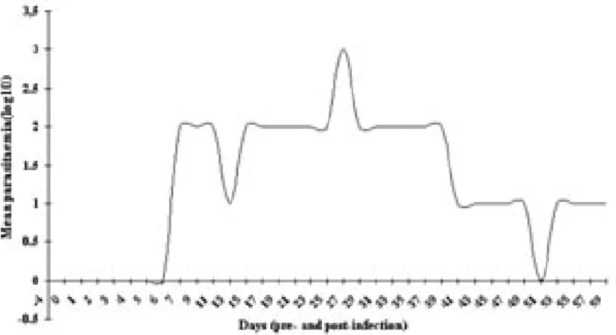
Parasitaemia. All infected goats developed parasitaemia. The pre-patent period varied between 7 and 11 days and showed a waved pattern of presence and absence of parasites characteristics of trypanosomes infection in animals (Fig. 1).
Rectal temperatures. There was a rise in mean rectal temperatures between day 5 and 7 post-infection in the infected buck with a peak mean of 40.08 0C by day 5. Thereafter
the temperature continues to fluctuate throughout the period of investigation.
Packed cell volume. The PCV of the infected goats was observed to be significantly (P<0.05) lower than that of control goats from day 11 up to day 47 post-infection when a gradual recovery in PCV started (Fig. 2)
Fig. 1. Mean parasitemia in T. evansi infected Savannah Brown bucks
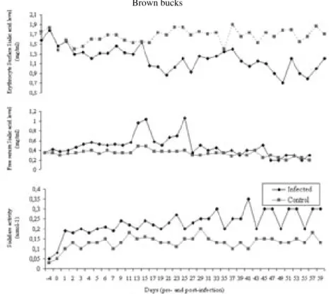
Fig. 4. Sialidase activity, free serum sialic acid and erythrocyte surface sialic acid concentrations in T. evansi infected and control Savannah Brown bucks
Fig. 3. Mean (± SD) haemoglobin concentration in T. evansi infected and control Savannah Brown bucks
Haemoglobin concentrations. The mean haemoglobin concentration declined in the infected group from 8.94 ± 0.21 g/dL on day 0 to 6.50 ± 0.70 on day 33 post-infection. Statistically significant difference (P<0.05) occurred between the infected and control group on days 11, 13, 21, 33, 35, and 35 post-infection (Fig. 3)
Erythrocyte surface sialic acid. A gradual decline in the mean erythrocyte surface sialic acid concentrations was observed starting on day 5 post-infection. However a significant drops in mean erythrocyte surface sialic acid occurred on days 21 (0.87 ± 0.31 mg/mL) and 27 (0.93 ± 0.12 mg/mL). Statistically significant difference (P<0.05) in the mean erythrocyte surface sialic acid concentrations occurred between the infected and control goats on day 5 and between days 17 and 27 post-infection (Fig. 4).
Serum sialic acid concentrations. Elevated levels of mean serum sialic acid concentrations were observed on day 15 (0.96 ± 0.11 mg/mL) and day 17 (1.04 ± 0.4 mg/mL) post-infection. Similarly an elevation occurred on day 27 (1.07 ± 0.06 mg/mL) post-infection. Statistically significant (P<0.05) differences between mean free serum sialic acid concentrations in infected and control goats occurred on days 15, 17, and 27 post-infection (Fig. 4).
Serum sialidase activity. Mean sialidase activity in infected goat increased minimally, whereas such changes did not occur in control goats. Similarly significant (P<0.05) differences occurred between mean sialidase activity in infected and control goats on days 21, 23, 27, 33, and 37 post-infection. It was also found that in some cases the activity of sialidase coincided with the decrease in erythrocyte surface sialic acid and increase in free serum sialic acid concentrations (Fig. 4).
Discussion
The observed clinical features in this study were dullness, emaciation, staggering gait, recumbency and anaemia, which were also recorded by LOSOS (1980) in cattle and by STEPHEN (1986) in horses.
The pre-patent period in this study varied between 7 and 11 days, as earlier reported by VERMA and GAUTAM (1978) on T. evansi-infected buffalos and cow calves and by
STEPHEN (1986) on T. evansi-infected goats. However, it appeared longer than was reported by AUDU et al. (1999) on T. evansi-infected Yankasa sheep. The intensity of parasitaemia ranged between 1-plus (+) and 3-plus (+++), with the highest levels of parasitaemia being recorded during the early phase of infection. This pattern was also reported by ANOSA and ISOUN (1980) on T. evansi-infected small ruminants.
The mean temperatures of infected goats rose for a few days post-infection and then dropped to remain within the normal range throughout the period of the study, thus agreeing with the previous report of VERMA and GAUTAM (1978) on T. evansi-infected buffalos
and cow calves, even though most of their experimental animals were afebrile during the course of the disease. In this study, there was no correlation between parasitaemia and rise in rectal temperature as parasites were detected even during the afebrile period, further agreeing with the reports of VERMA and GAUTAM (1978) on T. evansi-infected buffalos and cow calves.
The T. evansi-infected goats developed moderate levels of anaemia, as evidenced by a gradual fall in PCV and haemoglobin concentrations. The observed decreases in PCV and haemoglobin concentrations coincided with the fluctuating parasitaemia, which suggests that living trypanosomes were responsible for the progressive development of anaemia, as previously observed by OGBADOYI et al. (1999). It was also found in this study that there was a gradual increase in serum sialidase activity, with the activity coinciding in some cases with the decrease in erythrocyte surface sialic acid and the increase in free serum sialic acid concentrations. This suggests that T. evansi produced sialidase, cleaving red cells sialic acid into plasma, further supporting the report that T. evansi infection decreased the sialic content of erythrocyte membranes and increased sialic acid content in plasma (WALIA et al., 1996). More support comes from the observation, in this study, of the decrease in erythrocyte surface sialic acid, which coincided with the increased sialidase activities. Furthermore, free serum sialic acid was found to have increased with the appearance of the trypanosomes, with the highest value being recorded during the peak of parasitaemia and highest sialidase activities by T. evansi. Therefore, the decline in erythrocyte surface sialic acids with the increase in free serum sialic acid may be readily explained. However, the decline in erythrocyte surface sialic acid did not produce a significant increase in free serum sialic acid in T. vivax-infected cattle (ESIEVO et al., 1982). It was suggested that the sialic acid removed from the erythrocytes surface was metabolized or detoxified as soon as it was produced (ESIEVO et al., 1982). Indeed, KRAEMER (1966) also suggested that the enzyme sialyl transferase, present in calf thyroid gland (SPIRO and SPIRO, 1968) regenerated sialic acid on the surface of the cells.
The finding of a very significant increase in free serum sialic acid coinciding with a significant decease in red cell surface sialic acid was contrary to the finding of ESIEVO et al. (1982) which appears to suggest low levels of or absence of sialyl tranferase in goats.
It has been shown that T. vivax produced neuraminidase enzyme both in vitro and in vivo (ESIEVO et al., 1982) in T. vivax-infected cattle and this enzyme has been reported to cleave-off erythrocyte surface sialic acid in cattle (ESIEVO et al., 1982). Therefore, the finding that reduced erythrocyte surface sialic acid occurred concurrently with an increase number of trypanosomes in circulation, accompanied by development of anaemia, was evidence that the trypanosomes might be producing neuraminidase, which in turn cleaves off erythrocyte surface sialic acid, thus rendering them more prone to phagocytosis, as observed previously by ESIEVO et al. (1982) in T. vivax-infected cattle. A similar pathophysiological
mechanism may be operational in the development of anaemia in Savannah Brown bucks as a result of T. evansi infection. It is reasonable to surmise that a similar or identical control measure targeted towards this pathogenesis of the anaemia in T. vivax infection in cattle may be applicable to T. evansi infection in goats.
Conclusion
The findings of this study have provided an insight into the role of loss of erythrocyte surface sialic acid in inducing anaemia in trypanosomosis. It is reasonable to postulate that as
T. evansi multiplied in number in the peripheral circulation of infected goats, they produced neuraminidase which cleaved off erythrocyte surface sialic acid, which caused damage to the cells and rendered them more prone to haemolysis by the reticuloendothelial system. This was also reported by ESIEVO et al. (1982) in T. vivax-infected cattle. In addition, the cleaved Sialic acid from the erythrocyte surfaces possibly created new antigenic sites on the surfaces of the affected erythrocytes, to which antibodies could have been produced against, which further rendered them more prone to erythrophagocytosis (PIROFSKY, 1969).
References
AMINOFF, D. (1961): Methods for the quantitative estimation of N-acetylneuraminic acid and their application to hydrolytes of sialomucoids. J. Biol. Chem. 81, 384 -392.
ANOSA, V. O., T. T. ISOUN (1980): Further observations on testicular pathology in T. vivax infection of sheep and goats. Res. Vet. Sc. 28, 151-160.
AUDU, P. A., K. A. N. ESIEVO, G. MOHAMMED, O. J. AJANUSI (1999): Studies of infectivity and pathogenicity of an isolate of Trypanosoma evansi in Yankasa sheep. Vet. Parasitol. 86, 185-90.
BARRY, G. T. (1959): Detection of sialic acid in various Escherichia coli strains and in other species of bacteria. Nature 183, 117-118.
BRATOSIN, O., J. MAZURIER, H. DEBRAY, M. LECOCG, B. BOILLY, C. ALONSO (1995): Flow cytofluorimetric and lyses of young and senescent human erythrocytes probed with lectins. Evidence that sialic acid control their life span. Glycocon. J. 12, 258-267.
CORFIELD, A. P., R. SCHAUER (1982): Occurrence of sialic acids. In: Sialic Acids Chemistry, Metabolism and Function. (Schauer, R., Ed.). Springer, Wien New York. pp. 5-50
DODGE, J. T., C. MITCHELL, D. J. HANAHAH (1963): The preparation and chemical characteristics of haemoglobin-free ghosts of human erythrocytes Arch. Biochem. Biophy. 100, 119-130 ESIEVO, K A. N., D. I. SAROR, A. A. ILEMOBADE, M. H. HALLAWAY, (1982): Variation
in erythrocyte surface and free serum sialic acid concentrations during experimental T. vivax infection in cattle. Res. Vet. Sci. 32, 1-5.
HOMES, P. H., F. W. JENNING (1976): Pathogenicity of Parasitic Infections. Academic press. New York, p. 199.
KRAEMER, P. M. (1966): Regeneration of sialic acid on the surface of Chinese hamster cells in the culture. I. General characteristics of the replacement process. J. Cell. Physiol. 68, 85-90. LOSOS, G. J. (1980): Diseases caused by Trypanosoma evansi, A review. Vet. Res. Comm. 4,
165-181.
OGBADOYI, E. O., A. I. UKOHA, E. K. KYEWALABE (1999): Anaemia in experimental African trypanosomiasis. J. Protozool. Res. 9, 55-63.
PARIS, J., M. MURRAY, F. McODINBA (1982): A comparative evaluation of parasitological techniques currently available for the diagnosis of African trypanosomiasis in cattle. Act. Trop. 39, 307-316.
PIROFSKY, B. (1969): Auto immunization and the auto immune haemolytic anaemias. 1st ed. The
William and Wilkins Co., Baltimore, U.S.A. pp. 418-430.
SCHALM, O. W., N. C. JAIN, E. J. CARROLL (1975): Veterinary Hematology, 3rd ed., Lea and
Febiger, Philadelphia. pp. 32, 370, 372, 379, 422, 606.
SCHAUER, R., J. P. KAMERLING (1997): Chemistry, biochemistry and biology of sialic acid, In: Glycoproteins. (Montrevil, J., J. F. G. Vigenthar, H. Shachter, Eds.). Elsevier Amsterdam. pp. 11, 241-400.
SCHAUER, R., S. KELM, G. REUTER, P. ROGGENTIN, L. SHAW (1995): Biochemistry and Role of Sialic Acids In: Biology of Sialic acids. Chapter 2. Rosenberg, A. (ed) Plenum Press New York. pp. 7-67.
SPIRO, M. J., R. C. SPIRO 1968): Glycoproteins biosynthesis studies on thyroglobulin. J. Biol. Chem. 243, 6520-6528.
STEPHEN, L. E. (1986): Trypanosoma (Trypanozoan) evansi (Steel, 1885) Balbiani, 1888. X in trypanosomiasis: A veterinary perspective, 1st ed., lome. (Stephen, E., Ed.), Pergamon Press
Oxford, England. pp. 184-215.
TRAVING, P., R. SCHAUER (1998): Structure, function and metabolism of sialic acids. C.M.L.S. Cell. Mol. Life Sci. 54, 1330-1349.
VARKI, A. (1992): Diversity in the sialic acids. Glycobiol. 2, 25-40.
VERMA, B. B., O. P. GAUTAM (1978): Studies on experimental Surra (Trypanosoma evansi infection) in Buffalo and cow calves. Indian Vet. J. 55, 648-653.
WALIA, P. S., I. S. KALRA, P. D. JUYAL, S. P. AHUJA (1996): Role of sialidase activity of Trypanosoma evansi in inducing anaemia and immunomodulation in buffalo calves. J. Vet. Parasitol. 10, 1-9.
WARREN, L.(1959): The thiobarbituric acids assay for sialic acids. J. Biol. Chem. 234, 1971-1975.
WARREN, L. (1963): Assay of sialic acids. Methods Enzymol. 6, 463-465.
Received: 9 April 2005 Accepted: 3 November 2006
.
SHEHU, S. A., N. D. G. IBRAHIM, K. A. N. ESIEVO, G. MOHAMMED: Uloga sijalične kiseline na površini eritrocita u nastanku anemije u jaraca Savannah smeđe pasmine. Vet. arhiv 76, 521-530, 2006.
SAŽETAK
U jaraca Savannah smeđe pasmine, u tijeku pokusne zaraze tripanosomom Trypanosoma evansi, određivana je koncentracija sijalične kiseline na površini eritrocita, zatim koncentracija slobodne sijalične kiseline u serumu te aktivnost sijalidaze. Svi invadirani mužjaci oboljeli su od tripanosomoze sa značajnim opadanjem srednje vrijednosti hematokrita do vrijednosti 19,50 ± 2,12%. Navedeni je pad uslijedio 33 dana nakon infekcije i bio statistički značajan u odnosu na kontrolnu vrijednost 25,75 ± 0,96. Srednja koncentracija hemoglobina također je pala u invadiranih mužjaka s najizraženijim padom od 6,50 ± 0,70 g/dL 33. dana nakon zaraze. Ta vrijednost bila je također značajno različita (P<0,05) u odnosu na kontrolnu skupinu jaraca (8,53 ± 0,46 g/dL). Petoga dana nakon infekcije, anemija je nastavljena postupnim opadanjem srednje koncentracije sijalične kiseline na površini eritrocita. Utvrđene su značajne razlike (P<0,05) u srednjoj koncentraciji sijalične kiseline na površini eritrocita između zaražene i kontrolne skupine 5. dana, te između 17. i 27. dana nakon infekcije. U usporedbi s kontrolnom skupinom, u zaraženih životinja utvrđen je signifikantan (P<0,05) porast koncentracije slobodne sijalične kiseline u serumu 15., 17. i 27. dana. Aktivnost sijalidaze bila je značajno (P<0,05) povišena 7., 9., 21., 23., 27., 33. i 37. dana nakon infekcije. Pojava anemije tijekom infekcije može se povezati s aktivnošću tripanosoma u krvnom optjecaju koji tvore sijalidazu (neuraminidazu), a time i cijepanje sijalične kiseline na površini eritrocita čineći ih podložnijima za fagocitozu u retikuloendotelijalnom sustavu.