The Presence of Mercury Resistant Bacteria in Sediment of
Gold Processing Plant at Waekerta Village of Buru District,
Maluku Province and Their Activity in Reducing Mercury
SARMAWATY KOTALA
1, RETNO KAWURI
2and IDA BAGUS WAYAN GUNAM
31Student of Magister Biology Science Udayana University, Bali - 80114, Indonesia. 2Biology Department, Faculty of Mathematic and Natural Sciences,
Udayana University, Bali - 80361, Indonesia.
3Agroindustrial Technology Department, Udayana University, Bali - 80361, Indonesia.
http://dx.doi.org/10.12944/CWE.9.2.07
(Received: April 21, 2014; Accepted: May 17, 2014)
ABSTRACT
Mercury was one of the heavy metal polute in environment and had the toxic characteristic to the living creatures. Golden mining in Waeapo subdistrict used mercury to extract the gold and exile the waste to the environment freely. Several precedented research showed that waste sediment of gold processing contains mercury resistance bacteria. Mercury resistance bacteria can be used as bioremediation agent because those bacteria can reduce mercury. Mercury resistance bacteria has mer operon which contained in plasmid. The goal of this research is to isolate mercury resistance bacteria which is able to grow on medium nutrient agar (NA) containing 500 ppm of HgCl2 and to analyze the capability in HgCl2 reduction in nutrient broth (NB) medium. Bacteria isolation was done by platting method on Nutrient Agar containing 10 ppm of HgCl2. Bacteria identification was done by kit Microgen TM GnA + B-ID System and to know bacteria capability in reducing mercury was done by CV-AAS (Cold Vapour Atomic Absorption Spectrophotometer). Result showed, that the bacteria found in this research were Bacillus sp and Aeromonas hydrophila. Both of these bacteria were able to reduce HgCl2 in the amount of 98,7% for Bacillus sp and 98,33% for Aeromonas hydrophila. In the future those bacteria can be use as bioremediation agent.
Key words: Mercury Resistant Bacteria, Bacillus sp, Aeromonas hydrophila, Gold Processing.
INTRODUCTION
Mercury utilizing in golden mining could produce waste, which contains mercury and causes environment pollution. Mercury belongs to heavy metal which is toxic to living creatures. Mercury can attack the arrangement of central nervous and causes memory loss, tremors and decreases motion capability. Poisoning causing destruction of a fetus has been detected. Miniamata desease in Japan is the example of mercury poisoning1,2.
Mercury as a pollutant in the environment need attention and problem solving. Mercury detoxification can be done chemically by precipitation,
coagulation, reverseosmosis, ion exchange resins and adsorption using activated carbon3,4.However, this process is relatively expensive and could cause new problems, namely the accumulation of these compounds insediment and aquatic organisms4.
from sediment of gold processing which able to grow on NA medium containing 500 ppm of HgCl2 and to analize the capability of reduction of HgCl2.
MATERIAL AND METHODS Research Materials
This study used a sample of sediment taken from a waste disposal site of gold processing in Waekerta Village, Maluku. Materials used in this study were Nutrient Agar (Merck),Nutrient Broth (Merck), and HgCl2.
Research Instrument
The instrument used in this research is a hot plate, autoclave, vortex, incubator, kit Microgen TM GnA + B-ID System, microscopes, spectrophotometers, CV-AAS, and laminar air flow cabinet.
Sampling
Samples were taken as much as 20% from 42 gold processing sites in Waekerta Village, so that 9 locations were choose to get the sediment samples. Samples taken from each location on 5 different points were then mixed into one. Land sample from mining land was used as comparative so that total of all samples become ten. Location of sampling sites in the village of Waekerta as shown in Figure 2.
Bacteria Isolation
Mercury resistance bacteria isolation was done by spread plate method6. Sedimen and soil sample were diluted in a series(10-1, 10-2and10-3) with saline solution(0.85% NaCl). From the 10-3dilution were taken 0,1 ml and spread on petri dishes containing selective media namely nutrient agar (NA) containing 10 ppm of HgCl2. Then incubated at room temperature for 3 days. Grown bacterial isolates with different colonies morphological characters were reisolated again to a new medium in order to get pure cultures and stored in an agar slant for further testing.
Mercury Recistance Bacteria Selections Bacteria selection is based on the ability of bacterial isolates grown in medium with various HgCl2 concentrations. Bacterial isolates were grown by streaking method on NA medium which
contain 25 ppm of HgCl2 and incubated at room temperature for 24 hours. If the isolates grow, then chese bacterial isolates were re-grown by streaking method on the NA medium added with HgCl2 with a higher concentration of 50 ppm, 100 ppm, 250 ppm, 400 ppm, 500 pp min order to obtains uperior isolates, which were able to live in the highest HgCl2 concentration. Purified isolates was stored in nutrient agar slant medium with a temperature of 20° C.
Mercury Resistance Bacteria Identification Parameters observed for identification of mercury-resistant bacteria are colony form on NA medium, Gram staining, and character physiology (biochemical test). Physiological characteristics were tested using Microgen™ kit GNA+B-ID System Identification (Microgen Bioproduct, UK).
Determination of Optimum Temperature on the Growth of Mercury Resistant Bacteria
To deter mine the optimum growth temperature, the bacterial isolates were grown on nutrient broth medium and incubated a variety of temperature is: 25°C, 30°C, 37°C, and 45°C. Cultures were incubated at this temperature for 24 hours. Further growth of the isolates was measured degree of turbidity with a spectrophotometer at a wavelength of 620 nm. Absorbance values of bacterial cells can be observed at a wavelength of 620 nm, each treatment was repeated 3 times.
Determination of Optimum pH on Mercury Resistant Bacteria Growth
To determine the optimum pH of growth, the bacterial isolates were grown in nurient broth with a pH of 5, 6, 7, 8, and 9. Cultures were incubated at the optimum temperature for 24 hours. Growth of isolates was measured with a spectrophotometer at a wavelength of 620 nm and each treatment was repeated 3 times.
medium at a concentration of 10 ppm HgCl2 and incubated at room temperature on arotaryshaker (100 rpm). Suspension culture absorbance values were measured at a wavelength of 620 nm. Absorbance measurements were started from 0 hour up to 72 hours with an interval of 4 hours. Obtained absorbance data was then conversed into the growth curve. On the x-axis is time and and on the y-axis is absorbance. The growth curve will be compared with the growth curve of bacteria in NB medium without HgCl2.
Mercury Reducing Bacteria Activity Test Mercury reducing bacteria activity test was carried outtolook at the ability of superior isolates in reducing Hg. In this testing phase bacterial isolates were grown in NB medium for 24 hours in 250 ml erlenmeyer, then isolated cells were washed using saline solution and the absorbance was measured using a spectrophotometer at a wavelength of 620 nm. Culture with absorbance value of 2 was taken 0.1 ml and grown in 50 ml NB medium containing a concentration of 100 ppm HgCl2, then incubated for 7 days on top shaker (100 rpm). Furthermore, bacterial cells were separated from the medium by using a membran filter with the size of 0.2 µm. Hg concentration remaining in the medium was measured by Cold Vapour NB Atomic Absorption Spectrophotometer (CV - AAS). In addition, NB medium containing 100 ppm of HgCl2 without inoculated with bacteria resistant to mercury was used as a positive control and NB medium without HgCl2 and mercury resistant bacteria was used as negative control. The principle of CV-AAS working is to change the mercury dioxide compounds into the mercury ion, mercury ion subsequently reduced to metallic mercury and the cold vapor atomic absorption of it was analyzed at a wavelength of 253.7 nm. Reagents used were SnCl2 reductant, H2SO4 + HCl acid solution (Rondonuwu, 2011).To determine the levels of mercury removal efficiency, this formula was used:
The data were analyzed qualitatively and quantitatively. Qualitatively is by describing the results of the characterization and identification of mercury-resistant bacterial isolates were able to reduce mercury. Quantitatively, on the pH test and growth curve measurement was done by measuring the number of bacterial cells through the absorbance. The data obtained was made in the form of a bar graph, but the growth curve in the form of a line graph using Microsoft Excel program.
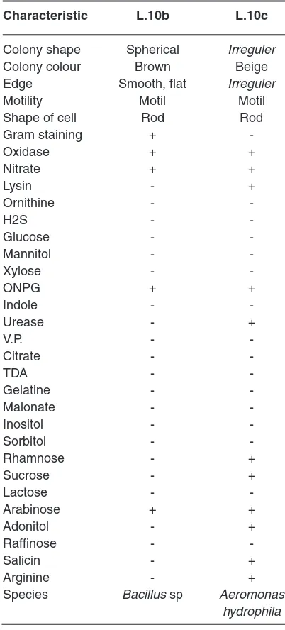
Table 1: Characteristic of L.10b and L.10c Characteristic L.10b L.10c Colony shape Spherical Irreguler
Colony colour Brown Beige
Edge Smooth, flat Irreguler
Motility Motil Motil
RESULTS AND DISCUSSION
Two isolates of mercury resistant bacteria capable of living NA medium containing 500 ppm of HgCl2 was found in gold processing sediment samples. The isolates were L.10b and L.10c. After identification, these isolates were identified as Bacillus sp and Aeromonas hydrophyla. Both macroscopic and microscopic forms of the isolate scan be seen in Figure 3 and Figure 4 as well as the character of each isolate are shown in Table1.
Bacillus sp was found as a mercury resistant bacteria in Japan and India7. In addition, Bacillus sp was also found in the Tondano river, Indonesia8. Bacillus cereus and Bacillus subtilis found in the Kalimas river Surabaya are also resistant to mercury9. Bacillus sp is more often
found as mercury resistant bacteria compared to Aeromonas hydrophila. Aeromonas hydrophyla has been found in gold mining sediment contaminated by Hgin Bandung, West Javaandable to grow at 550 mg/L HgCl210. In addition, some strains of A.hydrophila is found in sea water, fish, and waste water contaminated by heavy metals in Tunisia11.
Temperature is one of the environmental factors that influence the growth of bacteria. Bacillus sp. has the highest absorbance value (0.216) at 25°C and the lowest (0.118) at a temperature of 45°C. Aeromonas hydrophila has a high absorbance value (0.404) at 37°C and the lowest (0.224) at a temperature of 45°C (Figure 5). Temperature effect on bacterial growth because temperature affects the activity of enzymes in metabolism. The temperature affect the chemical reactions in the
Table 2: The Test Results in Reducing Mercury The concentration
of mercury Average Standard
Sample remaining in the efficiency deviation
medium (ppm) (%)
A 1,67 98,33 0,172
B 1,33 98,7 0,162
P 100 0 0
N 0 0 0
Description: A = treatment using bacteria Aeromonas hydrophila, B = treatment using bacteria Bacillus sp., P = positive control (NB medium containing 100 ppm of HgCl2), N = negative control (NB medium without HgCl2).
Fig. 1: Gold mining in Waekerta village(a) Gold processing, (b) Waste of gold processing in Environment
process of bacterial growth, growth rate, and the total amount of the growth of microorganisms12. Although the absorbance values of different bacteria are categorized both mesophilic bacteria. Mesophilic bacteria is a group of bacteria that can grow at a temperature of 20-45°C13.
Bacterial growth can be affected by various environmental factors, one of which is the pH of the medium. The degree of acidity of the medium affects the growth of Bacillus sp and A. hydrophyla. Bacillus sp grows optimally at pH 6 with a absorbance value of 0.106 and the absorbance values decreased when the pH of the medium increased (Figure 6).
In contrast to Bacillus sp,A. hydrophila has the highest absorbance value (0.192 ) at pH 7 and the lowest (0.11) at pH 5 (Figure 6). The degree of acidity affects the growth of bacteria because the pH affects the enzymes in the metabolism of bacteria. Enzyme activity will decrease if the pH is not appropriate, this is because the enzyme will be active in a proper state of ionization. The appropriate ionization conditions for different enzymes are also differ but generally ranges at pH 6-814. The enzyme can be denatured due to changes in pH. The enzyme works at neutral pH and will become inactive when the environment becomes very acidic or very alkaline15. Based on the growth ability in that pH range, Bacillus sp, and
Fig: 2: Map of Sampling Location: (a) Map of Maluku Province, Indonesia (source: Malukuonline. co.id); (b) Map of Buru District (source: informasi-maluku.Blogspot.com); (c) Map of Waeapo
Subdistrict, Waekerta Village (arrow) (source: minerthink.wordpress.com)
(a) (b)
A. hydrophila can be classified into the neutrophils bacteria. Neutrophil is a bacterial groups were able to grow at pH 6-814,15.
The growth of Bacillus sp and Aeromonas hydrophila in NB medium containing 10 ppm of HgCl2 and incubated for 3 days has not reached the stationary phase. The results obtained were different with control Bacillus sp, which reached stationary phase at 37th and A. hydrophila which reached the stationary phase at the 44th and death phase in the 68th hour (Figure 7). During the period of incubation with medium containing 10 ppm HgCl2, both of these bacteria were only able to reach the exponential phase. Bacillus sp achieve exponential phase at 68th and A. hydrophila at 48th hours (Figure 7). This is because the adaptation phase is long enough. This is due to HgCl2in the medium.In the
adaptation phase the synthesis of the new enzymes occurs, according to the media and the increase of cell numbers not found14. The length of the adaptation phase in medium containing HgCl2 occur in bacteria Ochrobactrum sp S79 and L6T2 isolates, wherein the second stationary phase of these bacteria occurs on day 4 to day 9 of incubation time16.
Aeromonas hydrophila and Bacillus sp resistant and able to reduce mercury levels of 100 ppm to 1.67 ppm for A.hydrophila and 1.33 ppm for Bacillus sp after incubated for 7 days. The results of mercury content remaining in the medium, were used to determine the efficiency of both bacteria in reducing mercury. Bacillus sp able to reduce mercury by 98.7%, where as A.hydrophila was 98.33% (Table 2). The ability of Bacillus sp and A.hydrophila in reducing mercury levels associated with a character
Fig. 4: (a) Aeromonas hydrophila, (b) microscopic, rod shape, 1000 x magnification
(a) (b)
Fig. 3: (a) Bacillus sp, (b) microscopic, rod shape, 1000 x magnification (arrow)
that is resistant to mercury. Bacterial resistance to mercury due to the mer operon contained in the plasmid4,5.
Mer operon consists of a wide variety of mergenes. Each bacterium has its own mergene
variations in the mer operon3. But the mechanism of bacterial resistance to inorganic mercury is almost the same in different bacteria species. This is due to the reduction of mercury from Hg2+ to Hg0 induced by mercuric ion reductase enzyme encoded by the mer operongenes Mer A2. Mercuric ion reductase formsa
Fig. 5: Graph the effect of incubation temperature on the growth of mercury-resistant bacteria (incubation period of 24 hours)
Fig. 6: Graph of the effect of pH on the growth of mercury-resistant bacteria
bond with Hg2+ and reduction occurs by the transfer of electrons through the flavin bond from NADPH into NADP, so that reduced Hg was formed, ie Hg05. The reduction of Hg2+ to Hg0 is away to remove oxidized mercury and to reduce mercury dissolved in a medium17.
Some bacteria of the Genus Bacillus are known to have a gene variation in meroperon. Bacillus megaterium and Bacillus macroides is abroad-spectrum mercury-resistant bacteria, whereas Bacillus cereus and Bacillus licheniformis are an arrow-spectrum mercury-resistant bacteria18. Bacteria which only has mercury reductase protein (MerA) is called by a narrow spectrum resistant bacteria, while broad-spectrum mercury-resistant bacteria are bacteria that have mercury reductase protein (MerA) and protein organo merkurilyase (MerB). Mer Bfunctions in catalyzing the termination of the mercury-carbon bond to produce organic compounds and ionic Hg in the form of salt thiols20. Bacillus sp and A.hydrophila found in
this study are not known the extent of the spectrum which is owned in reducing mercury.
Until now there has been no reports of mergene variations that are owned by Aeromonas hydrophila. However other species of the Genus Aeromonas are known variations in the mer operongenes.Aeromonas salmonicida has some mer genes in the mer operon, namely Mer A,Mer P, MerR, MerE, MerT, MerD, and MerB19. Aeromonas hydrophila is able to change the shape of the cells, from rod into a round shape after mercury exposure11.
ACKNOWLEDGMENT
The authors would like to express their aprreciation to the Head of Magister Biology Science and Udayana University Bali Indonesia for supporting this study. Appreciation is also send to Governor of Maluku province, Indonesia for the support in carrying out this research work.
REFERENCE
1. Chowdur y, S., Bala, N.N., Dhauria, P. International Journal of Pharmaceutical, Chemical, and Biological Science 2(4): 600-611 (2012).
2. UNEP. Global Mercury Assesment. Inter-Organization Programme for The Sound Management of Chemicals.Issued by UNEP Chemicals. Geneva, Switzerland. (2002) 3. Okoronkwo, N. E., Igwe, J. C., Okoronkwo, I.
J.African Journal of Biotechnology 6(4): 337 (2006)
4. D a s h , H . R . , D a s, S. I n t e r n a t i o n a l Biodeterioration and Biodegradation. 75 : 207-213 (2012).
5. Barkay, T., Susan, M. M., Anne, O. S. FEMS Microbiology Reviews. 27: 355-384 (2003). 6. Dubey, R.C., D.K. Maheshwari. Practical
Microbiology. S.Chand and Company LTD, New Delhi, 37 (2007)
7. Osborn, A.M., Kenneth, D.B., Peter, S., Donald, A.R. FEMS Microbiology Reviews. 19: 239-262 (1997)
8. Manampiring, A.E., Billy, J.K. Jurnal Ilmiah
Sains. 11 (1): 26-30 (2011)
9. Zulaika, E., Langkah, S., Agus, S. Journal of Basic and Applied Scientific Research, 2(7): 7263-7269 (2012)
10. Chaerun, S.K., Sakinah, H., Edy, S., Maelita, R.M. Microbiology, 6(2): 57-68 (2012) 2002. Microbial Life. Sinauer Associates Publishers,Massachusetts: 142: (2002) 14. Purwoko, T. 2009. Physiology of Microbe.
Penerbit Bumi Aksara, Jakarta, 234 (2009) 15. McKane, L., Judy, K. 1996. Microbiology
Essentials and Aplications Second Edition. McGraw-Hill, USA, (1996)
16. Imamuddin, H. Jurnal Ekosains 2(1); 26-32 (2010).
International Journal of Pharmaceutical, Chemical, and Biological Science. 2(4), 600-611 (2011).
18. Narita, M., Kazuyuki, C., Hiroshi, N., Hidenori, I., Chieh-Chen, H., Zen’ichiro, K., Simon, S., Ginro, E. FEMS Microbiology Reviews. 223; 73-82 (2003)
19. McIntosh, D., Michelle, C., Baijing, J., Frank,
A.F., Erin, M.P., Sarah, E.C., Zachary, B.Z., Ilana, C.G., Russel, D., Keith, A.J., Mike, B., Rachael, R. Journal of Antimicrobial Chemotheraphy. 61; 1221-1228 (2008) 20. Huang, C.C., Chien, M.F., Lin, K.H.