OTOMYCOSIS
|dolly
1
Otomycosis
Yan Edward, Dolly Irfandy
Otorhinolaryngology Head and Neck Surgery Department Medical Faculty of Andalas University/Dr. M. Djamil Hospital
ABSTRACT
Otomycosis is one of the common conditions encountered in a general otolaryngology clinic. The disease process a challenging and frustrating entity for both patients and otolaryngologists for it requires long term treatment and recurrence rate remains.
One case of otomycosis in a 41 years old woman is reported. The diagnosis was based on anamnesis, physical examination and KOH test. From laboratory examination, Aspergillus niger was isolated as etiologic agent. With the treatment of ear toilet and combination of Gentian violet an improvement was observed.
Keywords: Otomycosis, Aspergillus sp,Therapy ABSTRAK
Otomikosis adalah salah satu kondisi yang umum ditemukan di klinik THT. Penyakit ini merupakan tantangan dan menimbulkan rasa frustrasi bagi pasien dan dokter ahli THT. Hal ini disebabkan pengobatan yang memerlukan waktu lama dan rerata kekambuhan yang tinggi.
Dilaporkan satu kasus otomikosis pada seorang wanita umur 41 tahun. Diagnosis ditegakkan berdasarkan anamnesis, pemeriksaan fisik dan tes KOH. Dari pemeriksaaan laboratorium ditemukan Aspergillus niger sebagai penyebab. Dengan terapi pembersihan liang telinga dan obat oles telinga kombinasi gentian violet terdapat perbaikan.
Katakunci: Otomikosis, Aspergillus sp, Terapi
Korespondensi: dr. Dolly Irfandy: d_irfandy@yahoo.com
INTRODUCTION
Otomycosis is a fungal infection of the skin of the external canal. Although fungi may be the primary pathogens, they are usually superimposed on chronic bacterial infection of the external canal or middle ear.1,2,3 Although rarely life threatening, the disease process a challenging and frustrating entity for both patients and otolaryngologist for it frequently requires long term treatment and follow up, yet the reccurence rate remains high.4 Otomycosis or external otitis fungi are acute, subacute or chronic infections produced by yeasts and filamentous fungi that affect the squamous epithelium of the external auditory canal (EAC)3.
Although there has been controversy with respect whether the fungi are the true infective agents versus mere colonization species as a result of compromised local host immunity secondary to bacterial infection, most clinical and laboratory evidence to date supports otomycosis as a true pathologic entity with Candida and Aspergillus as the most common fungal species isolated.1-4
FREQUENCY
It has been estimated that cases of otitis externa was between 5-20 % of all otologic consultations.3 Otomycosis is a common pathology throughout the world. Its frequency varies according to different geographic zones from 9 to over 50 % of all patients with otitis externa,5 in relation to environmental factors (temperature, relative
humidity) and times of years.3 Moist and tropical environments provide the required milleu for fungal proliferation and increased in incidence may be contributed to increased sweat and environmental humidity altering surface epithelium of the EAC.5 Some studies found greater otomycosis frequency in women6,7
ANATOMY
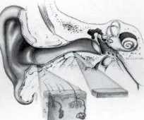
The external ear is composed of the auricle and external auditory canal. Both contain elastic cartilage derived from mesoderm and a small amount of subcutaneous tissue, covered by skin with its adnexal appendages. There is fat but no cartilage in the lobule.1,2 The external auditory canal (EAC) is typically 24 mm in length with a volume of 1-2 mL. The lateral third of the canal is made of fibrocartilage, whereas the medial two thirds are osseous.2
OTOMYCOSIS
|dolly
2
SKINThe EAC is lined by stratified squamous epithelium that is continuous with the skin of the pinna and the epithelial covering of the tympanic membrane. The subcutaneous layer of the cartilaginous portion of the canal contains hair follicles, sebaceous glands, and ceruminous glands, and is up to 1 mm thick. The skin of the osseous canal doesnt have subcutaneous elements and only 0.2 mm thick.
INNERVATION
Sensation to the auricle and external auditory canal is supplied from cutaneous and cranial nerves, with contributions from the auriculotemporal branches of the trigeminal (V), facial (VII), glossopharyngeal (IX), and vagus (X) nerves and the greater auricular nerve from the cervical plexus (C2-3). The vestigial extrinsic muscles of the ear, anterior, superior, and posterior auricular, are supplied by the facial nerve (VII).1
Figure 2. Microscopic view of the normal apopilosebaceous unit, demonstrating drainage of the secretions of the sebaceous and modified apocrine glands into the follicular canal of the hair follicle2
The ceruminous glands are modified apocrine sweat glands surrounded by myoepithelial cells; they are organized into apopilosebaceous units (Figure2). Cerumen prevents canal maceration has antibacterial properties and has normally acidic pH all of which contribute to an hospitable environment for pathogens 1,2
ETIOLOGY
In 80% of cases, the etiologic agent is
Aspergillus, whereas Candida is the next most frequently isolated fungus. Other more rare fungal pathogens include Phycomycetes, Rhizopus,
Actinomyces, and Penicillium.2 Aspergillus niger is usually predominant agent although A.flavus, A.fumigatus, A.terreus (filamentous fungi), Candida albicans and C.parapsilosis (yeast-like) are also common.3
Kumar (2005) studied otomycosis patients and isolated Aspergillus niger (52.43%), Aspergillus fumigates (34.14%), Candida albicans (11%), Candida pseudotropicalis (1.21%) and Mucor sp (1.21%). Some workers have reported other organisms as causative
isolates such as Penicillium sp and other known species of Candida such as C.parapsilosis, C.gulliermondi with varying percentages of isolation.8 Ahmad et al (1989) carried out a prospective study in 53 patients at ENT Department Faculty of Medicine University of Indonesia. They isolated Aspergillus species more frequently than Candida species.9
PATHOGENESIS AND PREDISPOSING FACTORS
Otomycosis is connected to the histology and physiology of EAC. This 2,5 cm long, 7-9 mm wide cylindrical canal is lined with a stratified keratinized squamous epithelium that continues along external side of tympanic membrane. At interior tympanic recess, medial to isthmus tends to accumulate remains of keratin and cerumen and it s difficult area to clean.3,10
Cerumen has antimycotic and bacteriostatic properties and is insect repellent. It is composed of lipids (46 to 73%), proteins, free amino acids and mineral ions it also contains lysozym, immunoglobulins and polyunsaturated fatty acids. Long chain fatty acids present in unbroken skin probably inhibit bacterial growth. Because of its hydrophobic composition, cerumen is capable to repelling water, made canal surface impermeable and avoid maceration and epithelial damage.3
The normal microorganisms found in EAC such as Staphylococcus epidermidis, Corrynebacterium sp, Bacillus sp, Gram-positive cocci (Staphylococcus aureus, Sterptococcus sp, non-pathogenic micrococci), Gram-negative bacilli (Pseudomonas aeruginosa, Escherichia coli, Haemophilus influenza, Moraxella catharalis etc) and mycelia fungi from Genus Aspergillus and Candida sp. This commensal microorganisms is not pathogen unless balance still remains between bacteria and fungi.3
Various factors influence transformation saprophytic fungi into pathogenic such as:
- Environmental factors (heat, humidity) commonly patients admitted at summer and autumn when it was hot and humid.3,4,6,11
- Changes in epithelial covering (dermatological diseases, micro traumas).
- Increase in pH level in EAC (bathing).3,4,11 Ozcan et al (2003) found that regular swimmer was reported as possible predisposing factor for otomycosis.3,4,6,8
- Qualitative and quantitative alteration cerumen (bathing). There appears to be little consensus with respect to predisposing factors for otomycosis. In addition, cerumen has been speculated to be supportive for fungal growth. 3,4,11
- Systemic factors (alterations in immunity, debilitating diseases, corticosteroids, antibiotics, cytostatics, neoplasia). Jackman et al (2005) reported ofloxacin may contribute to development of otomycosis.10
OTOMYCOSIS
|dolly
3
externa. A disrupted epithelial surface was agood medium for microorganism s growth. Epithelial damage also leads to decrease excretion from apocrine and cerumen glands which changes EAC environment became more suitable for microorganisms (normal pH 3-4).10
- Dermatomycosis can be a risk factor for recurrence because autoinoculation may be possible among parts of body.6-8
- Conditions and social habits. Female wearing traditional head covers were reported as predisposing factors for otomycosis. Traditional head coverings might increase humidity in ear canal and create ideal environment for fungal growth.6,7
CLINICAL FINDINGS
Symptoms of bacterial otitis externa and otomycosis are often indistinguishable. However pruritus is almost frequent characteristics for mycotic infections and also discomfort, hearing loss, tinnitus, aural fullness, otalgia and discharge.1-4,11
Otoscopy often reveals mycelia, establishing the diagnosis. The EAC may be erythematous and fungal debris may appear white, gray, or black. Patients have typically been tried on topical antibacterial agents with no significant response. The diagnosis can be confirmed by identifying fungal elements on a KOH preparation or by a positive fungal culture.1,2
The characteristics physical examination of fungal infection resembles that of common molds, with visible, delicate hyphae and spores (conidiophores) being seen in Aspergillus. Candida, a yeast, often forms mycelia mats with white character when it s mixed with cerumen they appear yellowish.5 Candidal infections can be more difficult to detect clinically because of its lack of characteristic appearance like Aspergillus such as otorrhea an not responding to aural antimicrobial. Otomycosis attributed to Candida is often identified by culture data.4 There appears to be no reported difference in presentation based on most prevalent organisms.5
LABORATORIUM EXAMINATION
Culture is rarely needed and does not an alter management.5 The fungi that produce otomycosis are generally saprophytic fungi species that abound in nature and that form a part of the commensal flora of healthy EAC. These fungi are commonly Aspergillus and
Candida. Aspergillus niger is usually the predominant agent although A. flavus, A. fumigatus, A. terreus
(filamentous fungi), Candida albicans and C. parapsilosis (yeast-like fungi) are also common.3
The morphology of the colony enabled us to distinguish between yeast-like and filamentous fungi. The majority of white creamy, smooth or rough colonies are yeasts or, very occasionally, the yeast-like phase of dimorphic fungi. Filamentous fungi tend to grow forming dusty, hairy, woolly, velvety or folded colonies that display a wide range of colors such as white, yellow, green, greenish blue, off-black, etc3
Ahmad et al (1989) in their study compared otomycosis diagnosis based on clinical examination and laboratory examination. They found no significant difference between those examinations and concluded generally that otomycosis can be diagnosed from clinical examination only.9
DIFFERENTIAL DIAGNOSIS
Otomycosis is occasionally difficult to distinguish from other form of otitis externa especially diffuse otitis externa. Mixed infection sometimes occurs.12 Kumar (2005) detected bacterial co infection among 44 cases of total 82 cases. Commonly isolated bacteria included negative coagulase staphylococci, pseudomonas sp. Staphylococcus aureus, E.coli and Klebsiella sp.8 Fungal infection may also develop in chronic suppurative otitis media.12
TREATMENT
Although multiple in vitro studies have examined efficacy from various antifungal agents. There is no consensus on most effective agent. Various agents have been used in clinic with variable success. Nevertheless, application of appropriate topical antifungal agents coupled with frequent mechanical debridement11 Usually results in prompt resolution of symptoms although recurrence or residual disease can be common.4
Many authors believe that important to identify causal agent in otomycosis cases in order to use an appropriate treatment. It s also recommended in chosen antimycotic should be based on susceptibility in identified species. However, others believe the most important therapeutic strategy is when we choose specific treatment for otomycosis based on efficacy and characterics drugs regardless of the causal agent. To date there is no FDA approved antifungal otic prescription for otomycosis treatment. Many agents with various antimycotic properties have been used and clinicians have stugle to identify most effective agent to treat this condition.11
Antifungal preparations can be divided into non specific and specific types. Non specific antifungals included acidic and dehydrating solutions such as:
- Boric acid is a medium acid and often used as a approved. Studies report an up to 80% efficacy.5,11
- Castellani s paint (acetone, alcohol, phenol, fuchsin, resocinol)
- Cresylate (merthiolate, M-cresyl acetate, propylene glycol, boric acid and alcohol).5,11
OTOMYCOSIS
|dolly
4
specifically for cases in humid environment withefficacy between 95.8% and 100%.5,11 Specific antifungal therapies consist of:
- Nystatin is a polyene macrolide antibiotic that inhibits sterol synthesis in cytoplasmic membrane. Many molds and yeasts are sensitive to nystatin including Candida species. A major advantage from nystatin is they are not absorbed in intact skin. Nystatin is not available as an ottic soluble for treats otomycosis. Nystatin can be prescribed as cream, ointment or powder. With efficacy rates up to 50-80%.5,11
- Azoles are synthetic agents that reduced concentration of ergosterol an essential sterol in normal cytoplasmic membrane.5,11
Clotrimazole is most widely used as topical azole. It appears to be one of the most effective agents for management in otomycosis with effective rate 95-100%. Clotrimazole has a bacterial effects and this is advantage when clinician treats mixed bacterial-fungal infections. Clotrimazole has no ototoxic effects and available as powder, lotion and solution.
Ketoconazole and fluconazole have a broad spectrum activity. Efficacy of ketoconazole reported 95-100% against Aspergillus species and Candida albicans. We can found as a 2 % cream. Topical fluconazole has been reported effective in 90% cases.5,11
Miconazole cream 2% has also demonstrated at efficacy rate up to 90%.5,11
Bifonazole is an antifungal agent and commonly used in 8 s. The potency of % solution is similar to clotrimazole and miconazole. Bifonazole and derivatives inhibited fungal growth up to 100%.5,11 Itraconazole is also has invitro and in vivo effects
against Aspergillus species.13
The ointment form has some advantages than ear drop formula because its remaining over ear canal skin for longer time. The ointment form may be safer in case of a perforated TM because it access into middle ear may be less due its high viscosity.5 Munguia and Daniel (2008) did not reveal any case reports of antifungals topical medication causing ototoxicity when used to treat otomycosis with an intact TM. Less data exists regarding safety for using ototopical medications in presence of tympanic perforation.11
Cresylate and gentian violet are known to be irritating to the middle ear mucosa.5 The use of cresylate ottic drops should be avoided in patients with MT perforations given its potential complications. Ho et al (2006) have observed transient sensorineural hearing loss associated with such use.4 In addition, gentian violet appears to be vestibulotoxic and incite middle ear inflammation in animal models and therefore it should be used with caution in the presence of open middle ear cleft.5 Common nonspecific preparations such as lactic acid and propylene glycol have been shown to elevate brain stem response thresholds in animal models and can be painful on application. A recent animal study showed no hair cell loss in presence of clotrimazole, miconazole, nystatin and tolnaftate. A conservative choice for therapy with an open TM is warranted, for example careful cleaning and a specific antifungal medication with a minimum of additives.5
The addition of oral antifungal is reserved for cases with severe disease and poor response to therapy, though it is rarely necessary. Ho et al (2006) believed oral antifungals are unlikely to succed in absence of adequate local care.4 It is important that the treatment besides being based on cures and the use of topical antimycotic drugs, be focused on restoring the physiology of the canal; that is to say, avoiding sudden maneuvers in EAC, taking care to avoid excessive appropriate medical or surgical treatment for otitis media, avoiding any situation that changes local homeostasis are all essential in order to bring about the definitive resolution of the disease.3
CASE REPORT
At February 1st 2010, 41 years-old woman (MR: 190328), came to ENT outward clinic M. Djamil hospital with chief complain pain and itchiness in her right ear since 4 weeks and worsen in last 5 days. She also felt fullness and hearing loss. There was a serous discharge came out from her right ear. She went to Primary Health care unit and they gave her ear drop and oral medication but she forgot name of the drugs.
After several days her complain did not decrease and she went to ENT outward clinic in M. Djamil hospital. She scratched her right ear canal with Q-tips but pain and itchy more worsen. No history of ear disease or treatment before. She was in healthy condition and not under certain treatment. She did not like swimming and did not have fluor albus or itchiness on part of her body. She wears head cover since 20 years ago and made from nylon stretch material for 8 hours a day. She works at humid environment (her room has air condition).
Figure 3. KOH test shown hyphae and filament
From physical examination at right ear, we found hyperemic at auditory canal and grayish-white debris with black specks and fine filaments in canal. Serous discharge was present. Tympanic membrane was intact and no abnormalities at her left ear. Tuning fork test shows Rinne negative on right ear and positive at left ear, Weber lateralization to right ear and prolonged Schwabach at her right ear. We scrap material from her right ear canal was positive for KOH test.
OTOMYCOSIS
|dolly
5
of ENT department on February 3rd 2010. She still feltitchiness and aural fullness but pain was already subsided. On physical examination, edema on right auditory canal decreased and minimal serous discharge was present and tympanic membrane was intact. The treatment was Gentian violet and ear toilet.
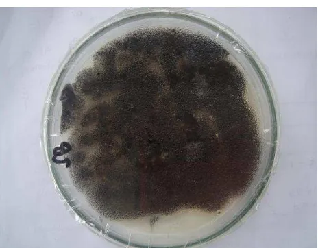
On February 5th 2010 the patient came and no felt itchy and aural fullness. We found there s no edema and discharge and the tympanic membrane was intact and no grayish white debris. The treatments were continued. On February 7th, 2010 she didn t have any complain and at her right ear canal we didn t found edema, no discharge and there isn t debris anymore. Laboratory examination was performed and specimen was taken from the debris. Microscopic examination revealed septate branching hyphae and spora. From culture examination with Sabouraud’s agar, after 7 days black powdery colonies grew and the microscopic examination of this colonies, revealed vesicles, sterigma and spore with conclusions equals to Aspergillus niger.
DISCUSSION
A 41 years-old woman, came to ENT outward department with chief complain pain, itchiness, aural fullness and hearing loss. Ho4 recorded that pruritus found 23% in cases, otalgia and otorrhea was 48% (in 63 patients). Hearing loss was found in 45% cases. Similar with Ozcan6 that found mostly cases had aural symptoms such as itching, otalgia, hearing loss, aural discharge and tinnitus. Otomycosis was found in all age groups and its seems similar between man and women.3,4,6,8,14 But in Turkey, Ozcan6 found 65 patient (74,7%) wears traditional head covers. Kumar8 found 29,26% wears turbans. This practice is associated with the prolonged covering of the external auditory canal which increases the humidity within the ear canal hence predisposes to otomycosis.15
The habit of cleaning the ears with feathers, matchstick and contaminated finger tips are known to encourage the inoculation and growth of the spores of fungus on the moist external auditory canal especially in patient with poor personal hygiene.15
Finding of grayish-white mass with black specks and fine filaments are typical for otomycosis. At microscopic examination we found septate hyphae and vesicles covered with sterigma and spora which is typical for genus Aspergillus.11 Direct microscopic as laboratory investigation was carried out for detection of fungal elements using KOH %, Gram s stain and PAS stain.8 This was confirmed by culture examination by Sabouroud agar where Aspergillus niger was isolated. Kumar8 found common fungal isolates from otomycosis patients, 43 cases (52,43%) same with Fasunla 15 found in 205 cases (48,35%). Ozcan6 in Turkey found 30 cases (44,8%). So Otomycosis is a recognized clinical entity in the tropical and subtropical regions.6,8,15 This fungi is frequently found as an agent in otomycosis and commonly non pathogen except when the environment is suitable for its growth.
Figure 4. Aspergillus niger’s colony on Sabouroud agar
In this patient, there was otalgia and narrowing auditory canal. According to Yassin (cited from Ahmad A et al9), otalgia and according to Youssef (cited Ahmad A et al9) narrowed auditory canal are usually found in otomycosis, except there was co infection with bacteria.
The patient was treated with Gentian violet. Gentian violet had been used to treat otomycosis as it is an aniline dye with effect as an antiseptic, antiinflamatory to reduce edema in auditory canal, antibacterial to treat mixed infection with bacteria and antifungal activity.
A standard regimen for otomycosis has not yet been established.6,8,11 There is no FDA (Food and Drugs Approval Bureau in United States) approved antifungal otic preparation for the treatment of otomycosis. Many agents with various antimycotic properties have been used and clinicians have struggled to identify the most effective agent to treat this condition. However, the use of few topical antifungals has persisted throughout time. In addition to topical therapy, the reviewed literature emphasized of aural hygiene in the treatment of otomycosis as intuitively ototopical medications works more following cleaning of secretions and debris.4,6,8,11
Ketoconazole and fluoconazole are azole antifungal agents that have a broad spectrum activity.11 Topical ketoconazole is our preferred antifungal agents for its efficacy against both Aspergillus and Candida species.4,11 Ketoconazole has shown an efficacy 95-100% in vitro against both species.11 Gentian violet is typically preferred as a weak solution in water. It has been used since the 1940s to treat otomycosis and it is an aniline dye with antiseptic, anti-inflamatory, antibacterial and antifungal activity. FDA approved its efficacy rate up to 80%11 The presence of mixed infection had been established in otomycosis such as Staphylococcus sp., Pseudomonas sp., Staphylococcus aureus, E.coli and Klebsiella sp.3,8,11
OTOMYCOSIS
|dolly
6
microtrauma and she wears head covers from nylonstretch material. This material cannot absorb the sweat well and increases temperature and humidity.6,7 Patient was suggested to switch her head covers to other materials which can absorb the sweat. The duration of treatment ranges from days to years. In follow up an improvement was observed. A good education can help to eliminate the predisposing factors and restoring physiologic environment.
Otomycosis could be asymptomatic but if left untreated may lead to morbidity like hearing loss. In recent study 56 patients (14,81%) had various degrees of conductive hearing loss.15 Prognosis on this patient is good but follow up still need, may eded to observed reccurency.
REFERENCES