Research Report
Dental Journal
(Majalah Kedokteran Gigi)2016 June; 49(2): 71–75
Synergistic effect of the combination of
Cinnamomum burmanii,
vigna unguiculata, and papain exracts derived from carica papaya
latex against C. albicans biofilms degradation
Muhammad Luthfi,Indah Listiana Kriswandini,and Fitriah Hasan Zaba
Departement of Oral Biology
Faculty of Dental Medicine, Universitas Airlangga Surabaya-Indonesia
abstract
Background: Candidiasis is an opportunistic infection commonly occurs on host with immunodeficiency, organ transplantation, leukopenia, or radiation therapy. Biofilms are structures that protect C. albicans from antifungals treatments. C. albicans biofilms display multidrug resistance to antifungal agents. Purpose: This study aimed to know whether the combination of Cinnamomum burmannii, Vigna unguiculata, and Papain extracts derived from Carica papaya latex has inadequate inhibitory effects against C.albicans biofilms compared to the combination of Cinnamomum burmannii and Vigna unguiculata extracts. Method: C. albicans growing on SDA were dissolved in 1 McFarland of sterile aquadest. Micro-plate was filled with 180 µL of SDB, glucose 8%, and 20 µL of C. albicans. Suspension was incubated at 37oC overnight. Extracts were added and incubated for 24 hours. Then, each well was
washed with distilled water, and stained with crystal violet 0.1% for 15 minutes. Afterward, each well was washed with distilled water and immediately stained with acetic acid. After 15 minutes of staining, the suspension was transferred to a new well, then measured with micro-plate reader at 595 nm. Results: The combination of Cinnamomum burmanii and Vigna unguiculata extracts had adequate inhibitory effects which is equal to 60.75%. Inhibition increased to 72.09%, 79.06%, and 79.50% after Papain derived from Carica papaya latex was added on concentrations of 138 mg/mL, 276 mg/mL, and 552 mg/mL. Conclusion: The combination of Cinnamomum burmanii (0.25µg/mL), Vigna unguiculata (200 µg/mL), and Papain (276 µg/mL) extracts showed an optimum synergic inhibition for C. albicans biofilms.
Keywords: C. albicans biofilm; Cinnamomum burmannii; Vigna unguiculata; Papain
Correspondence: Muhammad Luthfi, Department of Oral Biology, Faculty of Dental Medicine Universitas Airlangga. Jl. Mayjend. Prof. Dr. Moestopo no. 47 Surabaya 60132, Indonesia. E-mail: m.luthfi7@yahoo.com
introduction
Opportunistic fungal infection has been discussed in this decade. The ability of fungi to be able to infect the host actually depends on the immune response of the host and the presence of xenobiotics. Opportunistic fungal infections are mostly caused by Candida species infections.1
Candida albicans (C. albicans) grows excessively in patients with low immune circumstances, such as human immunodeficiency virus (HIV) infection, organ transplantation, leukopenia, post-surgery, or radiation therapy. Thrush or oropharyngeal candidiasis is a fungal
infection found on the surface of the oral mucosa, therefore, the ability of C. albicans to form biofilms has a huge impact on its ability to cause disease.2
Results of a research on patients with HIV / AIDS showed that the prevalence of oral candidiasis from 2008 to 2009 in Cipto Mangunkusumo Hospital was approximately 80.8%, while in Dr. Hasan Sadikin Hospital about 27% and in H. Adam Malik Hospital about 28.7%. Similarly, McCullough said that 70-80% of oral candidiasis is caused by C. albicans.3
Laboratory diagnosis and treatment of diseases caused by Candida species, especially C. albicans, Furthermore,
Dental Journal (Majalah Kedokteran Gigi) p-ISSN: 1978-3728; e-ISSN: 2442-9740. Accredited No. 56/DIKTI/Kep./2012. Open access under CC-BY-SA license. Available at http://e-journal.unair.ac.id/index.php/MKG
has not given satisfactory results because of the resistance to common antifungal. C. albicans biofilms have more multidrug resistance to fluconazole, amphotericin B, flucytosine, itraconazole, and ketoconazole than C. albicans in free-cells.1
Biofilms, moreover, are structured microbial communities, which are bound to the surface and become embedded in an extracellular matrix polymer produced.4 Extracellular matrix provides a significant contribution to drug resistance in C. albicans biofilms. Extracellular matrix is composed of materials considered as causative factors of resistance. Biofilms cannot be penetrated by antifungal for C. albicans cells coated by β-glucans, chitin, and glycoproteins. The realization of this biofilm is a self-defense form of C. albicans against radical agents.4
In addition, Cinnamon (Cinnamomum burmannii) is known to have antimicrobial properties. Cinnamomum burmannii contains with sinamaldehid, eugenol, cinnamic acid, sesquiterpene, and proanthocyanidin, which have antimicrobial power. Extract of Cinnamomum burmannii has a minimum inhibitory concentration (MIC) of 0.33 mg/ mL against C. albicans.5
Papain derived from Carica papaya latex and fluconazole, furthermore, has synergistic action in inhibiting the growth of C. albicans. The result is a synergistic effect to degrade the cell wall of C. albicans. Papain is responsible as antifungal at a concentration of 138 μg/mL. Papain containing a specific content in the form of Nasetil-β -D-glukosaminidase and α-D-mannosidase is known to have a role in the degradation of C. albicans biofilm matrix .6
Tolo bean extract (Vigna unguiculata), moreover, is known to contain the β-1,3-glucanase enzyme. Vigna unguiculata at a concentration of 200 μg/mL has higher activity of β-1,3-glucanase enzyme that at a concentration of 3.152 U/mg.8 The combination of β-1,3-glucanase enzymes derived from snails and Cinnamomum burmannii extract is able to lyse C. albicans biofilm, and the result is the death of C. albicans biofilm cells, about 75%.5
Snails have β-1,3-glucanase enzymes, but the enzymes are unstable. It is reported that β-1,3-glucanase enzymes derived from snails and stored at room temperature has glucanase activity decreasing dramatically. Similarly, in the storage temperature of 24oC, the activity of β-1,3-glucanase also decreases.6 Enzymes from Vigna unguiculata have good stability and high glucanase activity.7
In short, Cinnamomum burmannii extract will degrade free albicans cells. 8 Extracellular matrix of C. albicans biofilms is composed of β-glucan (50-60%), mannoprotein (30-40%), and chitin (0.6 to 9%).4β 1,3-glucanase enzyme derived from Vigna unguiculata extract is able to hydrolyze the components of the extracellular matrix in the glucanase form.7 α-β-D-mannosidase derived from Papain is able to hydrolyze the components of the biofilm matrix in the form of mannoprotein, while N-acetyl-β Dglukosaminidase of Papain is able to hydrolyze components of the biofilm matrix in the form of chitin.6 In other words, the use of
Cinnamomum burmannii extracts has antimicrobial activity,
while the combination of Vigna unguiculata and Papain extracts derived from Carica papaya latex is synergistic in the extracellular matrix of the biofilms hydrolyzing C. albicans. This aim of this study was to know whether the combination of Cinnamomum burmannii, Vigna unguiculata, and Papain extracts derived from Carica papaya latex has inadequate inhibitory effects against C. albicans biofilms compared to the combination of Cinnamomum burmannii and Vigna unguiculata extracts.
materialsandmethods
This research is an experimental laboratory research with post-test-only control group design. The research was conducted at Rumah Sakit Khusus Infeksi (Hospital for Infection) Universitas Airlangga in October-November 2015. Samples used in this research were albicans. The number of samples used were determined by using Lemeshow formula, about seven samples.
The samples were divided into five groups. The control group consisted of C. albicans planktonics and C. albicans biofilms without being treated. Group I consisted of C. albicans planktonics and C. albicans biofilms treated with 0.25 mg/mL of Cinnamomum burmannii extract and 200 μg/mL of Vigna unguiculata extract. Group II consisted of C. albicans planktonics and C. albicans biofilms treated with 0.25 mg/mL Cinnamomum burmannii extract, 200 μg/ mL of Vigna unguiculata extract, and 138 μg/mL of papain extract. Group III consisted of C. albicans planktonics and C. albicans biofilms treated with 0.25 mg/mL Cinnamomum burmannii extract, 200 μg/mL of Vigna unguiculata extract, and 276 μg/mL of papain extract. Group IV consisted of C. albicans planktonics and C. albicans biofilms treated with 0.25 mg/mL Cinnamomum burmannii extract, 200 μg/mL of Vigna unguiculata extract, and 552 μg/mL of Papain extract.
This research was started with the preparation of Cinnamomum burmannii, Vigna unguiculata, and Papain extracts. The extracts of Cinnamomum burmannii and Vigna unguiculata were obtained from Balai Penelitian dan Konsultasi Industri (Research and Industry Consulting Center) in Surabaya together with aquadest as solvent. Meanwhile, papain extract was derived from Carica papaya latex obtained from SIGMA (P3X15-250). Next, 25 mg of Cinnamomum burmannii extract was dissolved in 100 mL of distilled water. Vigna unguiculata extract then was added in a concentration of 200 mg/mL, whereas Papain extract was added in concentrations of 138 mg/mL, 276 mg/mL, and 552 mg/mL.
C. albicans planktonics and biofilms were prepared by using microtiterdish assay method. C. albicans taken from SDA stock was used to make suspension in 1 McFarland of sterile distilled water (3 x 10⁷ CFU/mL). Microplates then were filled with 180 mL of SDB together with 8% glucose and 20 mL of C. albicans to grow C. albicans biofilm. Meanwhile, to grow C. albicans planktonics, Micro-plates
Dental Journal (Majalah Kedokteran Gigi) p-ISSN: 1978-3728; e-ISSN: 2442-9740. Accredited No. 56/DIKTI/Kep./2012. Open access under CC-BY-SA license. Available at http://e-journal.unair.ac.id/index.php/MKG
then were filled with 180 mL of SDB together and 20 mL of C. albicans without 8% glucose.
Afterwards, those micro-plates were incubated for 24 hours at 37°C. They then were washed with sterile aquadest before the extracts were added into them and incubated for 24 hours. After that, the micro-plates were washed again, and then stained with 0.1% crystal violet for 15 minutes. Those micro-plates were washed with sterile distilled water. They then were given with acetic acid for 15 minutes and transferred to a new ones. OD values were read using micro plate reader at a wavelength of 595 nm.10,11 Inhibition effect then was determined based on the values of OD. If OD values obtained were getting smaller, inhibition effect would be indicated to be high. The formula used to calculate the inhibitory activity was as follows:11
(Mean of OD595 Control – mean of OD595 Concentration)
×100% Mean of OD595 Control
The normality of data were tested using Kolmogorov Smirnov test, while the homogeneity of data were tested using Levenne’s Test. Finally, to identify the significance of the difference among the treatment groups, Post Hoc-Tukey HSD test was conducted.12
results
Absorbance levels of planktonic and biofilm cells were observed in the value of optical density (OD) using a microplate reader with a wavelength of 595 nm. OD value obtained then was comparable with C. albicans biofilm formation. To determine the growth of C. albicans biofilms, a preliminary experiment on the relation of C. albicans biofilm growth and incubation time was conducted, and the results showed that the optimal biofilm growth was at 24 hours.
After knowing the optimal incubation time for C. albicans biofilm to grow well, a combination of the extracts was used to inhibit the growth of C. albicans biofilms. This aimed to compare the effectiveness of the combination of Cinnamomum burmannii, Vigna unguiculata, and papain extracts in inhibiting the growth of C. albicans biofilms. In the control group, C. albicans planktonic cells was untreated with C. albicans biofilm. In this research, the concentration of Cinnamomum burmannii extract used was 0.25 mg/mL, the concentration of Vigna unguiculata extract was also the same or equal to 200 pg/ml, while the concentrations of papain extract were 138 μg/ml, 276 pg/ml, and 552 μg/ml. The results of OD values obtained were as follows (Table 1).
Based on Table 1 above, it can be seen that the means of OD values of C. albicans planktonics was 0.367, while the means of OD values of C. albicans biofilm was 0.688. The OD value of C. albicans biofilms was higher than
the OD values of C. albicans planktonics. The OD value of C. albicans biofilms dropped to 0.27 after treated with the combination of Cinnamomum burmannii and Vigna unguiculata extracts. The OD value of C. albicans biofilms decreased into 0.192 after treated with the combination of the Cinnamomum burmannii, Vigna unguiculata, 138 μg/mL of papain extracts. The OD value of C. albicans biofilms remained down to 0.144 after Papain concentration was increased to 276 mg/mL, and the OD value declined into 0.141 after the addition of papain concentration to 552 mg/mL. The comparison of the OD values of C. albicans planktonics and C. albicans biofilms in the control group and the OD values of C. albicans biofilms treated can be seen in the graph below (Figure 1).
Table 1. The OD values of C. albicans biofilms after the treatment
Treatment groups N Means of SD
OD values
Planktonic cells 7 0.367 0.530
Biofilms 7 0.688 0.174
CB +VU extracts 7 0.270 0.187
CB +VU + P138 extracts 7 0.192 0.304
CB +VU + P276 extracts 7 0.144 0.207
CB +VU + P552 extracts 7 0.141 0.149
Note: CB: Cinnamomum burmanii extract; VU: Vigna unguiculata extract; P138: 138 μg/mL of Papain; P276: 276 μg/ mL of papain; P552: 552 μg/mL of Papain.
11
P138: 138 μg/mL of P276: 276 μg/mL of P552: 552 μg/mL of
Figure 1. The graph of OD Values of C. albicans biofilmswith various treatment.
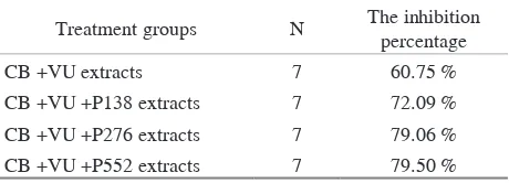
Table 2. The inhibition percentage of C. albicans biofilms
Treatment groups N The inhibition
percentage
CB +VU extracts 7 60.75 %
CB +VU +P138 extracts 7 72.09 %
CB +VU +P276 extracts 7 79.06 %
CB +VU +P552 extracts 7 79.50 %
Dental Journal (Majalah Kedokteran Gigi) p-ISSN: 1978-3728; e-ISSN: 2442-9740. Accredited No. 56/DIKTI/Kep./2012. Open access under CC-BY-SA license. Available at http://e-journal.unair.ac.id/index.php/MKG
The inhibition percentage of C. albicans biofilms obtained can be seen in Table 2. The inhibition percentage of the combination of Cinnamomum burmannii and Vigna unguiculata extracts against C. albicans biofilms in the control group was 60.75%. The inhibition percentage of the combination of Cinnamomum burmannii, Vigna unguiculata, and 138 ug/mL of Papain extracts was 72.09%. The inhibition percentage of the combination of Cinnamomum burmannii, Vigna unguiculata, and 276 mg/mL of Papain extracts was 79.06%. Meanwhile, the inhibition percentage of the combination of Cinnamomum burmannii, Vigna unguiculata, and 552 μg/mL of Papain extracts was 79.50%.
A statistical test was conducted on distribution of data in each group using the Kolmogorov-Smirnov test. The results of Kolmogorov-Smirnov test showed that the distribution of data in those treatment groups was normal because p-value in the treatment groups was greater than 0.05. After that, Levenne test was conducted to know the homogeneity of data. The results of Levenne test showed that p-value obtained was <0.05. It means that the variation of the data was not homogeneous. As a result, Kruskal Wallis test was performed. The results of Kruskal Wallis test showed that value obtained was > 0.05. It indicates that there were significant differences between each treatment group.
To know the differences of each treatment group, Post Hoc-Tukey HSD test then was carried out. The results of Post Hoc-Tukey HSD test showed that there was a significant difference between the group treated with biofilms and the treatment groups treated with CB + VU extracts, CB + VU + P138 extracts, CB + VU + P276, and CB + VU + P552 extracts. Similarly, there was also a significant difference between the group treated with CB + VU extracts and the groups treated with CB + VU + P276 and CB + VU + P552 extracts.
discussion
In the process of biofilm inhibition, there are some stages of the degradation of the elements of C. albicans biofilm biomass. Extracellular matrix is one of the elements composing the biomass of biofilms. One of the elements composing the extracellular matrix of the biofilms is glucan (50.60%). The hydrolysis mechanism of glucanase contained in Vigna unguiculata against C. albicans biofilm is related to glucan in the cell walls of fungi that can be utilized by glucanase enzyme as a substrate by cutting the glucose residues of non-reducing end of polymers or oligomers, resulting in forming a glucose monomer.13
Cinnamomum burmannii has several compounds that play a role in degradation of C. albicans cells, such as sinamaldehide and eugenol. The ability of sinamaldehide in inhibiting the growth of C. albicans due to the free 3-phenyl group that can bind to aspartic proteases in the wall of C. albicans cells and also bind to oxygen required
for the metabolism of C. albicans. These bounds can cause sinamaldehide inhibits the synthesis of enzymes on the wall of C. albicans cells and the metabolism of C. albicans cells, resulting in the death of C. albicans cells.8
Eugenol, is known to be lipophylic, which can penetrate between fatty acid chains and layers of bilayer membrane by altering the permeability of cell membranes. If the phenol compound interacts with the cell wall of C. albicans, there will be denaturation of proteins in the cells of C. albicans. The interaction causes a change in the balance of protein molecules, resulting in a change in the structure of the protein and triggering coagulation. Protein experiencing coagulation will lose its physiological activities that cannot function properly. Changes in the structure of proteins in C. albicans will cause increased permeability of the cells, so the cell growth is inhibited and then the cells will die, thus eugenol has an ability to reduce adherent and to inhibit metabolism of C. albicans biofilms. 14
Therefore, in the treatment group, the inhibition was adequate when C. albicans biofilms were treated with combination of Cinnamomum burmannii and Vigna unguiculata compared to the control group. It means that the combination of Cinnamomum burmannii and Vigna unguiculata extracts is able to inhibit C. albicans biofilms with the inhibition of 60.75%.
In addition, papain contains specific enzymes, namely
αD-mannosidase and N-acetyl-β-Dglucosaminidase hydrolyzing the extracellular matrix of the biofilms, such as mannoprotein and chitin. Glycosidase process of both the enzymes of Papain can occur by cutting the polysaccharide chain residues in the extracellular matrix of biofilms.6
In the other treatment group, moreover, the inhibition was adequate when 276 mg/mL of Papain was added to the combination of Cinnamomum burmannii and Vigna unguiculata used to treat C. albicans biofilms. Papain extract at that concentration could inhibit C. albicans biofilms with good inhibitory, increasing from 60.75% to 79.06%. It means that there was an increase in the inhibition of 18.31%.
The addition of papain extract at the concentrations ranging from 138 mg/mL to 276 pg/mL and 552 mg/mL did not show adequate inhibition. papain can be active when given activator since the enzyme contained in papain can be activated or inhibited. Compounds classified as activators of papain are cysteine, sufida and sulfite, as well as a chelator of heavy metals, such as EDTA and N-bromosuksinimida; whereas compounds classified as inhibitors of papain are PMSF, TLCK & TPCK, E-64, heavy metals, cystatin, and leupeptin.15 However, papain used in this research was not classified as an activator. Thus, OD values of the inhibition of C. albicans biofilms obtained were inadequate though the concentration of papain increased.
Another possibility is C. albicans can develop some mechanisms to overcome the existing antimicrobial agents by producing genetic mutation enzyme and transmission for new generation.16 In the process of extract administration,
Dental Journal (Majalah Kedokteran Gigi) p-ISSN: 1978-3728; e-ISSN: 2442-9740. Accredited No. 56/DIKTI/Kep./2012. Open access under CC-BY-SA license. Available at http://e-journal.unair.ac.id/index.php/MKG
the incubator temperature was 37°C. This possibility also becomes a factor causing the working of Papain on C. albicans biofilms not optimal. Concentration level (pH), furthermore, is also considered as a factor that can influence the effectiveness of the activity of the enzyme. The effectiveness of the enzyme showed a gradual increase with increasing pH from pH 3.5 to pH 7.5, whereas at pH 9 resulting in a decrease in the activity of papain.17 Meanwhile, in the treatment groups, PBS pH used as a solvent was 7. It can be concluded that the combination of 0.25 mg/mL of Cinnamomum burmannii, 200 mg/mL of Vigna unguiculata, and 276 mg/mL of papain extracts had an optimal and synergistic effect on the inhibition of C. albicans biofilms.
references
1. Darouiche RO, Mansouri MD, Kojic EM. Antifungal activity of antimicrobial-impregnated devices. Clin Microbiol Infect 2006; 12(4): 397-9.
2. Richard ML, Nobile CJ, Bruno VM, Mitchell AP. C. albicans biofilm defective mutants. Eukaryot Cell 2005; 4(8): 1493–502.
3. McCullough MJ, Savage NW. Oral candidosis and the therapeutic use of antifungal agents in dentistry. Aust Dent J 2005; 50(4 Suppl 2): S36-9.
4. Nett J, Lincoln L, Marchillo K, Massey R, Holoyda K, Hoff B, VanHandel M, Andes D. Putative role of beta-1,3 glucans in C. albicans biofilm resistance. Antimicrob Agents Chemother 2007; 51(2): 510-20.
5. Krishna KL, Paridhavi M, Patel JA. Review on nutritional, medicinal and pharmacologinal properties of papaya (Carica papaya Linn.). Natural Product Radiance 2008; 7(4): 364-73.
6. Giordani R, Siepaio M, Moulin-Traffort J, Régli P. Antifungal action of Carica papaya latex: isolation of fungal cell wall hydrolysing enzymes. Mycoses 1991; 34(11-12): 469-77.
7. Oliveira JTA, Barreto ALH, Vasconcelos IM, Eloy YRG, Gondim DMF, Fernandes CF, Freire-Filho FR. Role of antioxidant enzymes, hydrogen peroxide and PR-proteins in the compatible and incompatible interactions of Cowpea (Vigna unguiculata) genotypes with the Fungus Colletotrichum gloeosporioides. J Plant Physiol Pathol 2014; 2(3):2-8.
8. Erna F, Rostiny, Sherman S. Efektivitas minyak kayu manis dalam menghambat pertumbuhan koloni C. albicans pada resin akrilik. Journal of Prosthodontic 2010; 11(2): 19-23.
9. O’Toole, GA. Microtiter dish biofilm formation assay. J Vis Exp 2011; (47) pii: 2437.
10. Mahmoudabadi AZ, Zarrin M, Kiasat N. Biofilm formation and susceptibility to amphotericin B and fluconazole in C. albicans. J Microbiol 2014; 7(7): e17105.
11. Bakkiyaraj D, Nandhini JR, Malathy B, Pandian SK. The anti-biofilm potential of pomegranate (Punica granatum L.) extract against human bacterial and fungal pathogens. Biofouling 2013; 29(8): 929-37.
12. Kao LS, Green C. Analysis of variance: is there a difference in means and what does it mean?. J Surg Res 2008; 144(1): 158-70. 13. El-Katatny MH, Somitsch W, Robra KH, EI-Katatny MS, Gübitz
GM. Production of chitinase and β-1,3-glucanase by Trichoderma harzianum for control of the Phytopathogenic Fungus Sclerotium rolfsii. J Food Technol Biotechnol 2000; 38(3): 170-80.
14. Raja MRC, Srinivasan V, Selvaraj S, Mahapatra SK. Versatile and synergistic potential of eugenol: a review. Pharm Anal Acta 2015; 6(5): 367.
15. Dongoran DS. Pengaruh aktivator sistein dan natrium klorida terhadap aktivitas papain. Jurnal Sains Kimia 2004; 8(1): 30-5. 16. Nikaido H. Multidrug resistance in bacteria. Annu Rev Biochem
2009; 78: 119-46.
17. Omeje KO, Eze SO, Ozougwu VE, Ubani CS, Osayi E, Onyeke CC, Chilaka FC. Application of papain from paw paw (Carica papaya) latex in the hydrolysis of tiger nut (C.esculentus) proteins. Mitteilungen Klosterneuburg 2014; 64: 1-17.
Dental Journal (Majalah Kedokteran Gigi) p-ISSN: 1978-3728; e-ISSN: 2442-9740. Accredited No. 56/DIKTI/Kep./2012. Open access under CC-BY-SA license. Available at http://e-journal.unair.ac.id/index.php/MKG