e-ISSN: 2319-2380, p-ISSN: 2319-2372. Volume 11, Issue 2 Ver. I (February 2018), PP 12-16
www.iosrjournals.org
Morphological study of the respiratory system of bungo fish
(Glossogobius Cf. Aureus) from Lake Tempe, South Sulawesi,
Indonesia
Ade Andrew Samudera Pinontoan
1, Wahyuni
2, Andi Tamsil
3,
Dwi Kesuma Sari
1*1
(Study Program of Veterinary Medicine, Faculty of Medicine, Hasanuddin University, Makassar, South Sulawesi, Indonesia)
2(Disease Investigation Centre Maros, South Sulawesi, Indonesia)
3
(Faculty of Fisheries and Marine Sciences, Muslim University of Indonesia, Makassar, South Sulawesi, Indonesia)
Corresponding Author’s: Dwi Kesuma Sari
Abstract: Bungo fish (Glossogobius Cf. aureus) is native species from Tempe Lake, South Sulawesi. This study
was conducted with the aim to observe anatomical and histological of respiratory system of bungo fish. This study showed several characteristics such as the configuration of gills filament for the "V" shape without septum, operculum located adjacent to the pectoral, membrana branchialis converged with isthmus with a narrow slit, and short gills filters, however the fish did not have additional breathing organ and only depended on countercurrent flow system to obtaining oxygen. Histological observations showed some characteristics such as afferent arterioles at the end of the primary lamella and efferent arterioles at the base ofthe primary lamella, single layer of epithelial cells along the surface of the lamella, chloride cells between base of the secondary lamella, mucous cells at the base of secondary lamella, sustaining cartilage tissue at the base of the primary lamella, venous sinuses along the primary lamella which was forwarded to the secondary lamella through capillaries in the secondary lamella. Generally, respiratory system structure of the bungo fish was similar to the teleostei fishes.Lake Tempe is one of the big lakes located in South Sulawesi Province, Indonesia, precisely in Wajo Regency (70% of part areas), Sidrap Regency and Soppeng Regency. There are many fish species in the lake such as goldfish (Cyprinus carpio), nilem fish (Osteochillus hassellti), cork fish (Ophiocephalus striatus), Siamese gourami or snake-skin gouramy (Tricogaster pectoralis), bungo fish, fishpond (Helostoma temmincki), and nile tilapia fish (Oreochromis niloticus) [1]. One interesting thing is the bungo fish (Glossogobius Cf.aureus) is an indigenous species on Lake Tempe, but it does not get much attention either by researchers and local governments that lead to minimal conservation efforts. This can be seen by the lack of research on germplasm resources, conservation and efforts of local governments in the form of research and protection of populations and their habitats.
Water biota that can be developed as a cultivation commodity, both freshwater biota and marine biota, the number is still very much. Some species are endangered, while cultivation technology has not been mastered. For example, bungo fish or beloso fish, this fish is one of the important fish in Tempe lake, South Sulawesi which is increasingly pressed due to intensive catching, introduction and degradation of its habitat [1]. The information obtained shows no publication of the anatomical and histological features of the Glossogobius Cf. aureus, whereas the respiratory system is one of the systems present in the metabolism of living things.
II.
Materials And Methods
The study was conducted in December 2014 until January 2015. The location of the study at Anatomy and Histology Laboratory of Study Program of Veterinary Medicine Hasanuddin University and Pathology Laboratory of Disease Investigation Centre Maros. The material used is 6 bungo fish with average weight 12 gram. Bungo fish obtained from Lake Tempe by capture. Fish samples were then stored in formalin p.a 10%. The researchs were divided into 3 main procedures, namely the observation of anatomy, histology, and data analysis. In anatomy observation, fish are placed on fixation tray then photographed from 4 angles ie dorsal, ventral, lateral and anterior. For observation of the head and mouth cavities, the fish head is opened by cutting sagittally on the cranial area without cutting the gills and then being observed and photographed from the lateral direction. After that, a transversal cutting in the maxilla after the eye from the anterior angle of the fish is observed and photographed from the dorsal and anterior angles and the last transversal cutting on the mandible corresponding to the previous cut in the maxilla section and then observed and also photographed from the anterior angle. For further observation of the gills removed from the whole fish body and were observed from 3 angles of dorsal, ventral and lateral and then photographed.
The gill organs were separated from the sample and then fixed in a 10% formalin solution followed by dehydration with stratified alcohols, clearing with xylol and embeded in paraffin. The tissue in paraffin blocks were cut with a thickness of 4 μm using a microtome, then placed on the object glass and stored in an incubator with a temperature of 40oC for 24 hours. Samples were stained with Mayer's Hematoxylin Eosin (HE) for standard coloring to see the bungo fish structure. After staining, dehydration, cleansed in xylol, the samples were then mounting and ready for observation [6].
The observations were performed under a microscope, with 10x and 16x subjective lens magnification as well as an objective lens of 4x, 10x, and 40x. Photograph of the samples were done using a digital camera (Canon IXUS 80 IS, Japan). The observed portion is a gill composed of primary lamellae and sekuder, lacuna along with its constituent cells such as pillar cells, epithelial cells, mucus cells, undifferentiated cells, and chloride cells. Data analysis used is descriptive qualitative data analysis. In this method will explain the description of anatomy and histology in the respiration system of bungo fish.
III.
Results And Discussion
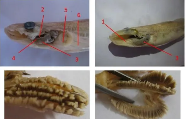
Fig. 1. Bungo fish with dorsal view observation(A), ventral (B), anterior (C) and left lateral view (D). 1. nostril; 2. os.operculare; 3. os.suboperculare; 4. os.preoperculare; 5. membrana branchialis; 6. radii branchialis; 7. pectoral fin; 8. os.interoperculare; 9. m.apparatus mandibula; 10. isthmus; 11. pelvis fin.
On observation of the head cavity no additional respiratory equipment was found to support the fish can breathe in the air. The number of arcus branchialis of bungo fish is 4 pairs. Gill filaments in bungo fish are in the form of a "v" letter without a septum or separator on the two pairs of filaments that exist along the branchial arcus. The bungo has a small, spiral-shaped gill filter which is paired along the branchial arcus and each pair forms like a loose pear along the first branchial arcus in the anterior section then becomes more closely arranged in the next branchial arcus to the fourth branchialicarcella at the posterior side (Fig. 2)
Fig. 2. Sagittal view of crania area of the gills of bungo fish. 1. Gills support muscles; 2. Gills screen; 3. gill filamen; 4. arcus branchialis; 5. pectoral fin support; 6. pectoral fin
Fig. 3. Bungo fish with transverse cuts in the transition area between the oral and pharyngeal cavities with anterior observation (A) and transverse cuts of the mandible with posterior observation (B). 1. gills; 2. cartilago
palate; 3. pectoral muscle; 4. septum separating pectoral muscle; 5. blood vessels; 6. hyoideum; 7. transitional septum on the pectoral fin muscle; 8. m. apparatus mandibulae; 9. muscles around the hyoideum
Fig. 4. Bungo fish with transverse cuts on the maxilla in the transition between the oral and pharynx with dorsal (A) and anterior (B) observations on bungo fish. 1. gills; 2. cartilago palate; 3. blood vessels;
4. gill support muscles
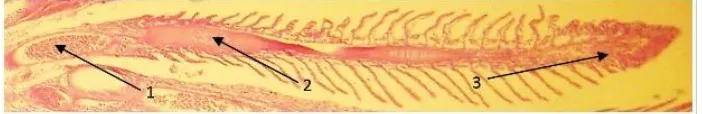
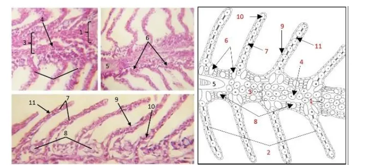
The bungo fish has afferent arterioles at the base of the primary lamellae and the efferent arterioles at the end of the primary lamella. There is a supportive cartilage network that supports primary lamella in bungo fish. The mucous cells of bungo fish in primary lamellae is located between each of the secondary lamellae bases. Chloride cells of bungo fish are located at right on the base of the secondary lamellae. The cells are associated with the respiratory system such as cells of the erythrocyte exist in every part of the lamella, the lamella primary erythrocytes flowing in the veins, called venous sinuses that connect afferent arterioles located at the base of the lamella primary and arterial efferent located at the end of the lamella primers and secondary lamella are present in the capillary lumen, then the outermost part is covered by epithelial cells (Figs. 5 and 6). According to other fishes, they are similarity with other fishes specially in theleost fishes [7].
Fig. 6. Histology of the gills of bungo fish, magnification: 400x sagittal pieces and the sketches. 1. primary lamella; 2. secondary lamella; 3. venous sinus; 4. erythrocytes present in venous sinuses; 5. primary lamella support cartilage; 6. chloride cells; 7. pillar cells; 8. mucus cells; 9. epithelial cells; 10. erythrocyte cells in the
capillary lumen; 11. lacuna
IV.
Conclusion
Based on the results of research conducted, it can be concluded that bungo fish only rely on gills as the main respiratory organs. Bungo fish have 4 pairs of gills with gill filament shaped "v" without septum. The histology structure of gills in bungo fish is similar to that of teleostei fish in general, namely primary lamella and secondary lamella consisting of mucus cells, chloride cells, supportive cartilage tissue at the base of primary lamella, pillar cells, and erythrocytes cells along the capillaries and venous sinus. The bungo fish has afferent arterioles at the base of the primary lamellae and the efferent arterioles at the end of the primary lamella. Generally, respiratory system structure of the bungo fish was similar to the teleostei fishes.
Acknowledgements
The authors would like to thank the Histology and Pathology Laboratory of Veterinary Medicine Program of Hasanuddin University Faculty of Medicine and Disease Investigation Centre Maros for doing the the research.
References
[1]. A. Tamsil. Study some characteristics of prespawning reproduction and possible spawning of bungo fish (Glossogobius cf. Aureus) at Lake Tempe and Lake Sidenreng South Sulawesi, Doctoral Diss., Bogor Agricultural University, Bogor, Indoensia, 2000
[2]. S.A. Omar. Iktiologi. (Fakultas Ilmu Kelautan dan Perikanan, Universitas Hasanuddin, Makassar, 2011) (ID)
[3]. G.S. Helfman, B.B. Collette, D.E. Facey, B.W. Bowen. The Diversity of Fishes, Biology, Evolution, and Ecology. (Willey-Blackwell, Hoboken, USA, 2009)
[4]. D.H. Evans, P.M. Piermarini, K.P. Choe. The Multifunctional Fish Gill: Dominant Site of Gas Exchange,Osmoregulation, Acid-Base Regulation, and Excretion of Nitrogenous Waste. Physiological Reviews, 85, 2005, 97–177.
[5]. P.C. Withers. 1992. Comparative Animal Physiology. (Saunders College Publishing, Londong, UK, 1992).
[6]. W.J. Bacha and L.M. Bacha LM. Colour Atlas of Veterinary Histology 3rd Ed. (John Willey & Sons, Ltd, Chichester, UK, 2012)
[7]. F. Takashima and T. Tibiya. An atlas of fish histology: normal and pathological features. (Kodansha, Japan, 1995).