R E S E A R C H A R T I C L E
Altered Level of Soluble fms-like Tyrosine Kinase 1 (sFlt1) and Hypoxia
Inducible Factor-1α (HIF-1α) in Normotensive Pregnancy and Preeclampsia
John Wantania
1,2,, Syakib Bakri
1,3, Karel Pandelaki
2, Maisuri Chalid
1,3 1Postgraduate Program in Biomedics, Hasanuddin University, Jl. Perintis Kemerdekaan Km.10, Makassar, Indonesia2Faculty of Medicine, Sam Ratulangi University, Jl. Kampus Unsrat, Manado, Indonesia 3Faculty of Medicine, Hasanuddin University, Jl.Perintis Kemerdekaan Km.10, Makassar, Indonesia
Corresponding author. E-mail: [email protected]
B
ACKGROUND: Preeclampsia is still a significantproblem worldwide. Of the many suggested mechanisms of its pathogenesis, the latest one is the balance of angiogenic factor and its relationship with
hypoxia factors. The objective of this study was to observe changes or dynamic process of soluble fms-like tyrosine kinase 1 (sFlt1) as anti-angiogenic factor and hypoxia-inducible factor 1-alpha (HIF-1α) as hypoxia marker in
normotensive pregnancy and preeclampsia in mid-term and full-term pregnancies.
METHODS: A cohort study was conducted on 36
normotensive subjects, first examination was conducted at 20-28 weeks of gestation. Then second examination was
conducted at the time of preeclampsia diagnosed or full-term pregnancy. Preeclampsia was characterized by hypertension
of systolic blood pressure ≥140 mmHg, diastolic blood pressure ≥90 mmHg, with two readings separated in 4-6 hours period, and/or proteinuria with urine dipstick of ≥1+ or ≥300 mg per 24 hours. Examinations of sFlt-1 and HIF-1α were done by enzyme-linked immunosorbent assay method. Statistical analysis was done using a significance
level of p<0.05.
RESULTS: Concentration of sFlt-1 was elevated in
normotensive pregnancy and preeclampsia. Higher sFlt-1 concentration elevation was seen in preeclamptic group comparing to normotensive group, although not significant. This finding was related to the fact that investigated
subjects were mostly developing mild preeclampsia merely. Comparing to normotensive group, preeclamptic group
had higher HIF-1α concentration-per-week elevation, but not significant. There was a positive correlation between
L
ATAR BELAKANG: Preeklamsia masihmerupakan masalah di dunia yang signifikan. Dari beberapa mekanisme patogenesis yang disarankan, keseimbangan faktor angiogenik dan hubungannya dengan faktor hipoksia merupakan mekanisme terbaru. Tujuan penelitian ini untuk melihat perubahan atau proses dinamis
pada soluble fms-like tyrosine kinase 1 (sFlt1) sebagai
faktor anti-angiogenik dan hypoxia-inducible factor 1-alpha (HIF-1α)sebagai penanda hipoksia pada kehamilan
normotensif dan preeklamsia pada waktu pertengahan dan akhir kehamilan.
METODE: Studi kohort dilakukan pada 36 subyek
normotensi, pemeriksaan pertama dilakukan pada usia kehamilan 20-28 minggu. Kemudian pemeriksaan kedua dilakukan pada saat didiagnosa preeklamsia atau saat akhir kehamilan. Preeklamsia ditandai dengan hipertensi dengan tekanan darah sistolik ≥140 mmHg dan/atau diastolik ≥90 mmHg, dengan pembacaan 2 kali pada rentang waktu 4-6 jam; proteinuria dengan dipstik urin ≥1+ atau ≥300 mg per 24 jam. Pemeriksaan sFlt-1 dan HIF-1α dilakukan dengan
metode enzyme-linked immunosorbent assay. Analisa
statistik menggunakan tingkat signifikan p<0,05.
HASIL: konsentrasi sFlt-1 mengalami kenaikan pada
kehamilan preeklamsia dan normotensi. Peningkatan konsentrasi sFlt-1 yang lebih tinggi terlihat pada grup preeklamsia, dibandingkan grup normotensif, meskipun tidak signifikan. Temuan ini berkaitan dengan fakta bahwa pada subyek yang diteliti, kebanyakan hanya mengalami preeklamsia ringan. Dibandingkan grup normotensif, grup preeklamsia memiliki peningkatan konsentrasi-per-minggu HIF-1α lebih tinggi, namun tidak signifikan. Terdapat
concentrations of sFlt-1 and HIF-1α, but not significant.
CONCLUSION: sFlt-1 concentration elevation was
correlated with preeclampsia. Therefore comparing to
averages, changes of sFlt-1 concentrations were more important. Concentrations of HIF-1α and sFlt-1 were
positively correlated.
KEYWORDS: sFlt-1, HIF-1α, preeclampsia, normotension
Indones Biomed J. 2013; 5(2): 121-8
Preeclampsia is a worldwide problem linked with
maternal and fetal mortality or morbidity. The incidence of
preeclampsia is approximately 1.8 to 16.7% of pregnancies, with the numbers varying between countries.(1) Based on
data of the World Health Organizations by a systematic
review of cases all over the world, 16% of maternal deaths
in developed countries, including the United States, are due to hypertension in pregnancy and its complications. This
figure outnumbers the figures of other major causes such as bleeding 13%, abortion 8% and sepsis 2%.(2)
In addition to the several dominant risk factors,
a number of theories have been developed related to pathomechanism of preeclampsia, e.g. gene, immunity,
oxidative stress, inflammation, hypoxia, angiogenic
imbalance and hormone. Although most of the theories
explain the initial problem of trophoblasts invasion, but until
now they have not been able to produce a concept that can give a thoroughly satisfactory answer, due to the seemingly
very complex interactions between one and the others.(3) Lyall, et al., have shown a decline in vascular
endothelial growth factor (VEGF) concentration in
preeclampsia compared with that in normal pregnancy.
VEGF level assessed in normal pregnant women was 12.89 pg/mL, compared with VEGF level in women with preeclampsia of 2.34 pg/mL. Decreased levels of VEGF in
patients with preeclampsia was associated with increased
soluble VEGF receptor released into the circulation, called soluble fms-like tyrosine kinase 1 (sFlt-1).(3)
sFlt-1 is a form of Flt-1 that has lost cytoplasmic
and transmembrane domains but still has a ligand-binding
domain. VEGF itself is a glycoprotein pro-angiogenic that works to increase proliferation, migration, and for survival
of endothelial cells in increased capillary permeability.
VEGF is a key signal used by the cells that are starved of oxygen (oxygen-hungry cells) to trigger the growth of blood vessels.(4)
Hypoxia inducible factor-1 (HIF-1) is an important
element used in the regulation of transcription of various
genes that appear in conditions with low oxygen level.(5) The etiology of the increased level of sFlt-1 is not clearly known until now, but it is suspected that hypoxia may
contribute to the increase of this anti-angiogenic factor. The objective of this study was to assess the dynamic
mechanism of sFlt-1 as an anti-angiogenic factor and HIF-1α as hypoxia marker in normotensive and preeclamptic
groups in mid-term and full-term pregnancy.
korelasi positif pada konsentrasi HIF-1α dan sFlt-1, namun tidak signifikan.
KESIMPULAN: Peningkatan konsentrasi sFlt-1 berkaitan
dengan preeklamsia. Oleh karena itu, dibandingkan rerata, perubahan konsentrasi sFlt-1 lebih penting. Terdapat korelasi positif antara konsentrasi HIF-1α dan sFlt-1 .
KATA KUNCI: sFlt-1, HIF-1α, preeklamsia, normotensi
Study Design, Subject Selection and Sample Collection
A cohort study was carried out during the year of 2012-2013 on 36 normotensive pregnant subjects. First examination was conducted at mid-term pregnacy, 20-28 weeks gestation. Second examination was conducted at
the time when preeclampsia was diagnosed or at full-term pregnancy if subjects remained normotensive. Multiple pregnancies and pregnancies with other complications were not included in this study. Our study was approved with
ethical clearance number DM.01.04/11.3/229/2012 from the Ethical Committee of Prof. Kandou Hospital. Informed
consents were obtained from all patients who were involved in this study.
Preeclampsia was characterized by hypertension with
two readings, separated by 4-6 hours period, with systolic blood pressure (SBP) ≥140 mmHg, diastolic blood pressure (DBP) ≥90 mmHg, and/or proteinuria by urine dipstick of ≥1+ or ≥300 mg per 24 hours after 20 weeks gestation.
Introduction
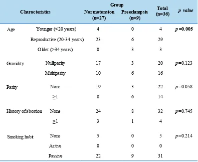
Younger (<20 years) 4 0 4 =0.005
Reproductive (20-34 years) 23 6 29
Older (>34 years) 0 3 3
Nulliparity 17 3 20 p=0.123
Multiparity 10 6 16
None 19 3 22 p=0.058
≥1 8 6 14
None 24 8 32 p=0.745
≥1 3 1 4
Smoking habit None 5 0 5 p=0.214
Active 0 0 0
Passive 22 9 31
Parity
History of abortion Gravidity
Age
Characteristics
Group
p
(n=27) (n=9)
Preeclampsia
Normotension (n=36)Total p value Table 1. Characteristics of subjects.
Table 2. Results of first and second examinations in normotensive group.
Median Min - Max Median Min - Max
sFlt-1 (pg/mL) 1,505.800 360 - 5,394 2,966.600 1,342 - 16,798 p=0.000
HIF-1α (ng/mL) 2.370 0.200 - 21.600 3.890 0.300 - 30.800 p=0.648
GA (week) 25 20 - 28 36 34 - 39 p=0.000
SBP (mmHg) 110 100 - 130 110 100 - 120 p=0.047
DBP (mmHg) 70 60 - 80 70 70 - 80 p=0.083
sFlt-1 / GA 60.510 13.300 - 192.600 81.960 37.600 - 466.600 p=0.029
HIF-1α / GA 0.110 0.010 - 0.900 0.110 0.010 - 0.790 p=0.118
1st examination (n=27) 2nd examination (n=27)
pvalue* Variable
*Wilcoxon test, GA: gestational age, Min: minimum, Max: maximum.
Venous blood was drawn using a serum separator tube and allowed to clot for two hours at room temperature before
centrifugation for 20 minutes at approximately 1,000g. Serum was immediately stored frozen in aliquots at -20oC
until assays for sFlt-1 and HIF-1α were performed.
Enzyme-linked Immunosorbent Assay (ELISA)
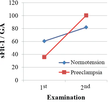
Figure 1. Changes of sFlt-1/GA ratio. GA: gestational age.
0
20
40
60
80
100
120
1
2
sF
lt
-1
/
GA
st
nd
Normotension
Preeclampsia
Examination
Examination
0
0.03
0.06
0.09
0.12
0.15
1
2
HI
F
-1
α
/
GA
Examination
Normotension
Preeclampsia
st
nd
Figure 2. Changes of HIF-1α/GA ratio. GA: gestational age.
Median
Min - Max
Median
Min - Max
sFlt-1 (pg/mL)
993.200
433 - 2184
3007.200
1473 - 6242
p
=0.008
HIF-1
α
(ng/mL)
3.400
0.000 - 15.300
4.930
0.200 - 17.600
p
=0.110
GA (week)
25.000
20 - 27
36.000
30 - 40
p=
0.008
SBP (mmHg)
120.000
100 - 130
140.000
140 - 160
p=
0.007
DBP (mmHg)
70.000
60 - 80
90.000
90 - 110
p=
0.007
sFlt-1 / GA
35.890
18.000 - 80.900
100.240
37.400 - 178.300
p=
0.015
HIF-1
α
/ GA
0.130
0.000 - 0.760
0.140
0.010 - 0.450
p
=0.953
Variable
1
p
value*
st
examination (n=9)
2
ndexamination (n=9)
Table 3. Results of first and second examinations in preeclamptic group.
*Wilcoxon test, GA: gestational age, Min: minimum, Max: maximum.
quantitative sandwich enzyme immunoassay technique. A
monoclonal antibody specific for sFlt-1 was pre-coated onto
a microplate. Standards and samples were pipetted into the
wells and any sFlt-1 present was bound by the immobilized
antibody. After washing away any unbound substances,
an enzyme-linked polyclonal antibody specific for sFlt-1 was added to the wells. Following a wash to remove any
unbound antibody-enzyme reagent, a substrate solution was added to the wells and color developed in proportion
to the amount of sFlt-1 bound in the initial step. The color
development was stopped and the intensity of the color was measured.
For HIF-1α detection, HIF-1α (Human) ELISA Kit (Abnova, Taipei, Taiwan) was used. Principally, the microtiter plate provided in this kit has been pre-coated with an antibody specific to HIF-1α. Standards or samples are
then added to the appropriate microtiter plate wells with a
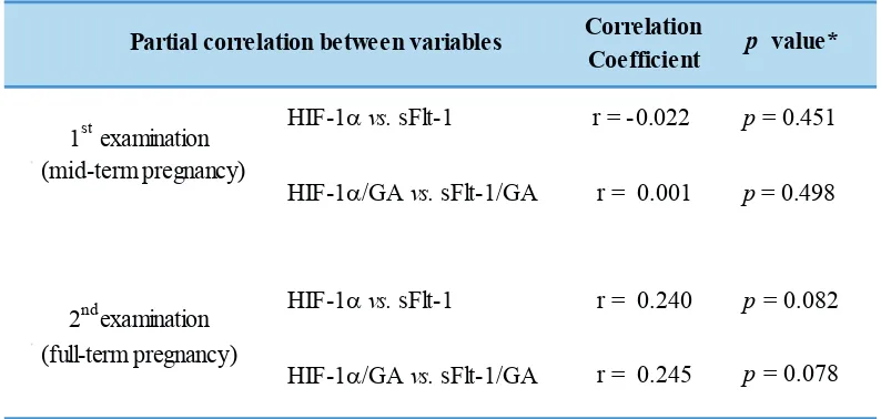
Table 4. Analyses of HIF-1α/GA and sFlt-/GA ratios in preeclamptic group.
HIF-1αvs. sFlt-1 r = -0.022 p = 0.451 HIF-1α/GA vs. sFlt-1/GA r = 0.001 p= 0.498 HIF-1αvs. sFlt-1 r = 0.240 p = 0.082 HIF-1α/GA vs. sFlt-1/GA r = 0.245 p= 0.078 1st examination
(mid-term pregnancy)
(full-term pregnancy)
Partial correlation between variables
2nd examination
Correlation
Coefficient p value*
*partial correlation test. GA: gestational age.
Results
The number of subjects involved at the first examination was 110, but only 40 subjects remained at the second examination. Among 40 subjects, only 36 subjects were eligible for complete laboratory assessment (Table 1). From 36 subjects, with 2 examinations (middle-term or 20-28 weeks gestation and at full-term pregnancies) for each subject, we collected 72 blood samples. All samples were processed and tested succesfully for sFlt-1 and HIF-1α concentrations (Table 2&3).
Table 1 shows that there was no significant differences
in the characteristics regarding gravidity, parity, abortion
history, and smoking habit between normotensive and
preeclamptic groups. Both groups comprised more subjects at the prime reproductive age, nulliparity, no history of
abortion and passive smokers. None of the younger group
developed to preeclampsia, while all subjects in the older group developed to preeclampsia. Statistical analysis
showed significant difference.
Differences of sFlt-1 and HIF-1α concentrations
according to gestational age (GA) were observed in
normotensive and preeclamptic groups. Analyses of HIF-1a and sFlt-1 concentrations changes at first and second
examinations for normotensive (Table 2) and preeclamptic (Table 3) groups are shown. There were significant elevation of sFlt-1 and ratio of sFlt-1/GA from first to second examinations for both normotensive and preeclamptic groups. However, no significant changes of HIF-1a and
ratio of HIF-1a/GA from first to second examinations
for both normotensive and preeclamptic groups. SBP
showed significant inclination for both normotensive and preeclamptic groups from first to second examinations. Meanwhile DBP increased significantly for preeclamptic group from first to second examinations.
Figure 1 shows increase of sFlt-1/GA ratio for both normotensive and preeclamptic groups from first to second examinations. Despite the fact that at first examination, sFlt-1/GA ratio was higher in normotensive than preeclamptic groups, sFlt-1/GA ratio of preeclamptic group was later
increased steeply and reached higher ratio than the one of
normotensive group at second examination.
Figure 2 shows a slight increase of HIF-1α/GA ratio in preeclamptic group from first to second examinations, for HIF-1α and Avidin conjugated to horseradish peroxidase
(HRP) is added to each microplate well and incubated. Then
a 3,3',5,5'-tetramethylbenzidine (TMB) substrate solution is added to each well. Only those wells that contain HIF-1α,
biotin-conjugated antibody and enzyme-conjugated Avidin
will exhibit a change in color. The enzyme-substrate reaction
is terminated by the addition of a sulphuric acid solution and the color change is measured spectrophotometrically.
Statistical Analysis
Statistical analysis was conducted using SPSS version 11.5 (IBM Corp., Armonk, NY), with significance level set at
We found a significant increase of preeclamptic risk
as related to age. Preeclamptic group was found to be
dominated by maternal age 35 years and above.
Conde-Agudello, et al., found that the relative risk of maternal age
≥35 years was 1.67 (95% CI 1.58-1.77).(6) Another report
shows the largest number of primipara with preeclampsia
is in category from 20 years (p<0.01). Meanwhile for the multipara, preeclampsia is most commonly developed in
age between 31-35 years.(7)
Increased preeclamptic risk in younger subjects with nulliparity was not found in this study, likely due to limited
number of the sample size. Rudra, et al., stated that increase
of preeclamptic risk in multiparity seemed to be related to hypoxia at the maternal-fetal surface.(8)
To observe the changes of sFlt-1 and HIF-1α, a
cohort-time series observation was designed. When only
normotensive group were observed (Table 2), it turned out that sFlt-1 concentrations and sFlt-1/GA ratios were increased significantly. This suggests that during pregnancy, sFlt-1 could increase normally along with GA. It was
reported that in normotensive pregnancy, the concentration
of sFlt-1 is considered stable until 2 months before the end
of pregnancy when it may show increased concentration.
(9) On the other hand, we also found significant increase of sFlt-1 concentrations and sFlt-1/GA ratios in the group that developed preeclampsia (Table 3). It has been shown that increased concentration of sFlt-1 in subjects who developed
preeclampsia seemed to be obvious at the second trimester,
although placental growth factor (PlGF) and VEGF might have started to crash at the end of the first trimester.(9)
According to Romero, et al., in patients who develop preeclampsia during pre-term and full-term pregnancy,
sFlt-1 levels were significantly higher that can be seen at 7 weeks, or even 11 weeks before the clinical diagnosis.(10) The changes of sFlt-1 concentrations in normotensive and preeclamptic groups were shown in Figure 2. Although at first
examination, sFlt-1/GA ratio was higher in normotensive than preeclamptic groups, sFlt-1/GA ratio of preeclamptic
group was later increased steeply and reached higher ratio
than the one of normotensive group at second examination. Park, et al., compared the concentration of sFlt-1 in normal pregnancy, mild preeclampsia, and severe preeclampsia.
The results showed that in normal pregnancy, median: 441 pg/mL, range: 58-1,959 pg/mL, mean±SD: 531±371 pg/mL; mild preeclampsia, median: 334 pg/mL, range: 60-3,375 pg/mL, mean±SD: 750 ± 908 pg/mL; severe preeclampsia, median: 762 pg/mL, range 261-3,309 pg/mL, mean±SD: 962±688 pg/mL. Median of sFlt-1 concentration was higher, although not significantly (p=0.07) in severe preeclampsia
compared with mild preeclampsia, while no significant differences were seen also between sFlt-1 concentration
in mild preeclampsia and those in normotensive patients (p=0.84).(11) This finding can be explained due to results in this cohort study that almost all study subjects developed into mild preeclampsia. This is in contrast to the study result of Chaiworapongsa, et al., who found that an increase of
sFlt-1 was correlated with disease severity.(12)
According to Lam, et al., preeclampsia does not
develop in all women with high sFlt-1 or low PlGF, but can also occur in women with lower sFlt-1 and higher PlGF with not yet explainable mechanism. It was suspected that the
enhancement of "soluble" factor occurred due to placental
hypoxia. These findings were similar to our current results showing that sFlt-1 concentration in preeclamptic group were lower than sFlt-1 level in normotensive group.(4)
Thomas, et al., suggested the possible involvement of sFlt-1
variants that significantly increased in preeclamptic subjects compared with normotensive subjects.(13)
In the normotensive pregnancies (Table 2), the HIF-1a concentration and HIF-1a / GA ratio did not change
significantly. These results suggested that in normotensive subjects, no hypoxic conditions were resulted that might
endanger the mothers and fetuses.
sFlt-1 concentrations were lower at first examination and later became higher at the second examination which showed that sFlt-1 concentration at first examination could
not be used as a reference for the possibility of preeclampsia. A number of studies also showed no differences were found between the groups of normotensive and mild preeclampsia in mid-term. This suggests that elevated
concentration of sFlt-1, in addition to the onset, also seem to
be related to severity of preeclampsia, therefore the increase was slower. The study of Unal, et al., showed that in severe
preeclampsia sFlt-1, the level might not show changes in
Discussion
meanwhile no increase of the ratio was observed in
Conclusion
In this study higher concentration of sFlt-1 were found in preeclampsia. Changes in the levels of sFlt-1 were more important than the average levels. Even the changes of HIF-1α level were greater in preeclamptic group, but it have not been proven as an established circulating marker of hypoxia for preeclampsia. HIF-1α levels tended to be positively correlated with sFlt-1.
References
1.Osungbade KO, Ige OK. Public Health Perspectives of Preeclampsia in Developing Countries: Implication for Health System Strengthening. J Pregnancy. 2011: 48109.
2.Khan KS, Wojdyla D, Say L, Gülmezoglu AM, Van Look PF. WHO analysis of causes of maternal death: A systematic review. Lancet. 2006; 367: 1066-74.
3.Lyall F, Bellfort M. Pre-eclampsia: etiology and clinical practice. Cambridge University Press: United Kingdom. 2007.
4.Lam C, Lim KH, Karumanchi SA. Circulating Angiogenic Factors in
the Pathogenesis and Prediction of Preeclampsia. Hypertension.
2005; 46: 1077-85.
5.Zagorska A, Dulak J. HIF-1: the known and unknown of hypoxia sensing. Acta Biochim Pol. 2004; 51: 563-78.
6.Conde-Agudelo A, Belizan JM. Risk factors for pre-eclampsia in a large cohort of Latin American and Caribbean women. Br J Obstet Gynecol. 2000; 107: 75-83.
7.Jasovic-Siveska E, Jasovic V, Stoilova S. Previous pregnancy history, parity, maternal age and risk of pregnancy induced hypertension. Bratisl Lek Listy. 2011; 112: 188-91.
8.Rudra CB, Williams MA, Schiff MA, Koenig JQ, Dills R, Yu J. A prospective study of maternal carboxyhemoglobin and preeclampsia risk. Paediatr Perinat Epidemiol. 2010; 24: 35–44.
9.Grill S, Rusterholz C, Zanetti-Dällenbach R, Tercanli S, Holzgreve W,
Hahn S, et al. Potential markers of preeklamsi-a review. Reprod
Biol Endocrinol. 2009; 7: 70.
10.Romero R, Nien JK, Espinoza J, Todem M, Fu W, Chung H, et al. A longitudinal study of angiogenic (placental growth factor) and
anti-angiogenic 13(+6) weeks of gestation. Prenat Diagn. 2008; 28: 1110–5.
11.Park CW, Park JS, Shim SS, Jun JK, Yoon BH, Romero R. An elevated maternal plasma, but not amniotic fluid, soluble fms-like tyrosine kinase-1 (sFlt-1) at the time of mid-trimester genetic amniocentesis is a risk factor for preeclampsia. Am J Obstet Gynecol. 2005; 193: 984–9.
12.Chaiworapongsa T, Romero R, Espinoza, J, Bujold E, Kim Y M, Goncalves LF, et al. Evidence supporting a role for blockade of the
vascular endothelial growth factor system in the pathophysiology of
preeclampsia. Am J Obstet Gynecol. 2004; 190: 1541-7.
13.Thomas CP, Andrews JI, Raikwar NS, Kelley EA, Herse F, Dechend
R, et al. A Recently Evolved Novel Trophoblast-Enriched Secreted
Form of fms-Like Tyrosine Kinase-1 Variant Is Up-Regulated in Hypoxia and Preeclampsia. J Clin Endocrinol Metab. 2009; 94: 2524-30.
14.Unal ER, Robinson CJ, Johnson DD, Chang EY. Second-trimester angiogenic factors as biomarkers for future-onset preeclampsia. Am J Obstet Gynecol. 2007; 197: 2111-4.
15.Perni U, Sison C, Sharma V, Helseth G, Hawfield A, Suthanthiran M, et al. Angiogenic factors in superimposed preeclampsia: a longitudinal study of women with chronic hypertension during pregnancy.
Hypertens. 2012; 59: 740-6.
16.Levine RJ, Maynard SE, Qian C, Lim KH, England LJ, Yu KF, et al.
Circulating Angiogenic Factors and the Risk of Preeclampsia. N Engl J Med. 2004; 350: 672-83.
17.Haggerty CL, Seifert ME, Tang G, Olsen J, Bass DC, Karumanchi SA,
et al. Second trimester anti-angiogenic proteins and preeclampsia.
Pregnancy Hypertens. 2012; 2: 158-63.
18.Peng M, Ding Y, Yu L, Zhou C. Change of HIF-1α protein expression in the second trimester.(14) Similar results were also obtained
in the study of Perni, et al., where at 20 weeks GA,
sFlt-1 concentration of normotensive group were in the range of 2,076 ± 2,452.1, while in the group with late-onset preeclampsia, were in the range of 1,426.6±1,132.2.(15)
According to Levine, et al., the average (mean)
concentrations of sFlt-1 increased approximately 5 weeks
before the onset of preeclampsia. This study did not observed
sFlt-1 concentration increase up to the age of 29 weeks GA
in subjects who subsequently developed to preeclampsia.
At the time of clinical onset, mean concentration of sFlt-1 was approximately 4,382 pg/mL.(sFlt-16) Haggerty, et al., also concluded that the changes i.e. increase of sFlt-1 in the
second trimester was associated with 39% increased risk of preeclampsia (95%, CI = 3%, 86%).(17)
In our current results of HIF-1a concentration and
HIF-1a/GA ratio, no significant difference between
normotensive and preeclamptic groups was observed at
both first and second examinations. This is probably due to another marker that precedes the other markers especially sFlt-1. Markers in fluctuating circulating levels or those that exist in a long period cannot be equated with the condition
in the placenta. Peng, et al., examined the expression of
HIF-1α in placental bed and found the highest expression
was in severe preeclampsia followed by mild preeclampsia.
(18) According to Olyeymanlou, et al., HIF-1a seems to
induce s-Flt1 in placental explants.(19) Furthermore Tal,
et al., stated that over-expression of HIF-1α raised s-Flt1 and soluble endoglin (s-Eng) level and eventually led to the manifestation of preeclampsia, namely hypertension and
proteinuria.(20)
Akhiles, et al., found that concentration of HIF-1α in prenatal blood samples in preeclampsia were higher than those in postnatal period. Mean percentage reduction of
the placenta bed and concentration of vWF in maternal peripheral
blood of pre-eclampsia. J Central South Univ (medical science).
2010; 35: 134-9.
19.Soleymanlou N, Jurisica I, Nevo O, Ietta F, Zhang X, Zamudio S, et al.
Molecular Evidence of Placental Hypoxia in Preeclampsia. J Clin Endocrinol Metab. 2005; 90: 4299-308.
20.Tal R, Shaish A, Barshack I, Polak-Charcon S, Afek A, Volkov A, et al. Effects of hypoxia-inducible factor-1alpha overexpression
in pregnant mice: possible implications for preeclampsia and
intrauterine growth restriction. Am J Pathol. 2010; 177: 2950-62. 21.Akhilesh M, Mahalingam V, Nalliah S, Ali RM, Ganesalingam M,