Dental Research, Dental Clinics, Dental Prospects
Original Article
Comparison of Microleakage and Thickness of Resin Cement in
Ceramic Inlays with Various Temperatures
Homayoun Alaghemand1 • Faezeh Abolghasemzadeh2*• Farzaneh Pakdel3 • Reza Judi Chelan4
1
Dental Material Research Center, Associate Professor, Department of Operative Dentistry, Faculty of Dentistry, Babol University of Medical Sciences, Babol, Iran
2Assistant Professor, Department of Operative Dentistry, Faculty of Dentistry, Babol University of Medical Sciences, Babol, Iran 3Assistant Professor, Department of Oral Medicine, Faculty of Dentistry, Tabriz University of Medical Sciences, Tabriz, Iran
4DentalMaterial Research Center, Member of Research Committee of Medical Students, Student of dentistry , Faculty of Dentistry , Babol University of
Medical Sciences, Babol, Iran.
*Corresponding Author; E-mail: faezeha64@yahoo.com
Received: 8 January 2013; Accepted: 14 July 2013
J Dent Res Dent Clin Dent Prospect 2014;8(1):45-50 | doi: 10.5681/joddd.2014.008 This article is available from: http://dentistry.tbzmed.ac.ir/joddd
© 2014 The Authors; Tabriz University of Medical Sciences
This is an Open Access article distributed under the terms of the Creative Commons Attribution License (http://creativecommons.org/licenses/by/3.0), which permits unrestricted use, distribution, and reproduction in any medium, provided the original work is properly cited.
Abstract
Background and aims.
Microleakage is still one of the major problems of composite-based restorations. This studycompared the microleakage and thickness of resin cement in ceramic inlays with various temperatures.
Materials and methods.
Class V cavities were prepared on the buccal and lingual aspects of thirty human molars withocclusal margins in enamel and gingival margins in dentin (3 mm wide, 5 mm long and 2 mm deep). Laboratory-made
inlays (LMI) were used for buccal cavities, and CAD/CAM inlays (CMI) were used for lingual cavities. All the cavities
were divided into six groups (n=10): 1) LMI at-5°C; 2) LMI at 50°C; 3) LMI at room temperature (25°C); 4) CMI at -5°C;
5) CMI at 50°C; 6) CMI at room temperature (25°C). Inlays were bonded to cavities in a pulp pressure- and
temperature-simulating device. After thermocycling and dye penetration, the teeth were divided into two mesiodistal halves. Amount of
dye penetration and film thickness were measured under a stereomicroscope and analyzed with Kruskal-Wallis, Wilcoxon
and Spearman's correlation tests ( = 0.05).
Results.
There were no statistically significant differences in leakage between different inlay temperatures (P > 0.05). Themean cement thickness in laboratory-made inlays (gingivalmargin, 83.7 ± 11 and occlusal margin, 84.7 ± 19) was greater
than that in CAD/CAM inlays (gingival margin, 69 ± 16 and occlusal margin, 84.7 ± 16). No correlation was found
be-tween cement thickness and microleakage either in enamel or dentin for any of the ceramic systems.
Conclusion.
Differences in inlay temperature had no effect on microleakage. CAD/CAM inlays had lower cementthick-ness than laboratory-made inlays, but this was not related to their microleakage.
Introduction ls have now become the bedrock of cosmetic dentistry. Ceramic inlays have less leakage and seat better than composite inlays. Bond strength of resin cements to etched ceramic inlays is stronger and more durable than composite inlays. Resin cement is the only recommended material for cementation of ceramic inlays. This cement restricts microleakage, increases the strength of the restoration and in short term reinforces residual tooth structure.1
In recent years, many improvements have been made in mechanical and physical properties of composite resins, but microleakage is still one of the major problems of composite-based restorations. It seems using composite resins without polymerization shrinkage is the main key to placement of a leakage-free restoration.2
Nowadays, it has been proven that composite's flow at the free surface can reduce the shrinkage stress of composite in bonded surface and reduce restoration microleakage. Reducing the rate of polymerization provides more time for composite to flow at free surface and reduce the shrinkage stress. Temperature of composite resin during setting is one of the factors that influences the amount of polymerization shrinkage stress. Low temperature causes reduction in the degree of conversion and polymerization rate. In addition, thermal expansion of cooled composite resin when placed under the oral cavity temperature can compensate polymeriza-tion shrinkage to some extent.3
Many studies have investigated the effect of tem-perature on microleakage of direct composite resin restorations, but none of them paid attention to the effect of different temperatures of ceramic inlays on microleakage of room-temperature resin cement.
4-6
Since the amount of film thickness was different in the laboratory-made and CAD/CAM inlays,7,8 we used both types of restoration to evaluate the effect of film thickness on microleakage of resin cement. Therefore, the aim of this in vitro study was to com-pare the microleakage and thickness of resin cement in ceramic inlays with various temperatures. The null hypothesis was that temperature of ceramic inlay and cement thickness would not affect microleakage.
Materials and Methods
Thirty caries-free freshly extracted human molars stored in saline solution were selected for this study. For disinfection, the teeth were immersed in 0.05%
thymol solution for no longer than 6 months.9 Stan-dardized Cl V cavities (5 mm in length, 3 mm in width, and 2 mm in depth) were prepared on the buccal and lingual aspects of each tooth, with the gingival wall extending beyond cemento-enamel junction, while the occlusal walls extended to the enamel. An impression was made from the buccal cavities with additional silicone material (Elite HD+, Zhermack, Rovigo, Italy) and sintered feldspathic porcelain inlays (Ceramco, Dentsply, USA) were made for each cavity according to manufacturer’s instructions. CAD/CAM (CEREC3D, Sirona Dental Systems, Germany) feldspathic inlays (CEREC® Blocs, Sirona Dental Systems, LLC, Germany) were made for lingual cavities. The cavities were divided into six groups (n=10):
1. Sintered feldspathic inlays cooled to −5°C
2. CAD/CAM feldspathic inlays cooled to
−5°C
3. Sintered feldspathic inlays warmed to 50°C
4. CAD/CAM feldspathic inlays warmed to
50°C
5. Sintered feldspathic inlaysat room tempera-ture (25°C)
6. CAD/CAM feldspathic inlays at room
tem-perature (25°C)
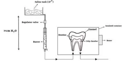
Pulp Pressure and Temperature Simulator
A device was madefor simulation of oral conditions, which sent water (37°C) with 35 cmH2O pressure into the pulp chamber. Prior to the bonding process, air of the chamber was kept at 37°C. The device con-sisted of an insulated container in which inlays were bonded to the teeth. The chamber temperature was controlled by a heater and a temperature-regulating device (thermocouple) at the same temperature as the oral cavity (37°C). To simulate the pulpal pressure, a burette was used. The burette was connected to a normal saline tank (the normal saline temperature was kept at 37°C with a heater and thermocouple) and by a regulator valve; the water flow was adjusted so that the water height was 34–40 cm. From the end of the burette, a narrow pipe came to the insulated chamber and was connected to the apex of tooth root with an 18-gauge needle. The apex of each root was dilated so that the needle could enter and was con-nected to the apex; therefore, water could enter from a root to the pulp chamber and pass through the other root (Figure 1).
Inlay Bonding Procedure
The inlays were etched for 90 seconds with 9.5% hydrofluoric acid (HF) (Ultradent Dental Co, South Jordan, Utah, USA) followed by rinsing with water for 30 seconds and drying with an air stream. One-bottle silane coupling agent (Ultradent Dental Co, South Jordan, Utah, USA) was applied over the etched region. The inlays were placed in a freezer at -5°C temperature at least for 5 minutes before the bonding process for cooling and were placed in a hot air oven at 50°C temperature at least for 5 minutes before the bonding process for warming. Panavia F2.0 resin cement (Kuraray Dental Co, Okayama, Japan)was used to bond inlays. One drop from each bottle of Panavia ED primer was mixed and applied to tooth surface as bonding agent according to manu-facturer's instructions. Before bonding a thin layer of unfilled resin (Margin Bond, Coltene/Whaledent, Switzerland) was applied to inlays.10 The inlay was bonded to the tooth while the tooth was connected to the pulp pressure- and temperature-simulating de-vice. Resin cement was cured for one minute with an LED light-curing device (Valo,Ultradent Dental Co, South Jordan, Utah, USA). The light intensity was 1000 mW/cm2.
Thermocycling
After the bonding procedure, the samples were
thermocycled for 500 cycles11 at 5°C/55°C water baths with 20 seconds of dwell time and 10 seconds of transfer time for each.
Microleakage Test and Cement Film Thickness Measurement
After thermocycling, the apices of the teeth were sealed with composite resin. All the surfaces of the teeth, except for 1 mm around the margins of each restoration, were sealed with 2 coats of nail varnish.
The teeth were immersed in 50 wt% silver nitrate for 2 hours in a dark place. Then the samples were rinsed under running water and immersed for 4 hours in developing solution under fluorescent light and
then flushed under running water.12 Then the
samples were mounted in epoxy resin and sectioned buccolingually from the middle of restorations. Each half was observed under a stereomicroscope (×40 magnifications) and scored for the degree of dye penetration at the occlusal and cervical walls:
0: No dye penetration
1: Dye penetration into the enamel or extending to half of the cervical wall
2: Dye penetration beyond the dentinoenamel junction or to more than half or the entire cervical wall
3: Dye penetration into the pulpal wall13
After taking digital images from the samples with a stereomicroscope (×40 magnification) (SZX12, Olympus America, Melville, NY, USA) the cement film thickness was measured with Analysis LL Starter software (Inet Soft Technology Corp., New Jersey, USA).
Data was analyzed by SPSS 18. Kruskal-Wallis test was used to compare leakage between different temperatures. Wilcoxon signed ranks test was used
Figure 1. Pulp pressure and temperature simulator. Table 1. Composition of materials used for this study according to manufacturer’s data
Material Manufacturer Type Composition
Kuraray Co., Dual-cure ED Primer A: HEMA, 10-MDP, 5-NMSA ED Primer B: 5-NMSA Paste A: 10-MDP, Bis-GMA, filler,
benzoyl peroxide, photoinitiator
Panavia F2.0
Osaka, Japan adhesive system
Paste B: Bis-GMA, filler, Sodium fluoride, amine
Margin Bond Coltene/Whaledent, Switzerland Unfilled resin BISGMA:35-40 % TEGDMA: 20-25 %
Silane Ultradent Dental Co, South
Jor-dan, Utah, USA Porcelain primer Methacryloxy propyltrimethoxy silane:5-15% Ethanol:87%
ultradent Dental Co, South Jordan, Utah, USA
Porcelain etchant Hydrofluoric Acid: 9.5%
Hydrofluoric Acid
HEMA: 2-Hydroxyethyl methacrylate
to compare the mean cement film thickness of enamel and dentinal margin of CAD/CAM and labo-ratory-made inlays and compare microleakage of dentinal and enamel margins of different types of inlays at different temperatures. Spearman's correlation coefficient was used to evaluate the relation between resin cement film thickness and microleakage at enamel and dentinal margins at all the temperatures. Statistical significance was set at 5% (P<0.05).
Results
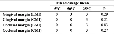
Table 2 shows the means of microleakage scores at occlusal and gingival margin of the samples. Kruskal-Wallis test did not reveal any significant differences in microleakage between the groups (P>0.05).
Wilcoxon test showed significant differences be-tween the mean cement film thickness of laboratory and CAD/CAM inlays (P<0.05). The minimum ce-ment film thickness belonged to CAD/CAM inlays (occlusal margin: 63.4±16, gingival margin: 69±16), and maximum cement film thickness belonged to laboratory inlays (occlusal margin: 84.7±19, gingival margin: 83.7±11).
Wilcoxon test showed that the difference in micro-leakage between gingival and marginal margins was statistically significant only in laboratory and CAD/CAM inlays at 25°C (P<0.05). The difference of mean microleakage was not statistically different at enamel margins between different temperatures (P>0.05). The difference of microleakage between gingival margins of laboratory inlays and CAD/CAM inlays was statistically significant only at 50°C and the difference of microleakage between the occlusal margins of laboratory inlays and CAD/CAM inlays was statistically significant only at -5°C.
Spearman’s correlation coefficient showed no sig-nificant relation between resin cement film thickness and microleakage at enamel and dentinal margins at all the temperatures (P<0.05).
Discussion
In this study, there was a slight decrease in micro-leakage in low- and high-temperature groups but this relationship was not significant statistically.
Microleakage studies use different methods. In this study dye penetration method was used because of its simple procedure and easy and accurate ability of observation by digital imaging.14 Several dye solu-tions are available for microleakage tests but silver nitrate dye penetration may be a particularly de-manding test of marginal seal because silver ions are smaller than the bacteria that usually live in the oral cavity.15 Therefore, in this study silver nitrate dye was used.
Many studies have investigated the effect of differ-ent temperatures of composite on microleakage. Some studies have shown that cold insert causes re-duction in composite resin microleakage.2,16,17 As-mussen18 found that the filling could be cooled through a certain temperature range without mar-ginal gap being formed.
Other studies have shown less microleakage with warmed composite resin.19,20 dos Santos et al4 found that warm composite resin (54°C and 60°C) had no effect on microleakage when LED light-curing sys-tem was used, but with QTH light-curing syssys-tems they found less microleakage.
Some studies have shown that different tempera-tures have no effect on microleakage.3,5,6,21 Daronch et al22 found no significant difference between the microleakage of warmed composite resin at 140°F, room temperature composite resin and flowable composite resin except for enamel margin of class V cavities; at room temperature composite resin micro-leakage was less than that of flowable composite resin.
To date, no research study has evaluated the effect of cooling or heating ceramic restorations on resin cement microleakage; only Morais et al23 evaluated the effect of heating the resin cement and ceramic restorations on bond strength. Since cooling the resin increases cement viscosity, interfering with restora-tion seating, and warming could increase its viscos-ity, resulting in difficulties in cement handling, in this study inlays were warmed or cooled instead of the resin cement.
Different studies have used different temperatures for warming and cooling composite resins but the exact reason for selecting these temperatures has not been mentioned in their studies. Since very different temperatures have been used in other studies we used -5°Cand 50°Cfor warming and cooling inlays. Inlay bonding procedure is more time-consuming Table 2. Comparison of microleakage at occlusal and
gingival margins between CAD/CAM and laboratory-made inlay groups
Microleakage mean -5°C 50°C 25°C P
Gingival margin (LMI) 0 3 3 0.29
Gingival margin (CMI) 0 0 3 0.21
Occlusal margin (LMI) 0 0 3 0.83
than direct composite resins. Therefore, in this study we used some higher and lower temperatures than other studies to compensate temperature changes occurring during inlay bonding to achieve results that could be extended to in vivo conditions. To be sure of reaching of inlays to exact temperatures, the inlays were kept for 5 minutes at 5°C and 50°C. Since there were no references in the literature, this time was selected by authors' experience.
In vitro microleakage studies have not simulated pulpal pressure.24 In this study, we used pulp pres-sure and a temperature device for simulating in vivo conditions to achieve more reliable results. Many studies have shown that heating composite resin in-creases free radical movement, increasing the degree of conversion, polymerization shrinkage and the re-sultant stress.25,26 Also, one of the effects of tempera-ture on materials is thermal expansion and contrac-tion. Warmed composite resin shrinks thermally when placed in the oral cavity temperature. On the other hand, higher temperatures can increase com-posite resin flowability and its adaptation to cavity walls. In theory, thermal expansion of cold compos-ite resin due to warming under oral temperatures can reduce polymerization-induced gap but cooling in-creases viscosity and causes problems during resto-ration seating.3 Because of these different influences of heating and cooling, different studies have shown different results.
Minimum marginal gap in indirect restorations can be achieved with low film thickness. Adaptation of indirect restorations is one of the most important cri-teria of clinical success.27 In this study, cement film thickness was used for measurement of marginal in-tegrity. A major concern about CAD/CAM restora-tion has been their pre-cementarestora-tion marginal fit. However, improvements in CEREC hardware and software have led to improvements in marginal fit.28
Ellingsen and Fasbinder29 compared the
pre-cementation fit of CERED2 crowns with CEREC3 crowns. CEREC3 had more marginal accuracy than CEREC2 in all areas. In the present study, like a study by Romao et al,30 the cement thickness was smaller in CAD/CAM inlays. This result may sug-gest that marginal integrity and accuracy of CAD/CAM system is not a concern anymore and these systems have acceptable accuracy for clinical use.
In this study, like that by Romao et al,30 no relation was found between microleakage and film thickness. It can be expected that better marginal adaptation can cause lower volume of resin cement and its re-sultant problems.31 Resin cement
polymerization-induced stress occurs in closed environments that can have negative effects on bond strength. Magni-tude of this force depends on the ratio of bonded to non-bonded surfaces (C-factor). Therefore, in theory, it seems that there is an inverse relationship between the thickness of the resin and microleakage. Alester et al32 showed that a thin layer of resin (50–2700 µm) increases polymerization stress but on the other hand they reported that resin cement layer was too thin and even small deformation in the substrate could release polymerization shrinkage stress. It seems these two factors (increasing the polymeriza-tion shrinkage stress because of thin layer of resin cement and stress releasing due to substrate deforma-tion) can affect each other and can be the reason for the results achieved in the present study.
According to the results of the present study, dif-ferent temperatures of inlays and cement thickness had no significant effect on restoration microleakage. But in this study we changed the inlay temperature. Maybe changing resin cement temperature can di-rectly affect microleakage. Further studies are re-quired to investigate the effect of resin cement tem-perature on inlay microleakage.
Conclusion
Within the limitations of this in vitro study, it can be concluded that heating or cooling of inlays had no significant effect on microleakage. CAD/CAM-made restorations had lower cement film thickness than laboratory-made restorations, resulting in a slight decrease in leakage, but this decrease was not statis-tically significant.
References
1. Summitt JB, Robbins JW, Hilton TJ, Schwartz RS, dos
Santos J Jr. Fundamentals of Operative Dentistry, 3rd ed. Illinois : Quintessence, 2006:516.
2. de la Torre-Moreno FJ, Rosales-Leal JI, Bravo M.
Ef-fect of cooled composite inserts in the sealing ability of resin composite restorations placed at intraoral tem-peratures: An in vitro study: Oper Dent 2003;28:297-302.
3. Moazami SM, Falah M. Effect of different composite
resin temperatures and different light exposure pat-terns on microleakage of composite resin restorations. Journal of Mashhad dental school 2004;28:105-10.
4. dos Santos RE, Lima AF, Soares GP, Ambrosano GM,
Marchi GM, Lovadino JR, etal. Effect of preheating resin composite and light-curing units on the micro-leakage of Class II restorations submitted to thermo-cycling. Oper Dent 2011;36:60-5.
5. Karaarslan ES, Usumez A,Ozturk B,Ata Cebe M.
pre-heating procedures on microleakage of class V resin restorations. Eur J Dent 2012;6:87–94.
6. Sabatini C, Blunck U, Denehy G, Munoz C. Effect of
Pre-heated Composites and Flowable Liners on Class
II Gingival Margin Gap Formation. Oper Dent
2010;35:663-71.
7. Keshvad A, Hooshmand T, Asefzadeh F, Khalilinejad
F, Alihemmati M ,Van Noort R. Marginal Gap, Internal Fit, and Fracture Load of Leucite-Reinforced Ceramic Inlays Fabricated by CEREC inLab and Hot-Pressed Techniques. J Prosthodont 2011;20:535-40.
8. Aboushelib MN, Elmahy WA, Ghazy MH. Internal
adaptation, marginal accuracy and microleakage of a pressable versus a machinable ceramic laminate ve-neers. J Dent. 2012;40:670-7.
9. Carvalho AA, Moreira FCL, Cunha LM, de Moura
SM, de Souza JB, Estrela C, etal. Marginal microleak-age of class II composite resin restorations due to re-storative techniques. Rev Odonto Ciênc 2010;25:165-9.
10. Hooshmand T, van Noort R, Keshvad A. Bond
dura-bility of the resin-bonded and silane treated ceramic surface. Dent Mater 2002;18:179-88.
11. International Organization for Standardisation
(ISO).Dental materials-testing of adhesion to tooth structure.ISO/TS 11405:2003(E).
12. Mousavinasab SM, Atai M, Alavi B. To Compare the
Microleakage Among Experimental Adhesives Containing Nanoclay Fillers after the Storages of 24 Hours and 6 Months. Open Dent J 2011;5:52–7.
13. Radhika M, Sajjan GS, Kumaraswamy BN, Mittal N.
Effect of different placement techniques on marginal microleakage of deep class-II cavities restored with
two composite resin formulations. J Conserv Dent
2010;13:9-15.
14. de Almeida JB, Platt JA, Oshida Y, Moore BK,
Cochran MA, Eckert GJ. Three different methods to evaluate microleakage of packable composite in class II restoration. Oper Dent 2003;28:453-60
15. Roberson TM, Heymann HO, Swift EJ JR.
Stur-devant’s Art and Science of Operative Dentistry, 5th ed. St Louis: Mosby Elsevier; 2006. p. 264.
16. Suresh Kumar BN, Savadamoorthi KS,
Karpagavinay-agam K. Sealing ability of class V resin composite res-toration with cooled composite inserts—an in vitro study. IJCD 2011;2:103-8.
17. Elsayad I. Cuspal movement and gap formation in
premolars restored with preheated resin composite. Oper Dent 2009;34:725-31.
18. Asmussen E. The effect of temperature changes on
adaptation of resin fillings: I. Acta Odontol Scand 1974;32:161-71.
19. Choudhary N, Kamat S, Mangala TM, Thomas M.
Ef-fect of pre-heating composite resin on gap formation at three different temperatures. J Conserv Dent 2011 ; 14: 191–5
20. Wagner WC, Aksu MN, Neme AL, Linger JB, Pink
FE, Walker S. Effect of Pre-heating Resin Composite on Restoration Microleakage: Oper Dent 2008;33:72-8.
21. Briso ALF, Sundefeld RH, Afonso RL, Paterno FA,
Sundefeld MLMM. Effect of refrigeration of resin ma-terials on the occurrence of microleakage in class II restorations: Brazilian Dental Science 2007;10:6-12.
22. Daronch M, Rueggeberg FA, Hackman S, Multon M,
De Goes MF.Effect of Composite Preheating on
Mi-croleakage: Class I and V. Baltimore Convention Cen-ter Exhibit Hall E-F Abs: 0604. 2005 March [cited 2012 July 10]. Available from: http://iadr.confex.com/iadr/2003SanAnton/techprogra m/abstract_27111.htm
23. Morais A, dos Santos AR, Giannini M, Reis AF,
Rodrigues JA, Arrais CA. Effect of pre-heated dual-cured resin cements on the bond strength of indirect restorations to dentin. Braz Oral Res 2012;26:170-6.
24. Gharizadeh N, Kaviani A, Nik S. Using electric
cur-rent during dentin bonding agent application and its ef-fect on microleakage under simulated pulpal pressure conditions. Dent Res J (Isfahan) 2010;7:23-7.
25. Rueggeburg A, Freedman G. Clinical benefits of
pre-warmed composites. Private Dentistry 2003;8:111-4.
26. Truillo J, Stansbury Y. Thermal effects on composite
photo polymerization monitored by real-time NIR. J Dent Res 2003;82:Abs:0819.
27. Schmid-Schwap M, Graf A, Preinerstorfer A, Watts
DC, Piehslinger E, Schedle A. Microleakage after thermocycling of cemented crowns—A meta-analysis. Dent Mater 2011;27:855-69.
28. Summitt JB, Robbins JW, Hilton TJ, Schwartz RS, dos
Santos J Jr. Fundamentals of Operative Dentistry, 3rd ed. Illinois : Quintessence, 2006:533.
29. Ellingsen LA, Fasbinder DJ. In vitro evaluation of
CAD/CAM ceramic crowns [abstract 2460]. J Dent
Res 2002; 81:A-331.
30. Romão W Jr, Miranda WG Jr, Cesar PF, Braga RR.
Correlation between microleakage and cement thick-ness in three Class II inlay ceramic systems. Oper Dent 2004; 29:212-8.
31. Audenino G, Bresciano ME, Bassi F, Carossa S. In
vitro evaluation of fit of adhesively luted ceramic inlays. Int J Prosthodont 1999;12:342-47.
32. Alester D, Feilzer AJ, de Gee AJ, Davidson C.L.