* Received for publication: March 29, 2005; accepted: September 15, 2005 INTRODUCTION
Draf II sinusotomy is subdivided into IIA and IIB [1]. Draf IIA, described by Stammberger as “uncapping the egg” or by May and Schaitkin [2] as the Nasofrontal Approach II, results in complete exposure of the frontal ostium after removal of any frontal cells occluding the frontal ostium and recess. Draf IIB is similar to the Nasofrontal Approach III [2] and comprises drilling out the frontal sinus floor between the lamina papyracea laterally and the nasal septum medially.
The frontal sinus recess os is a complicated 3-dimensional structure affected by the agger nasi cells, the frontal cells, the beak, the anteromedial frontal sinus floor, the attachment of the most anterior middle concha, and the nasal septum. Furthermore, these structures vary greatly between individuals, thereby constituting a challenge to the surgeon. All these struc-tures are covered by a mucous membrane, which plays an important role in the normal physiology and healing process of the frontal sinus and nose.
The beak is an important and consistent landmark, especially in revision endoscopic sinus surgery (ESS), when the typical
land-marks are absent. In revision ESS, the frontal ostium, even when blocked, can be located by exposing the prominent white bone of the beak and by following its superomedial aspect into the frontal os. The location of the beak is constant, thus making it important for localization of the frontal ostium when a nagi-vation system is not available. Anatomical remnants such as the axilla of the middle concha are valuable for easy and precise location of the frontal os.
High resolution CT scans (with axial and coronal planes), are mandatory for a safe and successful approach to the frontal sinus. The sagittal plane should be added when revision ESS is risky.
We present our experience with primary and revision endoscop-ic Draf II frontal sinusotomy.
MATERIAL AND METHODS
Assaf Harofeh Medical Center, Zerifin, Israel is a tertiary hospi-tal affiliated to the Tel Aviv University, and is located in the suburbs of Tel Aviv. The survey included the files and comput-ed tomography (CT) scans of 25 patients who were operatcomput-ed on
The endoscopic Draf II frontal sinusotomy:
non-navigated approach*
E. Eviatar, U. Katzenell, S. Segal, N. Shlamkovitch, L. Muallem Kalmovich,
A. Kessler, M. Vaiman
Department of Otolaryngology-Head and Neck Surgery, Assaf Harofeh Medical Center, Zerifin, affiliated to the Sackler Faculty of Medicine, Tel Aviv University, Israel
Objective: Endoscopic endonasal Draf II frontal sinusotomy is indicated for a variety of pathologies such as mucocele and non-responsive chronic frontal sinusitis. However, this approach is challenged and controversial. The objectives were to evaluate the advantages, disadvantages, indications, and rate of complications of this approach, without the use of a navigation system.
Methods: The files and computed tomography (CT) scans of 25 patients who underwent endoscopic endonasal Draf II sinusotomy at Assaf Harofeh Medical Center between 1999 and 2002 were reviewed.
Results:Thirty-one frontal sinuses were operated on and follow-up was between 18 and 62 months (average 30.3). Twenty-two sinuses (71%) had previous surgery. The Draf II proce-dure was used in 3.7% of all cases during the survey period. The most frequent indication for surgery was inflammation (48%) followed by mucocele (28%). In all but 2 sinuses (93%), the frontal floor between the lamina papyracea and the middle concha was drilled out. Twenty-four patients (96%) were successfully ventilated. No major complications were noted.
Conclusions:The Draf II approach can be used safely and successfully without a navigation system, including cases of revision endoscopic sinus surgery. Correct interpretation of the sur-gical field and a CT scan are crucial for success. Careful patient selection is essential for this procedure.
Key words: endoscopic; navigation; frontal sinusotomy; Draf II; beak
using the Draf II sinusotomy procedure between 1999 and 2002. All cases of Draf II sinusotomy were operated on by the senior author (E.E) without the use of a navigation system. During this period, 662 patients underwent endonasal endoscopic surgery (EES) for various reasons. Prior to surgery, all patients under-went axial and coronal plane CT scans. Reconstruction of the sagittal planes was performed in all patients who underwent revision surgery. All were operated on under general anaesthe-sia.
Surgical technique
We used the 0°, 30° and 45° angled rigid endoscopes and the two-handed technique [3] through the same nostril of the involved frontal sinus. No cannulation was applied to the frontal sinus before drilling out the floor of the sinus and no trephination was carried out to locate the frontal ostium. A non-guarded bayonet BinAir drill with a diamond bur was used. The frontal os was dissected with the aid of a cutting punch, sparing as much mucosa as possible. In primary ESS, the Agger nasi and ethmoid bulla were always resected in order to enable bet-ter visualization and instrumentation of the frontal recess and os. This allows for more conservative drilling of the beak and the frontal os with the use of angled endoscopes and drills. When this technique is not used, a large portion of the beak must be resected for os and recess visualization with a 0° endo-scope or microendo-scope. The drilling or punching out of the frontal floor was generally performed between the lamina papyracea and the middle turbinate unless this was considered insufficient in order to achieve the surgical goal. At the end of surgery, a 1-cm Merocel tampon was applied between the middle concha and the lamina papyracea for a period of 24 hours. In the last 3 cases, Quixil (a second generation biological glue) was used for haemostasis in place of the nasal tampons.
Follow-up was conducted every 3-6 months, using a flexible fiberoptic endoscope for suction and lavage of the sinuses. If the latter failed, probing was performed under direct vision using a rigid endoscope. CT scan was performed in cases when visualization of the frontal sinus by an endoscope and probing failed and after tumor removal.
RESULTS
The 25 patients comprised 12 women and 13 men. The age ranged from 10 to 63 years (mean 38.1 years). The follow-up period was between 18 and 62 months (average 30.3 months). Tables 1 and 2 summarize the presenting and postoperative symptoms according to pathology groups. Thirty-one frontal sinuses were involved, 11 patients had involvement of the left frontal sinus, 8 had involvement of the right frontal sinus, and 6 had bilateral involvement. During the years 1999 and 2002, the Draf II Sinusotomy procedure was used in 3.7% of all endoscop-ic sinus surgeries conducted at out department (25/662). Table 3 summarizes the pathologies and previous surgery in the frontal sinuses. The most frequent indication for surgery was inflam-mation (48%): 10 suffered from chronic sinusitis; 2 from allergic fungal sinusitis (AFS); mucocele (including pyomucocele)
Table 1. The presenting symptoms.
Pathology Exophthalmus Rhinitis/PND Headache Frontal Pain
Chronic sinusitis 0 5 9 6
Mucocele 2 1 6 6
Tumors 2 0 3 2
TOTAL 4 (16%) 6 (24%) 18 (72%) 14 (56%)
PND = post nasal drip
Table 2. The postoperative symptoms.
Pathology Exophthalmus Rhinitis/PND Headache Frontal Pain
Chronic sinusitis 0 1 3 0
Mucocele 0 0 0 1
Tumors 0 0 0 0
TOTAL 0 1 3 1
PND = post nasal drip
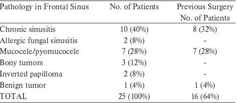
Table 3. The pathologies in the frontal sinuses.
Pathology in Frontal Sinus No. of Patients Previous Surgery No. of Patients
Chronic sinusitis 10 (40%) 8 (32%)
Allergic fungal sinusitis 2 (8%)
-Mucocele/pyomucocele 7 (28%) 7 (28%)
Bony tumors 3 (12%)
-Inverted papilloma 2 (8%)
-Benign tumor 1 (4%) 1 (4%)
occurred in 7 (28%) cases and 6 (24%) had soft or bony tumors. The bony tumors included 2 large osteomas and 1 fibrous dys-plasia. These 3 tumors necessitated endonasal cutting for evacu-ation through the nostril. Sixteen (64%) patients had undergone ESS in the past. The number of frontal sinuses that had under-gone previous surgery was even higher [22 (71%)], because 6 patients in the inflammation group had bilateral involvement.
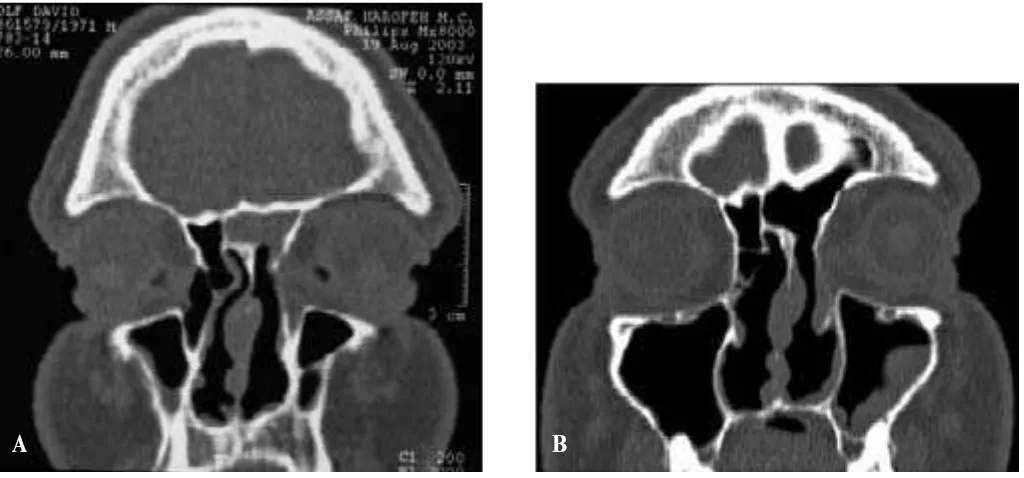
In all but 2 sinuses (93%), the frontal floor between the lamina papyracea laterally and the middle concha medially was drilled out, but not as far as the nasal septum (Figures 1A, 1B, 2A, 2B). Follow-up revealed that 24 cases (96%) of all frontal sinuses were ventilated. The patients who suffered from chronic sinusi-tis, including those with AFS, needed extended intensive topi-cal treatment, and systemic antibiotics and steroids to keep the
A
B
Figure 1A. Coronal CT scan of the left frontal sinusitis secondary to previous endoscopic sinus surgery.
Figure 1B. Coronal CT scan of the left frontal sinus after Draf II. The neo opening is between the lamina papyracea and the attachment of the middle concha.
Figure 2A. Coronal CT scan of the frontal sinuses. A mucocele secondary to previous ESS is seen in the left frontal sinus.
Figure 2B. Coronal CT scan of a patients who underwent a Draf II sinusotomy of the left frontal sinus. Note that the frontal sinus neo opening is between the lamina payracea and the nasal septum.
frontal sinus open and ventilated. Only 1 patient, a woman with a pyomucocele causing exophthalmus, double vision and headache, had recurrent disease for which revision surgery was considered. Although she was free of symptoms after surgery, the frontal sinus was completely opaque on CT scan. She refused revision surgery. Another woman with a mucocele sec-ondary to previous ESS conducted elsewhere complained of intermittent headaches 9 months after surgery. The frontal neo-ostium proved to be wide open endoscopically with normal mucous membrane of the frontal sinus walls. In the infection group, 1 patient had continuous nasal discharge and 3 others suffered from headaches. These patients did not suffer from frontal pain and their frontal sinuses were aerated. Two cases suffered from burning injury to the floor of the nostril as a
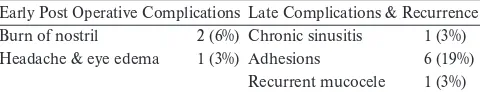
result of the friction heat created by the drill. Table 4 summa-rizes the early and late complications.
DISCUSSION
Frontal sinus pathology remains a challenge to the rhinologist [4]. Most surgeons prefer to manage frontal sinus pathology by the external approach. The improvement in endoscopic tools, such as angled microdebriders and giraffe through-cutting instruments, which are very helpful in surgical manoeuvres in the frontal sinus, make it easier for the surgeon to drill out the frontal sinus ostium. On the other hand, drilling in this narrow area and exposing bone may cause restenosis of the neo-ostium of the frontal sinus. Many techniques have been suggested to overcome the problem created by the anatomical complexity of the angled, narrow outflow of the frontal recess and ostium. There are several methods for locating the frontal os, such as trephination and irrigation, or using a wire probe as a guide [5-7]. McLaughlin et al. [8] described trans-septal frontal sinusoto-my, in which the septum is translocated to reach the frontal sinus ostium. In our experience, when none of the usual anatomical landmarks existed due to previous surgery, the frontal sinus os was located by exposing the smooth hard bone of the beak and drilling through it.
The beak is a semi-curved ridge on the lateral wall of the nasal space. Exposure of the beak and drilling out its medial-posterior aspect assists in locating the frontal ostium. We found this to be a very reliable and constant landmark in both primary and sec-ondary surgery.
In the present series, in 29 (93%) frontal sinuses the floor was drilled out between the lamina papyracea and the middle con-cha sufficiently to enable successful removal of the pathology. The frontal sinus floor had to be removed completely in only 2 patients. Nevertheless, we still consider this surgical method to
be more similar to Draf IIB sinusotomy than Draf IIA. However, with our technique, there is less drilling of the frontal ostium than the amount suggested by the Draf IIB procedure. Image-guided surgery may help in accurately positioning the surgical instruments, thus minimizing orbital and brain compli-cations, mainly in the absence of normal anatomical landmarks due to previous surgery, and in patients with anatomical vari-ants. However, it is obvious that navigation cannot replace com-plete anatomical understanding. Furthermore, the navigation system has certain disadvantages such as the high cost of a sys-tem that is only indicated in a small number of surgeries. Other disadvantages are radiation of the patient due to the need for 1-mm CT sections, and the need for accuracy of the system to be approximately 1-2 mm, which in this narrow area can be critical. We believe that all pathologies can be removed endonasally with the proper instrumentation and experienced surgeons. However, pathology located beyond the range of the instru-ments and the capability of the surgeon is a contraindication for endonasal frontal sinusotomy. We used Draf II successfully for the removal of large osteomas, even though this procedure is usually contraindicated in such cases [1]. The Draf II approach is also contraindicated for severe fractures involving the drainage pathway and in failed endonasal surgery for chronic frontal sinusitis [1]. In such cases, the external approach with obliteration is recommended [10]. However, Samaha et al. [9] did recommend revision surgery in cases of failed drillout surgery of the frontal sinus for chronic sinusitis. The Draf II procedure can be combined with other approaches such as the Lynch procedure, as in the case in the present series when pre-sented with a giant osteoma filling the frontal sinus and ostium. The anatomy and diameter of the ostium, the status of the mucous membrane of the frontal sinus and recess, and the nature of the pathology may have an impact on the outcome of frontal sinus endonasal surgery [11,12]. The healing process of the frontal sinus neo-os is dependent on the mucous membrane status. In cases of chronic sinusitis with or without polypoid degeneration, there is a higher rate of recurrent mucosal disease [13] and a lower rate of postoperative patency of the frontal os [14]. We believe that in cases when the mucuos membrane is healthy, such as in cases of tumors or mucoceles, there is a higher rate of patency [15]. In our experience, the favorable out-come in the infection group can be attributed to intensive post-operative cleansing and lavage, prolonged systemic antiobiotic and steroid treatment, and the relatively less traumatic surgical technique. We believe that the high success rate in our series can be attributed to a prudent selection of cases. The Draf II technique was used in only 3.7% of ESS surgery. There is no similar data from other centers with which to compare our results.
In the series conducted by Weber et al. [16] and Draf et al. [17], the success rates for the Draf II procedure were 70% and 61% respectively, for a longer follow-up period [51 months (mean) and 5 years, respectively]. A recent study by Weber et al. [18] showed similar results - 70.5% had a patent ostium in a follow-Table 4. Postoperative complications.
Early Post Operative Complications Late Complications & Recurrence Burn of nostril 2 (6%) Chronic sinusitis 1 (3%) Headache & eye edema 1 (3%) Adhesions 6 (19%)
up period of 12-98 months.
McLaughlin et al. [8] reported patency in all patients who underwent transseptal frontal sinusotomy (TSFS) during the follow-up period. They concluded that TSFS was especially suited for revision surgery in those patients with acquired frontal sinus stenosis.
On the other hand, Kikawada et al. [14] reported that the overall rate of patency of the frontal sinus neo-ostium was 42% in 12 frontal sinuses where the indications for Draf II were scarring and/or polyps. Metson et al. [15] reported 3 failures in 15 patients who underwent the unilateral procedure (Draf II proce-dure), and none in the bilateral drillout technique. Four in this series had mucocele and all were treated successfully by the Draf II procedure.
The success rate of the Draf II procedure in our series was 96% during a follow-up period of 30 months (average), which is high when compared to other series and is equal to the series of McLaughlin et al. [8], Metson et al. [15] and the series that used stents after surgery [19]. In our series, one patient who had a mucocele secondary to endoscopic sinus surgery continued to complain of headaches 9 months after surgery. Fiberoptic endoscopy revealed a wide-open frontal sinus ostium and nor-mal mucous membrane. The surgery in this patient was consid-ered by us to be successful. In the other patient operated on due to a pyomucocele causing exophthalmus, the frontal sinus was completely opaque 2 years following surgery, but without symptoms or exophthalmus. This was considered by us to be a failure. She refused revision endoscopic surgery. The follow-up period in the series presented by Weber et al. [16,18] and Draf et al. [17] was longer than in our series.
In a study by Chiu and Vaughan, ESS with surgical navigation was found effective and safe in revision frontal sinus cases [20]. Fifty-eight out of 67 patients (86.6%) who underwent revision ESS of the frontal sinus with navigation had a patent frontal recess and significant subjective improvement in symptoms. The average follow-up period was 32 months (range 24-48 months). Samaha et al. [9] found a higher, but non significant, success rate for the group that underwent image-guided frontal sinus drillout (83.1%) versus the non image-guided group (74.3%). Their series included a total of 100 patients with advanced chronic sinusitis. Our results demonstrate that the non-image guided approach can produce similar results. The complications in our series (Table 3) included 2 cases of first-degree burns to the nostril secondary to the friction heat created by the straight drill. As a consequence of this, we used the curved drills in the subsequent cases. No major complica-tions such as a CSF leak were detected in our series. Our results are similar to those of series that used a navigation system in surgery [9,10].
The advantages of the Draf II approach have been well estab-lished by many authors.
Although all cases presented here were operated on according to the triplanar CT scan (axial, coronal and sagittal views), surgi-cal navigation, which we did not use, might aid in achieving
bet-ter and safer localization of the frontal ostium, mainly in revi-sion surgery, and thus shorten the operative time.
In conclusion, non-navigated endoscopic Draf II sinusotomy is a safe procedure, including cases of previous surgery. It is essen-tial to carefully select the patients for Draf II sinusotomy, and fully understand the CT scan and anatomy. The beak is a reli-able, constant landmark in cases that have undergone previous surgery. The controversy surrounding Draf II sinusotomy, resulting from published data, is the high rate of restenosis. Follow-up is conducted mainly by means of a fiberoptic endo-scope, thus avoiding the use of radiation for repeated CT scans.
REFERENCES
1. Draf W (1992) Endonasal micro-endoscopic frontal sinus surgery: the Fulda concept. Op Tech Otolaryngol Head Neck Surg 2: 234-240.
2. May M, Schaitkin B (1995) Frontal sinus surgery: endonasal drainage instead of an external osteoplastic approach. Op Tech Otolaryngol Head Neck Surg 6: 184-192.
3. May M, Hoffman DF, Sobol SM (1990) Video endoscopic sinus surgery: a two-handed technique. Laryngoscope 100: 430-432. 4. Molony NC, Ah-See K, Rachmanidou A, Draf W (2000) A survey
of contemporary management of frontal sinus disease in the United Kingdom. Eur Arch Otorhinolaryngol 257: 247-250.
5. Friedman M, Landsberg R, Schults RA, Tanyeri H, Caldarelli DD (2000) Frontal sinus surgery: endoscopic technique and preliminary results. Am J Rhinol 14: 393-403.
6. Gallagher RM, Gross CW (1999) The role of mini-trephination in the management of frontal sinusitis. Am J Rhinol 13: 289-293. 7. Becker DG, Moore D, Lindsey WH, Gross WE, Gross CW (1995)
Modified transnasal endoscopic Lothrop procedure: further consid-erations. Laryngoscope 105: 1161-1166.
8. McLaughlin RB, Hwang PH, Lanza DC (1999) Endoscopic trans-septal frontal sinusotomy: the rationale and results of an alternative technique. Am J Rhinol 13: 279-287.
9. Samaha M, Cosenza MJ, Metson R (2003) Endoscopic frontal sinus drillout in 100 patients. Arch Otolaryngol Head Neck Surg 129: 854-858.
10. Weber R, Draf W, Kahle G, Kind M (1999) Obliteration of the frontal sinus - state of the art and reflections on new materials. Rhinology 37: 1-15.
11. Loury MC (1993) Endoscopic frontal recess and frontal sinus ostium dissection. Laryngoscope 103: 455-458.
12. Orlandi RR, Kennedy DW (2001) Revision endoscopic frontal sinus surgery. Otolaryngol Clin North Am 34: 77-90.
13. Kennedy DW (1992) Prognostic factors, outcomes and staging in ethmoid sinus surgery. Laryngoscope 102 (suppl 57): 1-18. 14. Kikawada T, Fujigaki M, Kikura M, Matsumoto M, Kikawada K
(1999) Extended endoscopic frontal surgery to interrupted nasofrontal communication caused by scarring of anterior ethmoid: long-term results. Arch Otolaryngol Head Neck Surg 125: 92-96. 15. Metson R, Gliklich RE (1998) Clinical outcome of endoscopic
surgery for frontal sinusitis. Arch Otolaryngol Head Neck Surg 124: 1090-1096.
16. Weber R, Draf W, Keerl R, Behm K, Schick B (1996) Long-term results of endonasal sinus surgery. HNO 44: 503-509.
17. Draf W, Weber R, Keerl R, Constantinidis J (1995) Current aspects of frontal sinus surgery. I: Endonasal frontal sinus drainage in inflammatory diseases of the paranasal sinuses. HNO 43: 352-357. 18. Weber R, Draf W, Kratzsch B, Hosemann W, Schaefer SD (2001)
Modern concepts of frontal sinus surgery. Laryngoscope 111: 137-146.
20. Chiu AG, Vaughan WC (2004) Revision endoscopic frontal sinus surgery with surgical navigation. Otolaryngol Head Neck Surg 130: 312-318.
E. Eviatar, MD
Department of Otolaryngology Assaf Harofeh Medical Center Zerifin 70300
Israel
Tel: 972-8-9779417 Fax: 972-8-9779421;
E-mail: [email protected]
SOCIETY NEWS
SOCIETY NEWS
FELLOWSHIPS IN RHINOLOGY
Sponsored by
THE EUROPEAN RHINOLOGIC SOCIETY
Available to Trainee-Surgeons from Eastern Europe
One place available on each of the following Courses in 2006-2007
International Course on in Functional-Aesthetic Nasal Surgery International Course in Functional Corrective Nasal Surgery Ulm, Germany, March 2006 Utrecht, The Netherlands, June 27-30 2006
Course Director: Prof.Dr. G. Rettinger Course Director: Dr. A.F. van Olphen
Language: English Language: English
International Course on FESS (Basic and Advanced Techniques) An Endoscopic approach to Rhinosinusitis Graz, Austria, September 07-09 2006 London, United Kingdom, October, 11-14, 2006 Course Director: Prof.Dr. H. Stammberger Course Directors: Prof. V.J. Lund and I.S. Mackay
Language: English Language: English
7th
International Course on Reconstructive and Aesthetic Surgery Fourth biennial International Milano Masterclass. Sisonal and of the Nose and Face “Around the Nose” Skull Base Endoscopic Microsurgery, Advanced Rhinoplasty and Nijmegen, The Netherlands, May 31, June 1, 2 2006 Pearls of Facial Plastic Surgery, Milano, March 23-27, 2007
Language: English Language: English
Sponsorship includes both registration fees and costs of accommodation. Application forms are available from the Secretary of the ERS.
Applications should be submitted at least three months before the course starts. Applications and/or queries should be directed to either the President or the Secretary of the European Rhinologic Society.
Prof. M. Önerci (President ERS), Department of Prof. G. Rettinger (Secretary General ERS), Department of Otorhinolaryngology, Hacettepe University, Sihhiye - Otorhinolaryngology, University of Ulm,
Ankara, Turkey Prittwitzstrasse 43, D-89070 Ulm, Germany