The journal homepage www.jpacr.ub.ac.id ISSN : 2302 - 4690
123
The Effect of KIO
3and KI Salt Towards Iodium Levels (I
2) in
Urine, Malondialdehyde (MDA) and The Histology of Thyroid
Gland of Goitrogenic Rat
Risman Heli,1*Chanif Mahdi1 and Sasangka Prasetyawan1
1Department of Chemistry, Faculty of Sciences, Brawijaya University
Jl. Veteran, Malang 65145, East Java Indonesia
*Corresponding author: [email protected]
Received 23 June 2014; Revised 20 August 2014; Accepted 10 September 2014
ABSTRACT
Goitrogenic substances can inhibit of iodine taking by the thyroid gland. Thus iodine concentration in thyroid gland will be low, and this phenomena is indicated by inflammation in the thyroid gland. Moreover, it can cause releaseing of an excessive amount of free radicals. This radicals, in the body, causes oxidative stress and also increase the levels of malondialdehyde (MDA). This is also as an indicator for lipid peroxidation and the decreasing of urinary iodine excretion levels (EIU). The treatment with KIO3 and
KI salt was intended to study the level of supplementation of iodine (I2) toward level of
MDA in serum and histological description of rat’s thyroid gland. The MDA levels was determined through TBA test (Thio Barbituric Acid), meanwhile the histological pattern of rat thyroid gland was determined by Hematoxylen -Eosin staining (HE). The results indicated both of KIO3 and KI salt significantly (p<0.01) reduced MDA level in the serum.
Treatment with KIO3 salt gave 33.62% while KI salt slightly higher (37,02%). In addition,
both of treaments displayed an recovering effect in thyroid gland.
Key word: Goitrogenic, Iodium, Malondialdehyde, KIO3 and KI salt therapy.
INTRODUCTION
Recent National Survey in Indonesia (1998) reported 53.8 million people live with iodine deficiency risk, 20 million suffer from goitre, 290 thousand estimated to suffer from creatine, and approximately 9 thousand infants born each year [1]. Thyroid disease in Americans country continuesly growth, and approximately 17,000 cases occured each year, and about 1,700 of them resulting death. A study in England from Whickham Study of the United Kingdom reported that goitre is suffered in 16% from the total population by people. In the Framingham study reported that the ultrasound examination of thyroid nodules found in older men over 60 years at 3%, while for 48-year-old woman recorded at 36% [2]. In America, majority cases of thyroid goitre caused by autoimmune and known as Hasimoto's disease. But, mostly the cause of goiter is due to iodine deficiency. It is estimated that about 200 million goitre among the 800 million people suffering from iodine deficiency. Whickham study reported the prevalence of goiter 26% of women, compared to only 7% of men or women genders dominated by a ratio of 4: 1 [2].
The journal homepage www.jpacr.ub.ac.id ISSN : 2302 - 4690
124 imbalance of radicals level in the body and lead to damaging of the cell membrane. This process can be identified by increasing of malondialdehid (MDA) level and other indicator for lipid peroxidation. Injecting or consuming of the antioxidant substances from outside the body can suppress elevation of MDA level in the body. Generally, treatment of iodine can affect by improving the level of iodine deficiency in the body. But, prolonged the iodine deficiency will interfere the process in formation of thyroid hormones. When the body has iodine deficiency, the concentration of thyroxine hormone in the blood is low. This condition stimulates the body to enhance the thyroid tissue. As a result is enlargement of thyroid gland, or is called a goiter [3]. Theoretically, therapy using substance contained iodine such as KIO3
and KI salt can increase iodine level, and this can be monitorred through urine iodine status, the iodine content in urine, lowering levels of malondialdehyde (MDA).
EXPERIMENT Animal treatment
All conditions of experiment and handling of the animals were conducted following the protocols approved by Ethical Clearences Committe in Brawijaya University (238-KEP-UB). A number of 20 rats (Rattus norvegicus) (female, body weight 125-200 g) were housed at room temperature in the animal house. This was gently care in the Laboratory of Cellular and Molecular Biology, Mathematics and Sciences Faculty, Brawijaya University Malang and were exposed to alternate cycles for 12 h light and dark. The rats were divided into four group, a healthy group, goitrogenic group, goitrogenic group treated with KIO3 salt and
goitrogenic group treated with KI salt. The goitrogenic rats groups were sonde injected with 1.75 mL KSCN in mouth and incubated for 3, 6, 9, 12, and 15 days. Then after 7 days, the goitrogenic rats group for divided into group treated with the KIO3 and the group treated with
KI salt. A dose level given oral was 80 mg/Kg of body weight (BW) for 14 days. After day 15, group of rats were dissected. Rats were killed by neck dislocation, and rat serum, urine and gland thyroid were taken. The serums were taken in abdomen section, heart. This was put into pakutener non-EDTA and left for three hours and centrifuged at 600 rpm for 15 min. This serum was further analyzed. Moreover, gland thyroid were also taken and the skin were slashed, and washed with 0.1% NaCl and immersed in 4% PFA for seven days.
Analysis of iodium level using Cerric Ammonium Sulfat (CAS) test
Urine samples 250 µg was pipetted and shaked up in a sealed test tube. Each of tube contain sample were added 0.75 mL of iodine. Ammonium persulfate 1.0 mL was also added. After that, all the sample tubes were heated in 91-98 °C for 60 min. The tubes were cooled down to room temperature, and 3.5 mL of arsenite (As2O3) was added followed by
homogenization for 15 min. Then, 4.0 mL cerric ammonium sulfate was dropped in each tube and mixed for 15 to 30 second. Then, each tubes was analysed using UV-Vis spectrophotometer at 420 nm with interval time 30 second.
Analysis MDA levels using TBA test
The journal homepage www.jpacr.ub.ac.id ISSN : 2302 - 4690
125 Histological analysis using Hematoxylen-Eosin (HE) staining method
A preparate of the rats thyroid gland was immersed in 1-3 xylol for 5 min, and was put into the various concentration of ethanol, from absolute ethanol 1-3, ethanol 95%, 80%, and 70% respectively for 5 min. This was soaked in aquadest for 5 min and put into hemotoxylen dyes for ± 10 min. Then, it was washed over flowing water for 30 min, and rinsed with aquadest before continued to coloring with eosin dye. Then, it was inserted to eosin alcohol for 5 min and soaked in the aquadest to release the excess of eosin. Moreover, in the dehydration process, the equipment was inserted in the graded ethanol 80%, 90%, and 95% to the 1-3 absolute of ethanol. Then, in the clearing process, it was completed by placing in the xylol 1, 2 and was further dried. The result preparate was mounted using entellan, and the dried and stained ultrathin sections were observed using a microscope (Olympus BX53) with a magnification of 100 times.
RESULTS AND DISCUSSION
Effect thiocyanate (KSCN) and level of iodium in urine
Direct observation from physical appearance of the treated rats indicated changing in the physiological bodies of rat suffering from goitre due to exposure to thiocyanate (KSCN). KSCN experimentally inhibits transportation of iodide ions in the body. Then, it will lower iodine content. Physical phenomena can be see also from mouse.
.
Figure 1. Normal rat (Healthty, A), rats exposed to KSCN (Goitrogenic, B), salt therapy rats with KIO3 (Therapy, C), and salt therapy KI rats (Therapy, D).
The journal homepage www.jpacr.ub.ac.id ISSN : 2302 - 4690
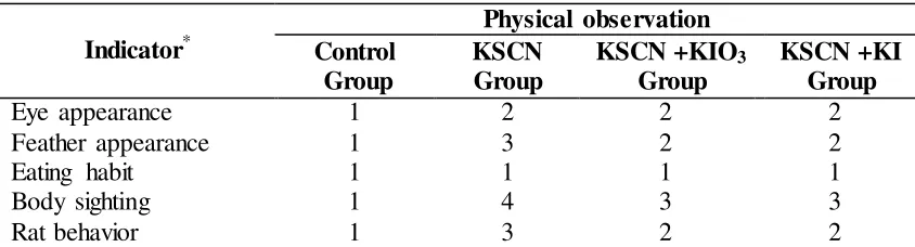
126 Table 1. Observation result on physiological alteration during observation on groups of
treated rats
*Note: Eye color: 1. Bright-red; 2. Not Bright-red; Feather: 1. Intact and clean; 2. Intact and dirty; 3. Fall out and dirty; Eating Habits: 1. a lot; 2. Medium; 3. Slightly; Behavior: 1. Normal; 2. Aggressive; 3. Idle siginificant differency above 99% (Figure 2).
Table 2. Urine Iodine Levels iodine such as monoiodothyrosine and diiodothyrosine. Both are part of the triglobulin located in each cell. These cause severe competition between serum thiocyanate and thyroxine to bind it, as a result of an increasing in serum thyroxine [4]. Salt administration using KIO3 and KI in mice on groups of goitrogenic rats reduces the level of iodine transport
The journal homepage www.jpacr.ub.ac.id ISSN : 2302 - 4690
127 hormogenesis, process in thyroid hormone. This can increase iodine, even thought cause inhibition of peroxide generation due to a increasing of I-intra-thyroidal content by supplementation of KIO3 and KI salt. Treatment with KIO3 and KI can also affect pituitary to
work harder secreting TSH and stimulate the thyroid gland to producing more thyroid hormone. As as result inititation of thyroid hyperplasia and hypertrophy (goiter).
Figure 2. Comparison of the iodine level in urine of healthy rats (Rattus novergicus), goitrogenic rats, and therapies rat with KIO3 and KI salt
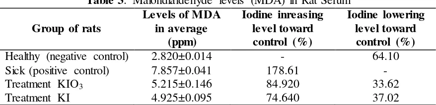
Levels of malondialdehyde (MDA) in rats serum
Measurement of malondialdehyde concentration conatined in serum of the treated groups of rat was tabulated in Table 3 and displaye as picture on Figure 3. It was recorded 0.095 ppm. Even though this concentration indicated reduction, however this concentration still much higher than that for group of healthy rats (2.820±0.014 ppm).
process of inhibition for transporting goitrogen thiocyanate (KSCN). Improvement number iodine in thyroid gland may also accelerate the activity of iodine in stimulating the secretion of thyroid hormone, triiodothyroxine (T3) and thyroxine (T4). This will increase TSH level in
The journal homepage www.jpacr.ub.ac.id ISSN : 2302 - 4690
128 Moreover, reducing MDA level means inhibit the free radical substances. This inhibition mechanism correlate to process in lipid peroxidation by both salts of KIO3 and KI
salt as antioxidant agent which counter act the free radicals in body. Iodine content in KIO3
and KI salt possibly was the resposible substance as antioxidant [5]. Malondialdehyde (MDA) which is the product of peroxidation as an indicator that a lipid disorder was occuring. Therefore, the high concentration of malondialdehyde in groups of goitrogenic rat serum was also an indication of the high levels of the adipose membrane tissue disorder.
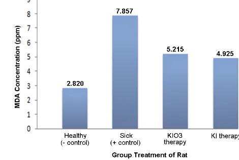
Figure 3. Comparison of the value of Serum levels of MDA in healthy rats (Rattus novergicuss),
goitrogenic rats, and therapies rat with KIO3 salt and therapies rat
with KI salt
Damaging of lipid membrane by free radical substances initiated by three stages; that are initiation, propagation, and termination. Initiation process is the process when a hydrogen atom is removed from the lipid molecules. Some compounds can react with hydrogen atoms forming hydroxyl radical (•OH), alkoxy (RO), peroxyl (ROO) and may also HO2 but not
including H2O2. Membrane lipids generally are phospholipid consist of unsaturated fatty
acids in which peroxidation is easily occur due to the issuance of methylene group (-CH2-) from the hydrogen atom contains only one electron. So, there are carbon atoms with no pair of electrone. The existence of a double bond in the fatty acid weakened the CH bonds on the carbon atom adjacent to the double bonds. It eased the transfer of a hydrogen atom [6]. When there is sufficient oxygen concentration lipid radicals, then it react with the oxygen to form a peroxyl radical (ROO•). This formation occurs in the propagation stage. At the termination, peroxyl radical (ROO•) attacks the other hydrogen atoms originating from other lipid molecules that are close by and produce lipid peroxides and peroxyl radicals or interact with other antioxidants [7]. This process causes the adipose membrane compliers cells dead, and thus damaging the adipose membrane.
Effect at KIO3 and KI salt on the histological features
Treatment of KIO3 and KI salt using dose of 80 mg/Kg BW theoretically can reduce
The journal homepage www.jpacr.ub.ac.id ISSN : 2302 - 4690
129 did not surround the thyroid follicles. The feature was not clear between thyrosit and parafolicular cells or cells itself where the position of thyrosit and parafolicular follicles along both outside and inside of the follicle. This damage is caused by the formation panus section, thus increasing the production of free radicals. These free radicals trigger the formation of antibodies by modifying the protein aggregates that activate phagocytic cells to cause inflammation or injure. The formation of antibodies itself againsts the auto-antigens or antigens from infectious genes (including goiter factors), and that lead to formation of immune complexes and lead to complex and activation of phagocytic [8].
Figure 4. HE Staining results on Goitrogenic Thyroid Gland Rat (Healthy rat, A), rats exposed to goiter KSCN (Sick rat, B). Salt therapy rats with KIO3 (Therapy, C), and salt
therapy with KI (Therapy, D). (Picture was recorded in 100x magnification, and arrows indicate the occurrence of structural changes in tyroid folicel, lumen, thyrosit and parafolicular thyroid cell on treatment).
Histology appearance from the thyroid gland of the goitrogenic rat got treatment with KIO3 and KI salt was displayed in Figure 4-C and D. Both picture gave features of thyrosit
The journal homepage www.jpacr.ub.ac.id ISSN : 2302 - 4690
130 appears on the body's immune response (TNF-α, IFN-γ,) causing the macrophage-mediated cell destruction occurs as well [9] [10]. This tissue damage was observed during of apoptosis on thyroid cells. It was indicated by irregularity pattern of cells, and there was thyrosit and parathyroid cells in the same featureon the thyroid follicles.
CONCLUSION
Goitrogenic rats treated with KIO3 and KI salt increase the levels of iodine (I-).
Treatment with KIO3 increase to 44.44% while therapy with KI reach to 56.79%. Moreover,
therapy with KIO3 and KI in rats exposed goitrogenic (KSCN) also decrease the
malondialdehyde (MDA) level to 33.62% and 37.02%, respectively. The histological features indicated imporvement on goitrogenic rat gland thyroid after theraphy with both substances.
ACKNOWLEDGMENT
The author would like to thank to Prof Dr Chanif Mahdi, MS who has been given the opportunity to be part of the research with KIO3 and KI therapy for defiency iodium in goitrogenic rats. The writer was also grateful to Dr. Akhmad Sabarudin and Dr Masruri who has assisted the completion of this manuscript.
REFERENCES
[1] Masjhur, J. S., Indonesian Journal of IDD, 2002, 1 : 29 - 33
[2] Hetzel, B.S.. An Overview of the Prevention and Control of Iodine Deficiency Disorder; in Hetzel, J.T Dunn and J.B. Stanbury (ed)., 2005, Elvesier Science Publisher, New York.
[3] Mulinda JR, 2005. Goiter. http://www.emedicine.com/MED/topic916.htm
[4] World Health Organisation (WHO), Assesment of Iodine Deficiency Disorders and
Monitoring Their Elimination. Second edition, 2001, WHO, New York.
[5] Lang, K. Die Rhodanbildung im Tierkorper, Biochem Z., 1933, 259: 243-256. [6] O’Dell JR. New Engl. J. Med.,1999, 340, 310-312
[7] Deaney CL, Feyi K, Forrest CM, Freeman A, Harman G, McDonald MS, Petrie A, Shaw SJ, Stone TW, Stoy N, Darlington LG. Res. Comm. Mol. Pathol. Pharmacol. 2000, 110, 1-2, 87-95.
[8] Bruch, C.G.and D.P Janet, AACN, 2002, 11, 543-551.