Emotion and consciousness
Naotsugu Tsuchiya and Ralph Adolphs
California Institute of Technology, HSS 228-77, Caltech, Pasadena, CA 91125, USA
Consciousness and emotion feature prominently in our personal lives, yet remain enigmatic. Recent advances prompt further distinctions that should provide more experimental traction: we argue that emotion consists of an emotion state (functional aspects, including emo-tional response) as well as feelings (the conscious experience of the emotion), and that consciousness consists of level (e.g. coma, vegetative state and wake-fulness) and content (what it is we are conscious of). Not only is consciousness important to aspects of emotion but structures that are important for emotion, such as brainstem nuclei and midline cortices, overlap with structures that regulate the level of consciousness. The intersection of consciousness and emotion is ripe for experimental investigation, and we outline possible examples for future studies.
Introduction
Sophisticated theoretical accounts of emotion[1–7]and of consciousness[2,3,8–12], together with recent data from functional imaging [13–24] and clinical populations[24– 31], provide an unprecedented opportunity for progress on these topics. We argue that both emotion and conscious-ness depend on neural representations of the subject’s own body, arising from structures in the brainstem and medial telencephalon that receive interoceptive information. This article focuses on domains where emotion and conscious-ness overlap and interact, and we suggest that each is necessary for aspects of the other. Future work requires not only more data but also further theoretical develop-ment of the concepts that are under investigation, which makes the intersection of emotion and consciousness a fruitful domain for collaborations among neuroscientists, psychologists and philosophers.
Clarification of terms
We all have an intuitive understanding of what ‘emotion’ and ‘consciousness’ mean, but making this usage suffi-ciently precise for scientific investigation requires further distinction (Figure 1). A clue to the problems lurking beneath the surface comes from observing that not every language has words for the particular aspects of conscious-ness or the particular emotions for which there are words in English[32].
We intend to sketch a usage that will describe normal, healthy human function, much of pathology, and one that will apply to many other mammals; however, we acknowl-edge that certain pathological states and conceivable thought experiments would require modifications. One
can begin by dividing emotion into two components: the conscious experience of the emotion (‘feeling’ [3]) and the behavioral, physiological and cognitive processes that specify the ‘emotion state’ (the functional aspects of an emotion[2]). The emotion state encompasses both evalua-tive components and attitudinal components (such as those stressed by action-tendency theories). The physiological and behavioral components of an emotion state provide criteria by which we typically attribute emotions to non-verbal animals. For instance, blushing, running away, screaming, laughing and crying are examples of this phys-iological aspect, an emotional response (the focus of this article), which also involves the brain: the neural responses that directly cause the bodily ones, as well as neural responses that serve to prepare the organism for beha-vioral engagement with the environment (red areas in
Figure 2). Thus, seeing a bear not only makes us scream
and run away but also triggers autonomic and motor-related neural activity that is the causal prerequisite for such bodily responses and increases attention, alertness and encoding of the stimulus into memory. Emotional responses and feelings can be dissociated: certain brain-stem lesions can produce emotional responses in the absence of normal feelings of those emotions[25].
‘Consciousness’ can also refer to two components that are commonly distinguished in the literature: the level (state) of consciousness (e.g. wakefulness, coma and dreamless sleep; green areas in Figure 2) and its content – whatever it is that we are conscious of (e.g. the scent of a perfume or the color of a rose). Whether we could be in a state of consciousness that has no content is an open question (conceivably, certain kinds of epileptic states or meditative states might qualify). The contents of our con-scious experience can be analyzed further into their phenomenal aspect and the access we have for knowing and reporting the content on the basis of its phenomenal aspect [9] (Figure 1). Normally, we have access to the properties of objects that we perceive in virtue of our phenomenal experiences of them.
Here, we follow the common view that emotion and consciousness emerge as a result of neuronal activity in the brain, but some accounts view emotions or conscious-ness as relationships between an organism and its environ-ment [12] (here, we acknowledge such relationships as contributing but not as constitutive; but see Ref. [33]). We also follow the common (but by no means universal) view that all mammals (and, plausibly, some other animals) have emotions and consciousness, although they do not have the same set of emotions nor the same elabo-rated contents of consciousness (or access to them) as do humans.
Opinion TRENDS in Cognitive Sciences Vol.xxx No.x
Corresponding author:Adolphs, R. ([email protected]). Available online xxxxxx.
TICS-565; No of Pages 10
Neural structures that regulate the level of consciousness
There are no completely reliable and consistent criteria for levels of consciousness, although there are standardly used clinical criteria for unconsciousness, such as unresponsive-ness, lack of voluntary movement and lack of any memory of conscious experience after recovery [34]. If the corre-lation between neurobiological descriptions and conscious-ness is sufficiently strong, the neurobiological data could be used as a diagnostic criterion. Indeed, a recent study used fMRI data to conclude that a patient in persistent vege-tative state (defined using conventional behavioral criteria
[34]) was nonetheless conscious[31].
Insight into the neural structures that are important for the level (state) of consciousness has come from classic and modern lesion studies as well as functional imaging studies, which show that consciousness is supported by a complex network that includes nuclei in the brainstem[12], parts of the thalamus[3], anterior cingulate, posteromedial cortex [14,35]and broad sectors of frontoparietal cortices
[8,36]that are all extensively interconnected (green areas in
Figure 2;Figure 3). A network of cortices on and adjacent to the medial wall of the hemispheres is activated more during wakeful rest than during perceptual and attentional engagement with the environment and likely to be crucial to the maintenance of consciousness[14](Figure 3a,b). The anterior and posterior cingulate cortices, medial and lateral parietal cortex and medial frontal cortex are activated when processing information related to self, emotion and internal monitoring[8,22]. This network shows strong connections among its components [37], as well as with association cortex and thalamic nuclei[35,38]. Several of these struc-tures point to a close association between consciousness and self-representation, an issue we take up further below.
Acute bilateral lesions of the anterior cingulate cortex result in akinetic mutism – an absence of volitional beha-vior and lack of engagement with one’s environment. (Typically, patients recover after some time and, although they often fail to show convincing evidence of being con-scious of their environment, they do exhibit wakefulness Figure 1. Defining components of emotion and consciousness.(a)We distinguish an emotion state from the emotion experience (feeling), although they are not mutually exclusive[2]. The feeling can have as content both the experience of components of the emotion state (such as bodily responses, a component that William James emphasized
[49]) and perception of a stimulus or memory that induced the emotion or towards which it is directed. The emotion state is also complex, but might rely on an integrated set of neural structures that coordinate it. Although we use ‘emotion state’ to refer to a broad time window within which an emotion occurs, all components are in fact dynamic processes that unfold in time. The action-tendency component of an emotion state might be similar to a motivational state and ‘wanting’, whereas the feeling component might be similar to ‘affect’ and ‘liking’, terms that are used in research on reward processing[60].(b)The most common distinction made in the literature is between the ‘state’ and the ‘content’ of consciousness. ‘State’ refers to the level of consciousness, such as wakefulness or coma, whereas ‘content’ refers to what it is we are conscious of.
[3].) The anterior cingulate cortex is activated by stimuli that trigger wakefulness and high arousal, such as pain
[38], and during trace conditioning[39], thought to be one diagnostic assay for consciousness[40](Box 1). It is also activated by a variety of interoceptive stimuli, such as air hunger (the sensation of insufficient oxygen[15]), and by the degree to which we are aware of our own emotions[16]. Both anterior [38] and posterior [41] cingulate cortices have been implicated in emotion. Thus, cingulate cortex stands out as a region that is important for expression, experience and motivational aspects of emotion, as well as more generally for conscious states (Figure 2).
Structures that are important for emotion are also important for the level of consciousness
In addition to being a possible content of consciousness, it has been argued that basic aspects of emotion are
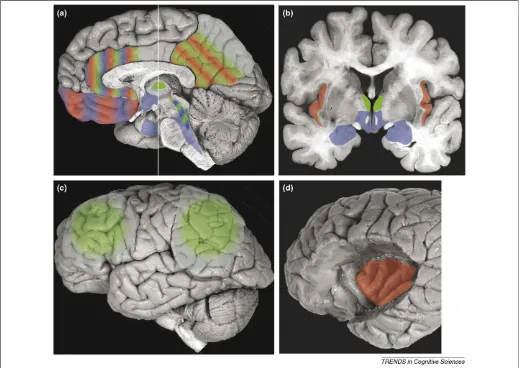
necessary for the level of consciousness in general. This basic emotional processing is thought to involve homeo-static regulation of the state of one’s own body and a representation of ‘self’ [3,42]. We should emphasize that this sense of ‘self’ is not the same as self-awareness in the reflective metacognitive sense, but the aspect of the state change in one’s own body related to the conscious experi-ence; for example, that it is one’s eyes through which a red rose is seen (modality-tagging) and that it is oneself who is seeing it (ownership). It is also distinct from the ability to have an explicit concept of one’s own body, as is assessed, for example, through mirror self-identification. Interocep-tive information is thought to constitute what, at the psychological level, has been termed a ‘core self’ [3,42], a grounding for our autobiography and the basis for our feeling of continuity (a more restricted and biologically based usage of the term ‘self’ than in many psychological Figure 2. Brain regions that are important for emotion state (blue), feeling of emotion (red) and level of consciousness (green). Other components of an emotion state, and the content of consciousness, are presumed to rely on more variable and distributed structures that would depend on the particular kind of emotion or conscious experience and, therefore, are not depicted here.(a)Sagittal view shows that the hypothalamus, amygdala, brainstem nuclei, including periaquedactal gray and parabrachial nuclei, orbitofrontal cortex and anterior cingulate cortex are important for the expression of emotion (blue). Anterior and posterior cingulate, including precuneus, and orbitofrontal cortex are important for the experience of emotion (red). Intralaminar thalamus and the ascending reticular formation are necessary for the maintenance of arousal and wakefulness – that is, the level (state) of consciousness[8,12]. The intralaminar thalamic nucleus is a diffuse structure, enlarged here for illustration purposes.(b)Coronal slice at the level of the white line in (a). The thalamus, hypothalamus and amygdala are shown again. Insular cortex (red) is an important structure for the experience of emotion [also shown in (d)].(c)Bilateral prefrontal and parietal cortices are broadly important for the level of consciousness (green)[36]. The figure omits other important central nervous system components of emotion, such as the rostral ventrolateral medulla (important for control of autonomic function) and components of the spinal cord itself, all of which contribute to substantial processing that is related to interoceptive and homeostatic information[46], and also parts of the nucleus accumbens and ventral pallidum that participate in reward and positive affect[4,60](See alsoFigure 3c).(d)The surface of the prefrontal cortex has been removed to reveal the insular cortex (red). Human brain images are adapted, with permission, from the Digital Anatomist Project at the University of Washington (http:// www9.biostr.washington.edu/da.html).
Opinion TRENDS in Cognitive Sciences Vol.xxx No.x 3
TICS-565; No of Pages 10
theories [43]). Because it represents homeostasis, it provides a sense of invariance that accompanies the ever-changing content of conscious experience. The struc-tures that are thought to be responsible for such self-representation include all those that represent interocep-tive information about the body, from brainstem nuclei through the thalamus to the cortex. The importance of these structures is compatible with the finding that primary sensory cortices can be robustly activated even in unconscious states[8,36].
The emotion state
Emotions are relatively ‘decoupled’, compared with reflexes, at both the sensory and the motor end. At the sensory end, they often require appraisal and evaluation, particularly in humans (although there are automatic components that are more reflex-like too); at the motor end, they generate motivation and disposition to behave (although, again, there are automatic and more reflexive components of emotional response). This is also a nexus where emotion and consciousness coincide functionally:
conscious emotion experiences might be required for the elaborate appraisals of situations that enable us to act intentionally and instrumentally. An illustration that emotions are not ordinary reflexes comes from the obser-vation that very different patterns of behavior can be produced by the same central emotional state: we can freeze or flee when afraid. These different behaviors, each adaptive in a different situational context, have also been shown to be dependent on different nuclei of the amygdala in animal studies.
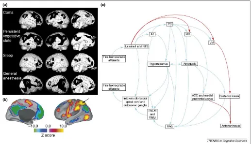
Following the classic experiments by Schacter and Singer[43], one view of emotion is parallel to the one we have sketched for consciousness. In this view, the state of emotion is often equated with nonspecific arousal, but the content of the emotion is thought to be determined by a host of other factors – perceptions of one’s own body state or action, evaluations and appraisals of the situation that evoked the emotion, inferences regarding the cause of or justification for the emotion, or awareness of action tendencies and motivation to behave in a particular way. This view is likely to make too strong a distinction Figure 3. Shared neural substrates for emotion and consciousness.(a)Data from functional imaging show that a frontoparietal network is compromised in coma, vegetative states, sleep and under anesthesia (black regions; but also see green regions inFigure 2). Abbreviations: F, prefrontal; MF, mesiofrontal; P, posterior parietal; Pr, posterior cingulate/precuneus. (a) reproduced, with permission, from Ref.[8].(b)In a quiet but awake resting state, there are two distinct networks of structures in the human brain that are either correlated (color-coded red to orange) or anti-correlated (blue to green) with the parietal cortex, indicated by the arrow. The blue–green network of structures might be particularly important for reflective and interoceptive processing and crucial to the level of consciousness and feeling emotions. (b) reproduced, with permission, from Ref.[14].(c)Anatomical structures that are important for feelings (emotion experience) involve homeostatic loops. A main circuit for efferent autonomic control arises from distributed cortical sectors in anterior and subgenual cingulate cortex, medial orbital cortex and insula, as well as from amygdala. These structures in turn project to paraventricular hypothalamus and periaqueductal gray (PAG) matter, from there to integrative centers in the medulla (RVLM, rostral ventrolateral medulla; VMM, ventromedial medulla) and the spinal cord, and then out to effector organs. Afferent autonomic processing occurs in part through dedicated sensory channels and involves brainstem nuclei, including the parabrachial nucleus (PB), the hypothalamus, thalamus (VM, ventromedial nucleus), and then again the same set of cortical regions. With the exception of the thalamus, there are homeostatic control loops at multiple levels, involving all structures in both afference and efference. Many of these structures are activated in a variety of experiments that involve emotion experiences[16,17,46], making them promising candidates for the neural correlates of emotion experience[3,46]. It has been proposed that primates possess a unique mapping of autonomic interoceptive information within the insular cortex that forms the substrate of conscious feelings[46]. The afferent limb is shown in the top row and the efferent limb in the bottom row; please note that only a subset of the connectivity is depicted in this figure for clarity. The red lines indicate pathways in primates thought to be more phylogenetically recent that provide a direct thalamocortical input, reflecting the physiological condition of the body. Abbreviations: A1, catecholaminergic cell groups A1; ACC, anterior cingulate; MD, medial dorsal nucleus; NTS, nucleus of the solitary tract. (c) modified, with permission, from Ref.[72].
(hence, we have not incorporated it into our taxonomy in
Figure 1) because arousal might be better thought of as one dimension of an emotion state and an integral part of the content of an emotion experience.
In this article, we emphasize structures that we believe are involved in processing all emotions, but there is sub-stantial literature on individual emotions. Although there is support for the disproportionate (but not exclusive) role of the amygdala in highly arousing emotions, perhaps particularly ones like fear, and the disproportionate (but again not exclusive) role of the insula in disgust, and
although there are case reports of very specific emotions that are elicited by electrical stimulation of discrete brain regions (such as premotor cortex for happiness [44] and parts of the basal ganglia for sadness[45]), localization in general seems to be distributed and complex. A large part of the complexity seems to arise from interactions between the way in which the emotion is induced, the particular kind of emotion and the gender of the subject[23]; no doubt other individual differences will also be important. How-ever, much of this literature is concerned primarily with emotion experience, perhaps the psychologically and personally most prominent aspect of emotion, and the one we turn to next.
Emotion experience
Where in the brain is the proximal neural substrate of emotion experience? In the 1930s, James Papez, in line with modern writers such as Damasio[3], suggested that emotional feelings arise from integration of sensory and homeostatic signals in cingulate cortex, foreshadowing Craig’s idea that a re-mapping of interoceptive signals in the anterior insula underlies conscious experience of emotion[46]. In general, more conceptually abstract, sym-bolic or cognitively mediated aspects of emotion experience seem to depend on more anterior sectors of prefrontal cortex, dorsal anterior cingulate cortex[16,47]and insular cortex.
The right anterior insular cortex is correlated with interoceptive awareness in a heartbeat-detection task
[13]. Additionally, and surprisingly, there was a corre-lation between gray-matter volume of the right anterior insular cortex and the subjective rating of visceral aware-ness, as well as self-report measures of anxiety. Of course, laboratory settings, such as a heartbeat-detection task, are rather artificial because we usually do not pay attention to, and might not be aware of, the interoceptive information that contributes to an emotion state and influences our feeling of the emotion. Conversely, we might be conscious of some somatic information without being conscious that it is part of an emotion state – this happens, to a patho-logical extent, in somatization disorders, where emotional distress is perceived as purely physiological. It has been hypothesized that individuals who have alexithymia (an inability to report and classify one’s own emotional feel-ings), while possibly sharing much of the normal sensory and motivational phenomenology, lack a higher-order abil-ity to conceptualize and categorize the nature of their experience or translate it into the appropriate words
[24,48]– a type of impaired access that can also apply in
pathological cases of basic visual consciousness (such as Anton’s syndrome) and prompting a diagnosis of ‘denial’ in these cases.
Is body-state information essential to emotion?
A somatic basis for feeling emotions was most famously proposed by William James, who proposed that emotion experience is merely our conscious perception of the phys-iological emotional response (and, moreover, that what differentiates different emotion experiences is just the different pattern of bodily responses)[49]. James’ the-ory implies that emotional response must precede and is
Box 1. Tools to study emotion experience as a window into consciousness
The most common way to measure emotion experience in humans is by self-report, and methods ranging from detailed questionnaires to analog scales have been well validated. As was extensively described by Charles Darwin, emotion states and emotion experi-ence are often accompanied by emotional responses that have come to serve a social communicative role and that might be relatively conserved across development and across mammalian species [60]. It has been suggested that researchers can take advantage of this when studying the neural correlates of conscious-ness in animals, babies and non-speaking patients[4,60]– aspects of their affective behavior, such as facial expression, can provide evidence of their conscious experience.
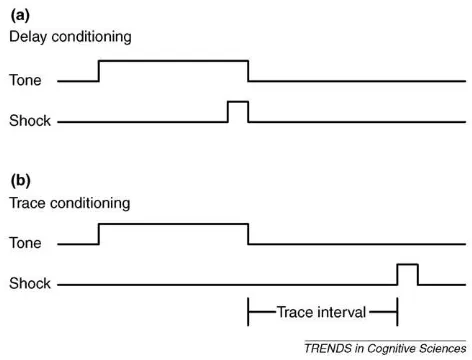
Another promising paradigm for examining emotion experience comes from tone-airpuff eyelid conditioning, where the conscious awareness of the conditioned stimulus–unconditioned stimulus (CS–US) contingency depends on their precise temporal relation-ship[40]; when the US and CS are separated by a short temporal gap (trace conditioning), the degree of conditioning is correlated with subjects’ awareness of the contingency, whereas when they overlap (delay conditioning), the degree of conditioning is indepen-dent of awareness (but also see Ref.[61]) (Figure Iin this box).
Fear conditioning with electrical shock is another paradigm for investigating the neural correlates of emotion experience. Investiga-tions using fear conditioning in humans[62,63]might establish the experiential aspects of trace conditioning, whereas investigations in rodents could offer a model system for using molecular biological techniques to transiently silence or activate specific, targeted neuronal populations in awake animals[39,64].
Figure I. Delay and trace conditioning. In classical conditioning, a previously neutral stimulus (CS), such as a tone, is repeatedly paired with an aversive stimulus (US), such as an airpuff to the eyelid or an electrical shock.(a)In delay conditioning, the CS co-terminates with the US.(b)In trace conditioning, the CS is followed by the US after a time interval. Human subjects can condition without becoming aware of the CS–US contingency in delay conditioning, but they need to become aware of the relationship in trace conditioning.
Opinion TRENDS in Cognitive Sciences Vol.xxx No.x 5
TICS-565; No of Pages 10
necessary for emotion experience, two ideas that have been widely criticized (see Ref.[7]for a review of the history of emotion theory and research). The problem with assuming that emotional response must precede emotion experience is that it inverts our intuitive causal order of these com-ponents; the problem with assuming that emotional response is necessary for emotion experience is that there seem to be cases of emotion experience in the absence of a physiological emotional response.
In many ways, the issue of causal order is similar to debates about dual-process models, such as classic argu-ments about the primacy of emotion versus cognition. One answer to these debates is to acknowledge that both kinds of process contribute at different points in time; both components overlap in time and each can influence the other. There can be rapid emotional responses, but that does not mean they terminate rapidly, nor does it preclude later emotion experience from modulating the responses
[50–52](Figure 4).
The issue of whether emotional response is required for emotion experience has focused on cases where either the motor component of emotional response or the perception of the emotional response (or both) is compromised. Patients who have locked-in syndrome due to brainstem lesions[53], complete spinal cord transections[26,27]and peripheral autonomic failure[18,28]seem to have largely normal (certainly not absent) emotion experience. Even though the motor responses and bodily sensations that would normally accompany emotion experience in these patients are severely compromised, they still have feelings. One reason these examples fall short of testing James’ ideas is that they are incomplete deafferentations of brain from body. Residual inputs from the vagus nerve, or from the cranial nerves innervating the face, might be sufficient to sustain largely normal emotion experience[3,54]. Another reason they fall short (one that James could not have imagined) is that central representations of body-state changes, even in the absence of contemporaneous bodily input, might suffice for emotion experience[3]– provided that there was normal emotion experience before the onset of the lesion that was sufficient to establish those central representations[28](see also red areas inFigure 2).
Is consciousness essential to emotion?
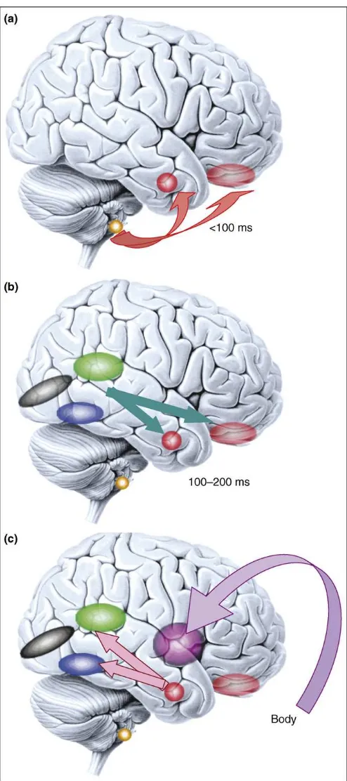
As we argued, feelings might have their basis in representations of the body, but we have no conscious access to most of the detailed neuronal processes that underlie emotion states and bodily homeostasis. The typical strategy to isolate the neural correlates of the contents of conscious experience is to contrast conscious and non-conscious pro-cessing of stimuli (Box 2), a strategy that has also yielded some purchase in the case of emotion experience. Fear conditioning to subliminal stimuli is a good example in which emotional responses occur without awareness of their triggering stimuli[55]. The presumed pathway that Figure 4. Microgenesis of emotional processing. Emotional responses span a
large temporal range (from 100 ms or less, to minutes).(a)Responses to emotional visual stimuli can occur rapidly in prefrontal cortex[50]or amygdala, in part mediated by subcortical inputs. Emotional response in the amygdala also influences early visual processing [51] and is modulated by volitional self-regulation[47,52].(b)At later time slices (100–200 ms), sensory cortices provide more detailed input to emotion-inducing structures like the amygdala. Two components that are important to face processing are shown: the superior temporal cortex (green), important for encoding dynamic information such as facial expression, and the fusiform gyrus (blue), important for encoding static information such as identity.(c)Once the emotional meaning of a stimulus has been evaluated by the brain, emotional responses are triggered in the body via projections from amygdala and medial prefrontal cortex to brainstem nuclei and hypothalamus (not shown), and are in turn represented in structures such as the insula. This figure emphasizes that what we refer to as an ‘emotion state’
throughout this article is in fact a complex set of processes that unfold at various points in time. Color key: black, primary visual cortex; blue, fusiform gyrus; green, superior temporal cortex; purple, insula; faint red, orbitofrontal cortex; solid red, amygdala; yellow, superior colliculus. Reproduced, with permission, from Ref.
[73].
Box 2. How to measure non-conscious processing
We restrict the term ‘unconscious’ to absence of the state of consciousness. When we are not awake or not dreaming, we are in a state of unconsciousness, which itself can be graded (ranging from dreamless normal sleep to persistent vegetative state, to deep anesthesia and coma). This use is distinct from the Freudian ‘unconscious’, which refers to contents of consciousness that cannot be accessed. We reserve the terms ‘non-conscious’ and ‘unaware’ for discussion of the contents of consciousness: when otherwise awake and conscious subjects are unable to report perceiving a stimulus, they are ‘unaware’ of the stimulus; when they are either unaware or when there is in fact no phenomenal conscious experience of that stimulus, the stimulus is processed ‘non-consciously’.
The most lenient criterion is to accept what subjects verbally report after an experiment. Although widely used (such as when obtaining reports after an fMRI session), this method is unsatisfactory because unattended items or task-irrelevant (implicit) features of stimuli might be inaccessible in subsequent recognition or recall tasks. Although attention and consciousness are usually tightly coupled, they are two distinctive processes [65]: when stimuli are presented foveally without any competing stimuli in the visual field, attention does not seem to contribute noticeably to visual awareness. A more stringent criterion for non-conscious processing is to ask subjects about their experience directly at the time the stimulus is processed. When
subjects deny seeing stimuli in verbal reports or ratings, the stimuli are said to be subjectively non-conscious. Although many studies that involve non-conscious states adopt this convention, the definition suffers from the possibility of criterion shifts: for the same subjective experience of visibility, some subjects might deny seeing a stimulus whereas others might report seeing it because their criterion of what to count as ‘seen’ differs[66].
The strictest procedure is to demonstrate null sensitivity (in any overt behavioral measure). For example, subjects can be given two alternative intervals (or locations), one of which contains the stimulus while the other does not. If they are at chance in detecting or discriminating one from the other, they are (objectively) unaware of the stimulus (our use of ‘subjective’ and ‘objective’ here refers to the method used, not to the nature of the conscious experience, which is always subjective in terms of its phenomenology). Several debates in the literature spring from these different methods for establishing non-conscious processing[67,68]. Recent studies have shown substantial brain activation or emotional responses to such objectively invisible stimuli (Figure Iin this box)[19]. Note that the method does not work in converse: above-chance behavioral discrimination does not necessarily demonstrate conscious aware-ness because patients who have blindsight would exhibit precisely such performance.
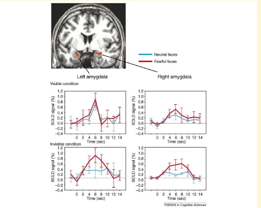
Figure I. Amygdala responses to invisible fearful and neutral faces. Bilateral amygdala responses to fearful faces (red) are independent of objective visibility, whereas the responses to neutral faces (blue) are modulated by visibility. During processing of stimuli that are not consciously perceived, the amygdala might couple with superior temporal sulcus[19]. Modified, with permission, from Ref.[19].
Opinion TRENDS in Cognitive Sciences Vol.xxx No.x 7
TICS-565; No of Pages 10
mediates this effect involves the superior colliculus, pulvinar thalamus and amygdala, which bypasses standard neocortical processing routes that are thought to be necess-ary for conscious detection, discrimination and identifi-cation of the stimulus [56]. Neuroimaging studies have found reliable activation in the amygdala (as well as fusi-form gyrus and superior temporal sulcus) in response to emotional visual stimuli of which subjects were completely unaware, using stringent psychophysical criteria to ensure lack of any access consciousness[19](Figure Iin Box 2). Relatedly, patients who have blindsight show modulation of amygdala activation on the basis of the emotional meaning of stimuli that they cannot consciously see[57]. Thus, as is the case for sensory stimuli more generally, there can be some degree of processing, and some neural responses, to emotionally salient stimuli that are presented under con-ditions where they are not consciously perceived. Non-con-scious stimuli can evoke emotion states.
It is not surprising to find that stimuli can, under certain circumstances, fail to lead to conscious experience, even though they can trigger emotional responses and even though they can modulate regional neural activation. But can there be a failure to experience the emotional responses themselves? Here, too, if they are sufficiently weak, the dissociation seems possible. Autonomic responses occur to subliminally presented stimuli, as in the case of subliminal fear conditioning described earlier, and subtle expressions on our own face occur in response to seeing others’ emotional expressions subliminally[58]and, in both cases, subjects can be unaware of their own emotional response.
But can emotion states that are not experienced at all still motivate behavior in the more general and flexible way that we suggested earlier was a hallmark of consciousness? One study[59]found that subliminally presented happy or angry faces could influence subsequent drinking behavior. Thirsty subjects who were shown the happy faces con-sumed twice as much beverage and rated its pleasantness higher than subjects who were shown neutral or angry faces. Importantly, presentation of the masked faces did not influence subjective ratings of the subjects’ feelings, so at least access consciousness of the induced emotion state was absent. In this case, non-conscious stimuli influenced motivation without affecting background feeling.
Another finding of interest came from a patient who had extensive bilateral lesions of the insula [29]and lost the ability to perceive taste. When presented with solutions of sugar, saline or lime juice, he described all of them as tasting ‘like pop’ and drank them indiscriminately. But when given a choice between contemporaneously pre-sented beverages, he showed a strong preference for the sugar solution. Despite no apparent conscious experience of the unpleasant salty or sour taste, nor any apparent emotional responses to it, he had strong motivational preferences based on the affective value of the taste when provided with the opportunity for a comparison. These last two examples show that behavior can be motivated by the affective value of stimuli that are not consciously perceived and that do not induce any conscious feelings of the emotion. Alternatively, these might be cases of phenom-enal consciousness without access consciousness.
Concluding remarks
What is the relationship between emotion and
consciousness? We have reviewed recent studies that address this question, by considering a role of brain
Box 3. Outstanding questions
One could examine across development and across phylogeny
the correlation between an elaborated self-representation and the capacity for a rich conscious experience. Do all invertebrates have explicit central interoceptive representations? Can this criterion be used to determine which species might be capable of conscious experience?
What is the relationship of moods (temporally extended
emo-tions) to consciousness? Neuronal activity during sleep, under anesthesia and during wakefulness has been studied at the neurochemical, electrophysiological and computational level. Positive or negative mood alters the distribution of neurochemical modulators. What is the electrophysiological consequence of this? Can one model the effects of mood in computational models of consciousness?
What is the role of language and symbolic thought in emotion
experience? The cognitive interpretation of a situation influences the emotion experience, but how and at what stage of processing? Is there an aspect of emotion experience that is relatively independent of thought and reflection, and an aspect that depends on it?
Are states, like thirst, an emotion? Our treatment here suggests
that states that are normally considered emotions, like anger and fear, share much in common with states that are normally not considered emotions, like thirst, hunger and pain. We believe that the evidence to date supports a broader concept of ‘emotion’ that links it to interoception on the one hand, and to motivated behavior on the other. But we also feel that this view leaves room for domains within such a broad notion of emotion, of Ekman-like ‘basic’ emotions, and of social–moral emotions, for example. What distinguishes social emotions, basic emotions and states like thirst and hunger seem like tractable questions that can be investigated at psychological and neurobiological levels.
In visual neuroscience, illusions have proved to be powerful tools
for dissociating particular content of consciousness under con-stant stimulus input (e.g. the Necker Cube, Rubin’s vase or binocular rivalry). Are there such illusions for emotion experi-ence? There are already some examples along these lines: choice blindness[69], the rubber-hand illusion[20], paradoxical heat[70], placebo effects[21]and hypnosis. These might be useful to add to the toolkit of affective neuroscience.
Is the amygdala necessary for feeling fear? Although one study
found that amygdala lesions do not alter self-report measures of experienced fear[71], detailed interview-based assessment sug-gests that, whereas patients might endorse largely normal concepts of fear experience, their actual behavior and experience are more consistent with an absence of experienced fear[30]. The amygdala might be important for attentional selection that is normally crucial for subsequent experiences of fear. To determine whether or not the amygdala participates directly in the neural correlate of fear experiences, further experiments are required: measuring neuronal activity in the amygdala while manipulating subjective fear experience or electrically stimulating the amygdala to characterize the causal relationship between amygdala activity and fear experience.
Charles Darwin described in detail the aspects of emotional
expression that serve a social communicative function, and phenomena such as emotional contagion and empathy demon-strate that emotional expression in others can induce emotions in ourselves. This topic has received considerable attention from simulation theories and from discoveries of mirror neurons and mirror systems in the brain. Should a broader conception of an emotion state, or even an emotion experience, be transpersonal and include more than one brain? Could a broader conception of consciousness be similarly distributed across multiple individuals within a group or society? These rather radical suggestions could be exciting future directions for social neuroscience[33].
structures that regulate both emotion state and the level of consciousness, by considering aspects of emotion that were independent of conscious awareness and by discussing what is known about the neural correlates of emotion and consciousness. In addition to arousal mechanisms in the brainstem and thalamus, emotional processing in cin-gulate cortices and other midline structures might be important for maintaining a sense of ownership, which might be necessary for any conscious experience.
If basic emotion processing is necessary for
consciousness, severe impairment in basic emotion pro-cesses should lead to compromised consciousness. A more neuroanatomically specific hypothesis would be that alteration to structures that represent physiological changes in one’s own body would alter or destroy conscious experience. Future experiments in animals might selec-tively lesion (perhaps reversibly) or electrically stimulate specific structures that are implicated in such processing: midline cortices, the insula, parts of the thalamus and interoceptive and homeostatic nuclei in the brainstem.
(SeeBox 3for Outstanding questions.)
We have suggested that interoceptive representations of changes in body state, together with motivations to behave in certain ways, underlie the content of emotion experi-ence, but this is not typically how they are experienced as emotions. Rather, in virtue of these interoceptive and premotor representations, we become aware of ourselves as subjects situated in a given circumstance (often social) and as agents who have the capacity to influence the circumstances and cope with the challenges that they pose
[5]. Thus, we see more psychological theories of emotion[2]
and more biological ones [3,4] as commensurate and suggest that different patterns of somatic and premotor representations give each emotion its unique flavor. Finally, we acknowledge that thoughts about and reflec-tions on the emotion influence how it is experienced (at least in humans), although this is a component beyond the scope of the present article.
Acknowledgements
We thank Christof Koch and three reviewers for helpful comments on the manuscript. This article is supported in part by funding from the Gordon and Betty Moore Foundation and the National Institutes of Health.
References
1 Russell, J.A. (2003) Core affect and the psychological construction of emotion.Psychol. Rev.110, 145–172
2 Lambie, J.A. and Marcel, A.J. (2002) Consciousness and the varieties of emotion experience: a theoretical framework.Psychol. Rev.109, 219– 259
3 Damasio, A.R. (1999)The Feeling of What Happens: Body and Emotion in the Making of Consciousness,Harcourt Brace
4 Panksepp, J. (2005) Affective consciousness: core emotional feelings in animals and humans.Conscious. Cogn.14, 30–80
5 Prinz, J. (2004)Gut Reactions,Oxford University Press 6 Rolls, E.T. (2005)Emotion Explained,Oxford University Press 7 Dalgleish, T. (2004) The emotional brain.Nat. Rev. Neurosci.5, 583–
589
8 Baars, B.J.et al.(2003) Brain, conscious experience and the observing self.Trends Neurosci.26, 671–675
9 Block, N. (2005) Two neural correlates of consciousness.Trends Cogn. Sci.9, 46–52
10 Koch, C. (2004) The Quest for Consciousness: A Neurobiological Approach,Roberts and Publishers
11 Chalmers, D.J. (1996) The Conscious Mind: In Search of a Fundamental Theory,Oxford University Press
12 Zeman, A. (2001) Consciousness.Brain124, 1263–1289
13 Critchley, H.D.et al.(2004) Neural systems supporting interoceptive awareness.Nat. Neurosci.7, 189–195
14 Raichle, M.E. and Gusnard, D.A. (2005) Intrinsic brain activity sets the stage for expression of motivated behavior.J. Comp. Neurol.493, 167– 176
15 Liotti, M.et al.(2001) Brain responses associated with consciousness of breathlessness (air hunger).Proc. Natl. Acad. Sci. U.S.A.98, 2035– 2040
16 Lane, R.D. et al. (1998) Neural correlates of levels of emotional awareness: evidence of an interaction between emotion and attention in the anterior cingulate cortex.J. Cogn. Neurosci.10, 525–535 17 Damasio, A.R.et al.(2000) Feeling emotions: subcortical and cortical
brain activity during the experience of self-generated emotions.Nat. Neurosci.3, 1049–1056
18 Critchley, H.D. et al. (2001) Neuroanatomical basis for first- and second-order representations of bodily states.Nat. Neurosci.4, 207– 212
19 Jiang, Y. and He, S. (2006) Cortical responses to invisible faces: dissociating subsystems for facial-information processing.Curr. Biol.
16, 2023–2029
20 Ehrsson, H.H.et al.(2004) That’s my hand! Activity in premotor cortex reflects feeling of ownership of a limb.Science305, 875–877 21 Petrovic, P.et al.(2002) Placebo and opioid analgesia – imaging a
shared neuronal network.Science295, 1737–1740
22 D’Argembeau, A.et al.(2005) Self-referential reflective activity and its relationship with rest: a PET study.Neuroimage25, 616–624 23 Wager, T.D. et al. (2003) Valence, gender, and lateralization of
functional brain anatomy in emotion: a meta-analysis of findings from neuroimaging.Neuroimage19, 513–531
24 Berthoz, S.et al.(2002) Effect of impaired recognition and expression of emotions on frontocingulate cortices: an fMRI study of men with alexithymia.Am. J. Psychiatry159, 961–967
25 Parvizi, J.et al.(2001) Pathological laughter and crying. A link to the cerebellum.Brain124, 1708–1719
26 Cobos, P.et al.(2004) Effects of spinal cord injuries on the subjective component of emotions.Cogn. Emot.18, 281–287
27 Nicotra, A.et al. (2006) Emotional and autonomic consequences of spinal cord injury explored using functional brain imaging.Brain129, 718–728
28 Heims, H.C.et al.(2004) Social and motivational functioning is not critically dependent on feedback of autonomic responses: neuro-psychological evidence from patients with pure autonomic failure.
Neuropsychologia42, 1979–1988
29 Adolphs, R.et al.(2005) Preferring one taste over another without recognizing either.Nat. Neurosci.8, 860–861
30 Tranel, D.et al.(2006) Altered experience of emotion following bilateral amygdala damage.Cogn. Neuropsychiatry11, 219–232
31 Owen, A.M.et al.(2006) Detecting awareness in the vegetative state.
Science313, 1402
32 Russell, J.A. (1991) Culture and the categorization of emotions.
Psychol. Bull.110, 426–450
33 Adolphs, R. Consciousness: situated and social. In Cambridge Handbook of Consciousness (Zelazo, P. and Moscovitch, M., eds), Cambridge University Press (in press)
34 Bernat, J.L. (2006) Chronic disorders of consciousness.Lancet367, 1181–1192
35 Parvizi, J.et al.(2006) Neural connections of the posteromedial cortex in the macaque.Proc. Natl. Acad. Sci. U.S.A.103, 1563–1568 36 Laureys, S. (2005) The neural correlate of (un)awareness: lessons from
the vegetative state.Trends Cogn. Sci.9, 556–559
37 Fox, M.D.et al.(2005) The human brain is intrinsically organized into dynamic, anticorrelated functional networks.Proc. Natl. Acad. Sci. U.S.A.102, 9673–9678
38 Vogt, B.A. (2005) Pain and emotion interactions in subregions of the cingulate gyrus.Nat. Rev. Neurosci.6, 533–544
39 Han, C.J.et al.(2003) Trace but not delay fear conditioning requires attention and the anterior cingulate cortex. Proc. Natl. Acad. Sci. U.S.A.100, 13087–13092
40 Clark, R.E.et al.(2002) Classical conditioning, awareness, and brain systems.Trends Cogn. Sci.6, 524–531
Opinion TRENDS in Cognitive Sciences Vol.xxx No.x 9
TICS-565; No of Pages 10
41 Maddock, R.J. (1999) The retrosplenial cortex and emotion: new insights from functional neuroimaging of the human brain.Trends Neurosci.22, 310–316
42 Damasio, A. (2003) Feelings of emotion and the self.Ann. N. Y. Acad. Sci.1001, 253–261
43 Schachter, S. and Singer, J.E. (1962) Cognitive, social, and physiological determinants of emotional state.Psychol. Rev.69, 379–399
44 Fried, I.et al.(1998) Electric current stimulates laughter.Nature391, 650
45 Bejjani, B.P.et al.(1999) Transient acute depression induced by high-frequency deep-brain stimulation.N. Engl. J. Med.340, 1476–1480 46 Craig, A.D. (2002) How do you feel? Interoception: the sense of the
physiological condition of the body.Nat. Rev. Neurosci.3, 655–666 47 Ochsner, K.N. and Gross, J.J. (2005) The cognitive control of emotion.
Trends Cogn. Sci.9, 242–249
48 Lane, R.D.et al.(1997) Is alexithymia the emotional equivalent of blindsight?Biol. Psychiatry42, 834–844
49 James, W. (1884) What is an emotion?Mind9, 188–205
50 Kawasaki, H.et al.(2001) Single-neuron responses to emotional visual stimuli recorded in human ventral prefrontal cortex.Nat. Neurosci.4, 15–16
51 Vuilleumier, P.et al.(2004) Distant influences of amygdala lesion on visual cortical activation during emotional face processing. Nat. Neurosci.7, 1271–1278
52 Schaefer, S.M.et al.(2002) Modulation of amygdalar activity by the conscious regulation of negative emotion.J. Cogn. Neurosci.14, 913– 921
53 Bauby, J.D. (1997)The Diving Bell and the Butterfly,Knopf 54 Bechara, A. (2004) The role of emotion in decision-making: evidence
from neurological patients with orbitofrontal damage.Brain Cogn.55, 30–40
55 Wong, P.S.et al.(2004) Event-related brain correlates of associative learning without awareness.Int. J. Psychophysiol.53, 217–231 56 Johnson, M.H. (2005) Subcortical face processing.Nat. Rev. Neurosci.
6, 766–774
57 Morris, J.S.et al.(2001) Differential extrageniculostriate and amygdala responses to presentation of emotional faces in a cortically blind field.
Brain124, 1241–1252
58 Dimberg, U.et al.(2000) Unconscious facial reactions to emotional facial expressions.Psychol. Sci.11, 86–89
59 Winkielman, P. and Berridge, K.C. (2004) Unconscious emotions.Curr. Dir. Psychol. Sci.13, 120–123
60 Berridge, K.C. (2000) Measuring hedonic impact in animals and infants: microstructure of affective taste reactivity patterns.
Neurosci. Biobehav. Rev.24, 173–198
61 Lovibond, P.F. and Shanks, D.R. (2002) The role of awareness in Pavlovian conditioning: empirical evidence and theoretical implications.J. Exp. Psychol. Anim. Behav. Process.28, 3–26 62 Carter, R.M.et al.(2003) Working memory and fear conditioning.Proc.
Natl. Acad. Sci. U.S.A.100, 1399–1404
63 Knight, D.C.et al.(2004) Neural substrates mediating human delay and trace fear conditioning.J. Neurosci.24, 218–228
64 Slimko, E.M.et al.(2002) Selective electrical silencing of mammalian neurons in vitro by the use of invertebrate ligand-gated chloride channels.J. Neurosci.22, 7373–7379
65 Koch, C. and Tsuchiya, N. (2007) Attention and consciousness: two distinct brain processes.Trends Cogn. Sci.11, 16–22
66 Kunimoto, C.et al.(2001) Confidence and accuracy of near-threshold discrimination responses.Conscious. Cogn.10, 294–340
67 Whalen, P.J.et al.(2004) Human amygdala responsivity to masked fearful eye whites.Science306, 2061
68 Pessoa, L.et al.(2006) Target visibility and visual awareness modulate amygdala responses to fearful faces.Cereb. Cortex16, 366–375 69 Johansson, P. et al.(2005) Failure to detect mismatches between
intention and outcome in a simple decision task.Science310, 116–119 70 Davis, K.D. et al. (2004) Perceptual illusion of ‘paradoxical heat’
engages the insular cortex.J. Neurophysiol.92, 1248–1251
71 Anderson, A.K. and Phelps, E.A. (2002) Is the human amygdala critical for the subjective experience of emotion? Evidence of intact dispositional affect in patients with amygdala lesions. J. Cogn. Neurosci.14, 709–720
72 Craig, A.D. (2003) Interoception: the sense of the physiological condition of the body.Curr Opin. Neurobiol.13, 500–505
73 Adolphs, R. (2002) Recognizing emotion from facial expressions: psychological and neurological mechanisms.Behav. Cogn. Neurosci. Rev.1, 21–61
![Figure 1. Defining components of emotion and consciousness. (a) We distinguish an emotion state from the emotion experience (feeling), although they are not mutuallyexclusive [2]](https://thumb-ap.123doks.com/thumbv2/123dok/3175730.1388606/2.612.52.569.49.446/figure-defining-components-emotion-consciousness-distinguish-experience-mutuallyexclusive.webp)